
Abstract
Purpose
To report the effects of aflibercept on eyes with large retinal pigment epithelial detachment (PED) associted with polypoidal choroidal vasculopathy (PCV).
Methods
We reviewed the medical records of patients with PEDs associated with PCV that were treated with aflibercept after intravitreal ranibizumab had failed.
Results
Three eyes of patients aged 72, 79, and 80 years were studied. Reflective material was seen in the PED along the outer surface of the retinal pigment epithelium (RPE) by spectral-domain optical coherence tomography (SD-OCT). A complete resolution of the serous PEDs was found after two aflibercept injections; however, all eyes had a fibrovascular PED. In addition, one eye developed a retinal hemorrhage and a recurrent PED just after the third injection of aflibercept. The visual acuity in this eye decreased from 10/20 to 2/20.
Conclusion
The reflective material below the outer surface of the RPE in serous PED suggests the presence of neovascularization. Intravitreal aflibercept could be considered for large PEDs in eyes with PCV but should be carefully applied.
Introduction
Intravitreal injections of pegaptanib or bevacizumab have been reported to be efficacious and safe treatments for retinal pigment epithelial detachments (PEDs) associated with occult choroidal neovascularization (CNV) secondary to age-related macular degeneration (AMD).Citation1 Aflibercept, a recombinant fusion protein, has a higher binding affinity for vascular endothelial growth factor (VEGF), and it has been shown to be beneficial for patients with AMD who were refractory to multiple injections of either bevacizumab or ranibizumab.Citation2,Citation3 In addition, a recent study showed that intravitreal aflibercept may be an effective treatment for serous PEDs in which bevacizumab and ranibizumab were not effective.Citation4
We report three cases of polypoidal choroidal vasculopathy (PCV) with a large PED that was refractory to ranibizumab but responded to intravitreal aflibercept. We present our spectral domain optical coherence tomographic (SD-OCT) findings, which provided some evidence on the development of the PED.
Case reports
Case 1
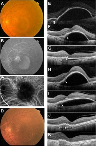
A 79-year-old man presented with a large serous PED. Fluorescein angiography (FA) displayed an occult CNV, and indocyanine green (IA) images showed a PCV. At the initial visit, his best-corrected visual acuity (BCVA) was 2/20 oculus sinister (OS). Three monthly injections of ranibizumab slightly flattened the PED and reduced the subretinal fluid (SRF), but a recurrence developed. Three additional monthly injections of ranibizumab (total of six injections) did not improve the PED, and there was an increase of the highly reflective material beneath the outer surface of the RPE in the SD-OCT images.
We switched to aflibercept and, after one intravitreal injection, the PED was slightly flattened; two additional injections of aflibercept were given, which flattened the PED over the hyper-reflective material observed by SD-OCT. The BCVA remained at 2/20 OS ().
Figure 1 Findings from case 1, a 79-year-old Japanese man with a large PED in the left eye with PCV.
Abbreviations: BCVA, best-corrected visual acuity; CNV, choroidal neovascularization; FA, fluorescein angiography; IA, indocyanine green; OS, oculus sinister; PCV, polypoidal choroidal vasculopathy; PED, pigment epithelial detachment; RPE, retinal pigment epithelium; SD-OCT, spectral domain optical coherence tomographic; SRF, subretinal fluid.
Case 2
A 72-year-old man presented with a large serous PED in his right eye associated with a PCV. His BCVA was 14/20 OD. He had received six ranibizumab injections over a 10-month period, but the PED worsened and the SRF recurred. Hyper-reflective materials were seen beneath the outer surface of the RPE in the SD-OCT images. The BCVA decreased to 8/20 OD.
Two monthly injections of aflibercept resulted in a complete resolution of the serous PED; however, the flattened PED contained fibrovascular material beneath the RPE layer in the SD-OCT images.
Case 3
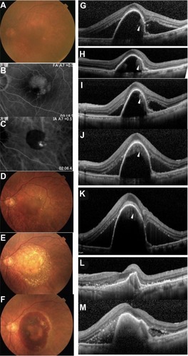
An 80-year-old man presented with a large serous PED. FA displayed an occult CNV, and IA images showed a PCV. His BCVA was 10/20 OS. Three monthly injections of ranibizumab slightly flattened the PED, but a recurrence was observed after 3 months. Three additional monthly injections of ranibizumab (total of six injections) failed to reduce the PED, and hyper-reflective materials were seen beneath the outer surface of the RPE in the SD-OCT images. We switched to aflibercept, and after the two additional monthly injections, an exudative CNV lesion was observed instead of the serous PED. OCT showed a loss of optical free space and highly reflective materials, suggesting the formation of fibrovascular PED. One day after the third monthly injection of aflibercept, a subretinal hemorrhage and recurrent PED were observed. BCVA was reduced to 2/20 ().
Figure 2 Images from case 3, an 80-year-old Japanese man with a large PED associated with a PCV in the left eye.
Abbreviations: BCVA, best-corrected visual acuity; CNV, choroidal neovascularization; FA, fluorescein angiography; IA, indocyanine green; OS, oculus sinister; PCV, polypoidal choroidal vasculopathy; PED, pigment epithelial detachment; RPE, retinal pigment epithelium; SD-OCT, spectral domain optical coherence tomographic; SRF, subretinal fluid.
Discussion
We found from three cases that intravitreal injections of aflibercept reduced or flattened large serous PEDs associated with PCV that were refractory to ranibizumab. Bird and MarshallCitation5 and Casswell et alCitation6 proposed that an interaction between the RPE and the Bruch membrane was central to the development of a PED, and they were not mutually exclusive.Citation7 Alternatively, GassCitation8 proposed that PEDs in eyes with AMD occurred by neovascular ingrowth with subsequent exudation from the new vessels.
It is well known that PCVs frequently accompany PEDs.Citation9 We found highly reflective materials within the serous PED beneath the outer surface of the RPE in all three cases. After aflibercept treatment, all of our cases had an apparent contracture of the accumulated material. This suggested the presence of fibrovascular tissues within the flattened PED. This is supported by the findings of Spaide,Citation10 who examined the internal structure of PEDs in eyes with AMD and found that PEDs frequently have reflective materials on the back surface of the RPE, suggestive of CNV. These findings can be explained by the hypothesis presented by GassCitation8 on PED formation.
We found a sudden subretinal hemorrhage and recurrent PED in case 3. Following anti-VEGF therapy, a contracture of fibrovascular tissue has been reported in eyes with proliferative diabetic retinopathy.Citation11 We suggest that aflibercept therapy may have enhanced fibrosis of neovascular tissues and caused a traction of the vessels, which then resulted in a sudden subretinal hemorrhage and a recurrence of the PED.
In conclusion, the reflective material below the outer surface of the RPE in serous PEDs suggests the presence of neovascularization. Intravitreal aflibercept could be considered for large PEDs in eyes with PCV but should be carefully applied.
Disclosure
The authors have no conflict of interest to declare in this work.
References
- AriasLTreatment of retinal pigment epithelial detachment with antiangiogenic therapyClin Ophthalmol2010436937420463807
- BakallBFolkJCBoldtHCAflibercept therapy for exudative age-related macular degeneration resistant to bevacizumab and ranibizumabAm J Ophthalmol20131561152223706500
- ChoHShahCPWeberMHeierJSAflibercept for exudative AMD with persistent fluid on ranibizumab and/or bevacizumabBr J Ophthalmol20139781032103523766432
- PatelKHChowCCRathodRRapid response of retinal pigment epithelial detachments to intravitreal aflibercept in neovascular age-related macular degeneration refractory to bevacizumab and ranibizumabEye (Lond)201327566366723558214
- BirdACMarshallJRetinal pigment epithelial detachments in the elderlyTrans Ophthalmol Soc U K1986105Pt 66746823310342
- CasswellAGKohenDBirdACRetinal pigment epithelial detachments in the elderly: classification and outcomeBr J Ophthalmol19856963974032408659
- SingermanLJStockfishJHNatural history of subfoveal pigment epithelial detachments associated with subfoveal or unidentifiable choroidal neovascularization complicating age-related macular degenerationGraefes Arch Clin Exp Ophthalmol198922765015072483142
- GassJDDrusen and disciform macular detachment and degenerationTrans Am Ophthalmol Soc1972704094364663679
- SaitoMIidaTNagayamaDCross-sectional and en face optical coherence tomographic features of polypoidal choroidal vasculopathyRetina200828345946418327139
- SpaideRFEnhanced depth imaging optical coherence tomography of retinal pigment epithelial detachment in age-related macular degenerationAm J Ophthalmol2009147464465219152869
- ArevaloJFMaiaMFlynnHWJrTractional retinal detachment following intravitreal bevacizumab (Avastin) in patients with severe proliferative diabetic retinopathyBr J Ophthalmol200892221321617965108