
Abstract
Oculocutaneous albinism is a group of congenital disorders caused by alterations of melanin biosynthesis. We report our findings in a patient with oculocutaneous albinism who presented with foveal hypoplasia and a foveal hemorrhage. A 48-year-old man noted a dark spot in the middle of the visual field of his right eye. He had depigmented skin, white hair, white eyebrows, and white cilia. He also had horizontal nystagmus and depigmented irides. His best-corrected visual acuity was 2/100 with −14.0 diopters in the right eye and 3/100 with −5.0 diopters in the left eye. Ophthalmoscopy showed diffuse depigmentation in both eyes and a foveal hemorrhage in the right eye. Optical coherence tomography showed the absence of a foveal pit in both eyes and a subretinal hyperreflective lesion corresponding to the foveal hemorrhage in the right eye. Fluorescein angiography showed that the retinal and choroidal vessels were relatively hypofluorescent because of the lack of a blocking effect of the pigments in the retinal pigment epithelium. Fluorescein angiography and indocyanine green angiography did not show any evidence of choroidal neovascularization in either eye. The foveal hemorrhage in the right eye spontaneously regressed and finally resolved at 3 months after onset. At the final examination, the patient reported that his vision had recovered. A foveal hemorrhage is a rare condition in an eye with foveal hypoplasia associated with albinism. The hemorrhage may be related to high myopia and also to the hypoplasia of the fovea associated with albinism.
Introduction
Albinism is a group of congenital disorders in which melanin biosynthesis is altered, and it affects approximately one in 4,000 people.Citation1 The condition can be broadly divided into two categories, ie, oculocutaneous albinism and ocular albinism. In oculocutaneous albinism, there is a reduction or absence of melanin in the hair, skin, and eyes, whereas in ocular albinism, the melanin deficiency is limited to the eyes.Citation2
The ocular manifestations associated with albinism include nystagmus, depigmented irides, foveal hypoplasia, macular transparency, strabismus, and high myopia.Citation3 However, there has been only one report of a foveal hemorrhage in eyes with albinism.Citation4 We report our findings in a patient with oculocutaneous albinism who had a foveal hemorrhage in an eye with foveal hypoplasia.
Case report
A 48-year-old man consulted our hospital because he had noted a dark spot in the middle of the visual field of his right eye of 2 weeks’ duration. Our examination showed that he had depigmented skin, white hair, white eyebrows, and white cilia (). He had horizontal nystagmus and depigmented irides. His best-corrected visual acuity was 2/100 with −14.0 diopters in the right eye and 3/100 with −5.0 diopters in the left eye. Ophthalmoscopy showed diffuse depigmentation in both eyes (). He was diagnosed with oculocutaneous albinism based on the systemic and ocular findings.
Figure 1 Findings in a 48-year-old Japanese man with oculocutaneous albinism. His best-corrected visual acuity was 2/100 with −14.0 diopters in the right eye.
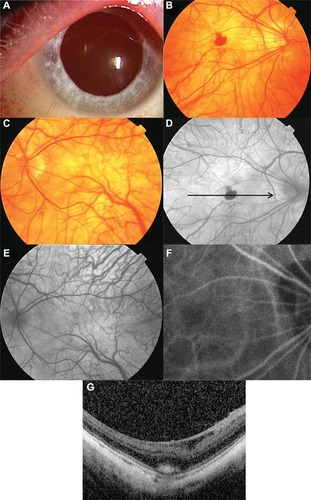
Ophthalmoscopy showed that his right eye had a foveal hemorrhage. Fluorescein angiography (FA) showed relatively hypofluorescent retinal and choroidal vessels because of the lack of a blocking effect by pigments in the retinal pigment epithelium (). FA also showed an area of hypofluorescence in the right fovea that corresponded with the foveal hemorrhage (). FA and indocyanine green angiography (IA, ) did not show any evidence of choroidal neovascularization (CNV) in either eye. We could not obtain good quality optical coherence tomographic images because of the nystagmus in both eyes. However, the blurred optical coherence tomographic images showed an absence of the foveal pit in both eyes and the presence of a subretinal hyperreflective lesion corresponding to the foveal hemorrhage in the right eye ().
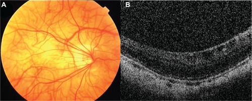
The foveal hemorrhage in the right eye spontaneously regressed and finally resolved at 3 months after onset (). The best-corrected visual acuity of his right eye did not change after resolution of the hemorrhage, but he reported that his vision was better.
Figure 2 Three months after onset. The patient’s best-corrected visual acuity has not improved.
Discussion
A foveal hemorrhage in oculocutaneous albinism is relatively rare. Shinno et al reported a case of foveal hemorrhage in oculocutaneous albinism,Citation4 and suggested that the foveal hemorrhage was due to CNV associated with high myopia. They came to this conclusion because FA and IA showed a hyperfluorescent lesion corresponding to CNV. However, in our case, FA and IA did not show CNV, although the patient had high myopia of −14.0 diopters accompanied by a staphyloma.
Oculocutaneous albinism is usually associated with foveal hypoplasia,Citation5 and we were able to demonstrate foveal hypoplasia by the absence of a foveal pit in the optical coherence tomographic images. It was interesting to detect a foveal hemorrhage in an eye with foveal hypoplasia. The anatomical structure of the fovea, Bruch’s membrane, and choriocapillaris in an eye with foveal hypoplasia may be different from that in normal eyes. However, a search of PubMed did not yield any previous publications mentioning the anatomical structure of the fovea in eyes with foveal hypoplasia.
Oculocutaneous albinism is often accompanied by high myopia. It is well known that a foveal hemorrhage in high myopic eyes without CNV is due to a rupture of Bruch’s membrane and the choriocapillaris complex. The incidence of a foveal hemorrhage has been reported to be approximately 3% in highly myopic eyes.Citation6 Our patient had a staphyloma with high myopia of −14.0 diopters, and this may have made the foveal tissues more fragile. Thus, we suggest that the foveal hemorrhage observed in our case was probably due to high myopia and it is likely that the foveal hypoplasia also contributed to the foveal hemorrhage.
In conclusion, a foveal hemorrhage is a rare condition in an eye with a foveal hypoplasia associated with albinism. The foveal hemorrhage in our case may be related to high myopia and also may be related to anatomical alterations associated with foveal hypoplasia.
Disclosure
The authors report no conflicts of interest in this work.
References
- MohammadSGottlobIKumarAThe functional significance of foveal abnormalities in albinism measured using spectral-domain optical coherence tomographyOphthalmology201111881645165221570122
- ChongGTFarsiuSFreedmanSFAbnormal foveal morphology in ocular albinism imaged with spectral-domain optical coherence tomographyArch Ophthalmol20091271374419139336
- RossiSTestaFGargiuloAThe role of optical coherence tomography in an atypical case of oculocutaneous albinism: a case reportCase Rep Ophthalmol20123111311722548044
- ShinnoMYumikoYShojiKChoroidal neovascularization in a case of oculocutaneous albinismRinsho Ganka200458712451249 Japanese
- ThomasMGKumarAMohammadSStructural grading of foveal hypoplasia using spectral-domain optical coherence tomography a predictor of visual acuity?Ophthalmology201111881653166021529956
- TokoroTExplanatory factors of chorioretinal atrophyTokoroTAtlas of Posterior Fundus Changes in Pathologic MyopiaTokyo, JapanSpringer Verlag1998