
Abstract
Endophthalmitis following pars plana vitrectomy is a very uncommon cause of endophthalmitis. Cases reported over the last decade show a decrease in incidence over time. To optimize visual outcome, early diagnosis and treatment are essential. In this review we report a summary of the incidence of endophthalmitis following vitrectomy, various risk factors for their occurrence, the microbiological profile and the visual outcomes post treatment.
Introduction
Endophthalmitis is characterized by severe inflammation of the ocular tissues and fluids. Acute-onset postoperative infectious endophthalmitis is the most frequent category and may be associated with severe visual loss. The incidence of endophthalmitis varies with the surgical procedure performed. The most reported categories of endophthalmitis are following cataract surgery/intraocular lens implantation and following intravitreal injections. A review of the literature indicates the incidence in these scenarios to vary from 0.07% to 0.4% for cataract surgery and from 0.038% to 0.065% for intravitreal injections.Citation1–Citation5 Endophthalmitis following pars plana vitrectomy (PPV) is an uncommon cause of endophthalmitis (). The reported rates of post-PPV endophthalmitis have been decreasing over the last decade. In this review, the incidence of post-PPV endophthalmitis, risk factors for occurrence, microbiological profile, and treatment outcomes in the sutureless PPV era are reported.
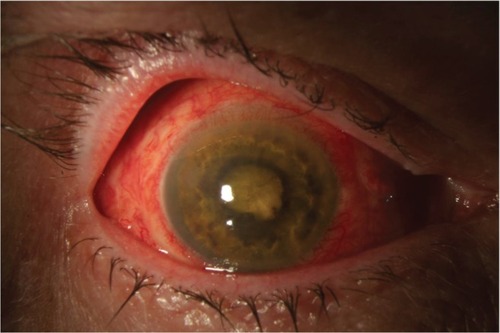
Figure 1 Anterior segment photograph of a patient with endophthalmitis following pars plana vitrectomy operated for rhegmatogenous retinal detachment. Cultures were positive for Bacillus cereus. Visual acuity was light perception after treatment.
Incidence
Endophthalmitis following PPV is relatively uncommon. Over the years, there have been various studies which reported incidences of endophthalmitis after PPV ().Citation6–Citation13 The incidence ranges between 0.03% and 0.14% for 20 G PPV. None of the cases had additional concurrent surgery.
Table 1 Incidence of endophthalmitis following pars plana vitrectomy in 20 G surgery
Incidence in transconjunctival sutureless vitrectomy
The first reported case of endophthalmitis in transconjunctival sutureless vitrectomy (TSV) surgery was in 2005 following a 25 G PPV ().Citation14 The vitreous sample was negative on Gram stain and culture. After intravitreal and oral antibiotics, vision increased from hand motion close to face to 6/6 at the 3-week follow-up visit.
Table 2 Incidence of endophthalmitis in transconjunctival sutureless vitrectomy
Older studies comparing 25 G with 20 G PPV generally reported higher rates in the 25 G group. Kunimoto and Kaiser compared endophthalmitis rates after 20 G and 25 G PPV in 2007 and reported 0.23% in 25 G cases as compared with 0.018% in 20 G cases, which was statistically significant.Citation15 Both the groups had final median visual acuity of counting fingers. Shaikh et al reviewed 129 consecutive cases of 20 G and 129 consecutive cases of 25G PPV in 2007 and reported two cases (1.55%) of endophthalmitis in the 25 G group and none in the 20 G group.Citation16 Chen et al in 2009 reported a nonsignificantly higher rate of endophthalmitis in 25 G (0.23%) compared with 20 G (0.03%) PPV.Citation17 Scott et al in a multicenter study 2008 reported a significantly higher rate of endophthalmitis in 25G TSV (0.84%) versus 20 G PPV (0.03%).Citation18 Of these, most cases (9/11) in the TSV group that developed endophthalmitis had straight incisions which may have been a possible risk factor.
However, other studies, and especially newer studies, have reported no significant difference in endophthalmitis rates between the two groups, perhaps reflecting advances in wound construction and improvements in surgical technique.Citation19–Citation21 There are also reports which show very rare occurrence of endophthalmitis post TSV.Citation22,Citation23
In a 2-year, prospective, nationwide surveillance study from the UK, the rate of endophthalmitis after PPV was 28 per 48,433 (0.058%). There were no statistically significant differences in the rates between small gauge TSV and 20 G PPV.Citation24 Culture positivity was reported in 60.7% of cases. Outcomes were generally poor, with 29.6% of eyes having final visual outcomes of light perception, no light perception, or evisceration.Citation25
Predisposing factors
There are certain factors that may increase the incidence of post-PPV endophthalmitis (). These include inadequate wound closure, postoperative hypotony, vitreous incarceration at a sclerotomy site, aqueous intraocular tamponade, additional concomitant intraocular procedures, and surgeon learning curve.
Table 3 Risk factors associated with endophthalmitis after pars plana vitrectomy
Inadequate wound closure and hypotony
In the first reported case of endophthalmitis following TSV, the authors speculated lack of adequate wound closure and wound leak to be possible contributory factors.Citation14 The hypotony from a sclerotomy leak may allow ingress of microorganisms into the vitreous cavity from the ocular surface.Citation26–Citation28 An optical coherence tomography study reported the benefits of a sutured sclerotomy over an unsutured one with respect to wound closure.Citation29 Other studies have reported ingress of India ink across unsutured sclerotomies while no ink was shown to enter the globe in cases of sutured sclerotomies.Citation30–Citation34
Sclerotomy site vitreous incarceration (vitreous wick)
Another potential risk factor for endophthalmitis may be vitreous incarceration at a sclerotomy site. Endoscopic evaluation of autopsied vitrectomized eyes have reported vitreous incarceration at the port sites.Citation34,Citation35 In an unsutured wound, this can allow communication of ocular surface bacteria to the inside of the globe by a “vitreous wick” phenomenon wherein microorganisms can migrate from the exterior into the vitreous cavity along a strand of vitreous prolapsing out of the unsutured sclerotomy wound.Citation36
Type of intraocular tamponade
In earlier reports,Citation15,Citation18 no intraocular gas or oil tamponade following PPV was used. It was hypothesized that balanced salt solution is likely to provide poorer wound integrity at the sclerotomy site compared with tamponade agents due to different surface tension properties. Higher surface tension properties of air, gas, and silicone oil may prevent fluid from egressing across the sclerotomy.Citation37 It is also suspected that air, gas, and silicone oil do not support bacterial growth as well as balanced salt solution.
Contamination of the vitreous cavity
Vitreous contamination rates by microorganisms are reported to be significantly higher during TSV than during 20 G surgery (22% versus 2.4%).Citation38,Citation39 Although the penetrating instruments in their study were not cultured, the most common organism isolated in their study was Propionibacterium acnes which is consistent with residual ocular surface flora. The lower incidences of bacterial contamination during 20 G surgery may be associated with less risk of surgical instruments directly contacting the conjunctiva.
Associated pharmacotherapy
Subconjunctival antibiotics are sometimes used at the conclusion of intraocular procedures. With emergence of TSV, many centers have done away with this practice. In addition, some surgeons use intravitreal triamcinolone, dexamethasone, or bevacizumab injections at the end of surgery. These additional injections, along with the practice of eliminating subconjunctival antibiotics, could potentially increase the risk of endophthalmitis.Citation18
Surgeon learning curve
As in any other ophthalmic surgery, vitrectomy also has a learning curve. In one study, surgeons beginning to transition from 20 G to TSV surgeries were reported to face higher rates of hypotony and wound leakage, which are potential risk factors for endophthalmitis.Citation27 A common challenge during the learning curve is a poorly constructed incision, which may lead to postoperative wound leakage.
Diabetes mellitus
There is a theoretical association between diabetes mellitus and an increased risk of endophthalmitis following PPV. As diabetics are relatively immunologically compromised, there is a potential for an increased rate of procedure-related infections. Diabetics are relatively more likely to have longer surgeries for proliferative diabetic retinopathy and associated complications such as traction retinal detachment. These surgeries may have multiple insertions and removal of instruments and may be combined with cataract surgery, increasing the risk of endophthalmitis. In most studies describing endophthalmitis, a substantial proportion of the patients were diabetic.Citation6,Citation8,Citation10–Citation12,Citation15,Citation17–Citation19,Citation40
Microbiological spectrum
Multiple studies have implicated a variety of microorganisms. The reported bacteria include coagulase-negative staphylococci, Pseudomonas species, Propionibacterium, enterococci, and Bacillus species. Most studies report that coagulase-negative staphylococci is the most common organism.Citation40–Citation44
Clinical features
Clinical features of endophthalmitis following PPV are similar to those of endophthalmitis following cataract surgery. Typically patients present acutely with a hypopyon and dense vitritis. Some cases, however, may demonstrate a delayed presentation, perhaps due to the lack of vitreous and consequent lack of severe posterior segment inflammation until later in the course of the disease. Possibly because of coexisting advanced vitreoretinal diseases, the visual outcomes after treatment of endophthalmitis following PPV are often poor.
Prophylaxis
As in any case of endophthalmitis, early detection and prompt treatment are important elements in having a successful clinical outcome. The following preoperative, intraoperative, and postoperative measures can be taken to theoretically reduce the incidence of post-PPV endophthalmitis.Citation34
Preoperative preparation
Preoperative preparation of lids and lashes with povidone-iodine 10% solution is highly recommended as it has been shown to reduce ocular flora in these areas. In patients with reported iodine allergy, chlorhexidine gluconate can be considered for the eye lids.Citation25 However, povidone-iodine 5% should be used directly on the ocular surface.
Usage of lid speculum and adhesive surgical drape should ensure that no eye lashes are exposed in the surgical field. If a few lashes stay exposed after draping, consider redraping or removing the exposed lashes. In patients with active ipsilateral eye lid infection (stye or severe blepharitis), elective vitreous surgery can be postponed until the inflammation is treated.
Intraoperative care
Some centers use antibiotics in the infusion fluid, but this is a controversial practice. At this time, there have been no reported studies comparing the incidence of post-PPV endophthalmitis in eyes with or without antibiotics in the infusion fluid.
During small-gauge TSV, it is recommended to displace the conjunctiva before making a trocar entry. After entry, the conjunctiva can be released to its original position. This ensures that the conjunctival entry and the scleral entry wound are not in the same line, which may reduce risks of bacterial entry into the vitreous cavity.
During small-gauge TSV, scleral incisions should be beveled and not perpendicular to the scleral surface. A beveled incision offers better wound closure, reducing risks of sclerotomy leak.
After small-gauge TSV cannula removal, it is important to check for wound leak or vitreous wicking. If the eye goes soft or there is egress of air bubbles, the sclerotomy should be sutured. One may consider suturing the conjunctiva and the sclerotomy separately in such cases.
Postoperative care
Complaints of pain and blurred vision should alert the surgeon to look for subtle signs of endophthalmitis. The Endophthalmitis Vitrectomy Study did not enroll patients with post-PPV endophthalmitis, so its results do not directly apply to these cases. Nevertheless, certain principles apply. Vitreous tap and inject is quick, inexpensive, and does not require access to an operating room or specialized vitrectomy equipment. Prior to antibiotic injection, a vitreous cavity sample should be obtained for culture and sensitivity. If the vitreous cavity is filled with gas or silicone oil, a sample from the anterior chamber can be taken. A negative microbiology report does not rule out endophthalmitis. In case of doubt, one can repeat the sampling and treat with a broad-spectrum intravitreal antibiotic combination.
Culture positivity
Various studies have demonstrated culture positivity rates in cases of post-PPV endophthalmitis (). Culture positivity is very varied across studies. On average, about 70% of all samples across studies have shown a positive microbiology culture report. In a large multicenter study, Cohen et alCitation8 had 16 culture-positive cases out of 18 cases (89%) of endophthalmitis post vitrectomy. Abi-Ayad et al have shown the lowest incidence of 29% (4/14 cases).
Table 4 Culture positivity rates in endophthalmitis after pars plana vitrectomy
Visual outcomes
Visual outcomes after treatment of post-PPV endophthalmitis are varied in the literature. In general, the outcomes are worse than that after cataract surgery, perhaps due to the underlying retinal pathology and associated poor visual potential. Although good visual outcomes have been reported in a few patients, most large studies show poor post-treatment visual gain ().
Table 5 Visual acuity outcomes in reported series of post-pars plana vitrectomy endophthalmitis after treatment
Conclusion
Endophthalmitis after PPV is a rare but potentially very serious event. The outcomes are often poor despite prompt and appropriate treatment. The risk was potentially higher in the initial years of TSV surgeries, which could be hypothesized to be due to leaking sclerotomies. However, most recent studies have reported very low rates.
Disclosure
SGS has been a consultant for Bausch & Lomb and Santen, and has received lecture fees from ThromboGenics. The other authors have no conflicts of interest to report in this work.
References
- FerroJFde PablosMLogronoMJPostoperative contamination after using vancomycin and gentamicin during phacoemulsificationArch Ophthalmol19971151651709046249
- AabergTMJrFlynnHWJrMurrayTGIntraocular ceftazidime as an alternative to the aminoglycosides in the treatment of endophthalmitisArch Ophthalmol19941218198285882
- KattanHMFlynnHWJrPflugfelderSCNosocomial endophthalmitis survey: current incidence of infection after intraocular surgeryOphthalmology1991982272382008282
- HughesDSHillRJInfectious endophthalmitis after cataract surgeryBr J Ophthalmol1994782272328148340
- McCannelCAMeta-analysis of endophthalmitis after intravitreal injection of anti-vascular endothelial growth factor agents. Causative organisms and possible treatment strategiesRetina20113165466121330939
- HoPCTolentinoFIBacterial endophthalmitis after closed vitrectomyArch Ophthalmol19841022072106607726
- AabergTMFlynnHWJrSchiffmanJNewtonJNosocomial acute-onset postoperative endophthalmitis survey. A 10-yr review of incidences and outcomesOphthalmology1998105100410109627649
- CohenSMFlynnHWJrMurrayTGSmiddyWEEndophthalmitis after pars plana vitrectomy. The post vitrectomy endophthalmitis study groupOphthalmology1995102702712
- ZhangSDingXHuJGaoRClinical features of endophthalmitis after vitreoretinal surgeryYan Ke Xue Bao2003193943 Chinese12852086
- EifrigCWScottIUFlynnHWJrSmiddyWENewtonJEndophthalmitis after pars plana vitrectomy: incidence, causative organisms, and visual acuity outcomesAm J Ophthalmol200413879980215531315
- JoondephBCBlancJPPolkinghornePJEndophthalmitis after pars plana vitrectomy: a New Zealand experienceRetina20052558758916077355
- MollanSPMollanAJKonstantinosCDurraniOMButlerLIncidence of endophthalmitis following vitreoretinal surgeryInt Ophthalmol20092920320518311476
- WykoffCCParrottMBFlynnHWJrNosocomial acute-onset postoperative endophthalmitis at a university teaching hospital2010150392398
- TaylorSRAylwardGWEndophthalmitis following 25-gauge vitrectomyEye2005191228122915543184
- KunimotoDYKaiserRSWills Retina ServiceIncidence of endophthalmitis after 20 and 25 gauge vitrectomyOphthalmology20071142133213717916378
- ShaikhSHoSRichmondPPOlsonJCBarnesCDUntoward outcomes in 25-gauge versus 20-gauge vitreoretinal surgeryRetina2007271048105318040243
- ChenJKKhuranaRNNguyenQDDoDVThe incidence of endophthalmitis following transconjunctival sutureless 25 vs 20 gauge vitrectomyEye20092378078418535597
- ScottIUFlynnHWJrDevSEndophthalmitis after 25 gauge and 20 gauge pars plana vitrectomy: incidence and outcomesRetina20082813814218185150
- ShimadaHNakashizukaHHattoriTMoriRMizutaniYYuzawaMIncidence of endophthalmitis after 20 and 25 gauge vitrectomy: causes and preventionOphthalmology20081152215222018930557
- HuAYHBourgesJLShahSPEndophthalmitis after pars plana vitrectomyOphthalmology20091161360136519576499
- ScottIUFlynnHWJrAcarNIncidence of endophthalmitis after 20-gauge vs 23-gauge vs 25-gauge pars plana vitrectomyGraefes Arch Clin Exp Ophthalmol201124937738020853005
- ParoliniBRomanelliFPrigioneGPertileGIncidence of endophthalmitis in a large series of 23-gauge and 20-gauge transconjunctival pars plana vitrectomyGraefes Arch Clin Exp Ophthalmol200924789589819280207
- HaasASeidelGSteinbruggerITwenty-three-gauge and 20-gauge vitrectomy in epiretinal membrane surgeryRetina20103011211619834355
- ParkJCRamasamyBShawSPrasadSLingRHA prospective and nationwide study investigating endophthalmitis following pars plana vitrectomy: incidence and risk factorsBr J Ophthalmol20149452953324420916
- WykoffCCFlynnHWJrHanDPAllergy to povidone-iodine and cephalosporins: the clinical dilemma in ophthalmic usageAm J Ophthalmol20111514621163372
- AylwardGWSutureless vitrectomyOphthalmologica2011225677520881440
- FujiiGYDe JuanEHumayunMSInitial experience using the transconjunctival sutureless vitrectomy system for vitreoretinal surgeryOphthalmology20021091814182012359600
- AcarNKapranZUnverYAltanTOzdoganSEarly postoperative hypotony after 25-gauge sutureless vitrectomy with straight incisionsRetina20082854555218398355
- ChenDLianYCuiLLuFKeZSongZSutureless vitrectomy incision architecture in the immediate post-operative period evaluated in vivo using optical coherence tomographyOphthalmology20101172003200920605215
- GuptaOPMaguireJIEagleRCJrThe competency of pars plana vitrectomy incisions: a comparative histologic and spectrophotometric analysisAm J Ophthalmol200914724325018947818
- SinghRPBandoHBrasilOFEvaluation of wound closure using different incision techniques with 23-gauge and 25-gauge microincision vitrectomy systemsRetina20082824224818301029
- ThompsonJTAdvantages and limitations of small gauge vitrectomySurv Ophthalmol20115616217221236459
- MeredithTAAntimicrobial pharmacokinetics in endophthalmitis treatment: studies of ceftazidimeTrans Am Ophthalmol199391653699
- KaiserRSPrennerJScottIUThe Microsurgical Safety Task Force: evolving guidelines for minimizing the risk of endophthalmitis associated with microincisional vitrectomy surgeryRetina20103069269920386097
- NagpalMWartikarSNagpalKComparisons of clinical outcomes and wound dynamics of sclerotomy ports of 20, 23, and 25 gauge vitrectomyRetina20092922523119202426
- ChenSDMohammedQBowlingBPatelCKVitreous wick syndrome – a potential cause of endophthalmitis after intravitreal injection of triamcinolone through the pars planaAm J Ophthalmol20041371159116015183823
- ChiangAKaiserRAveryREndophthalmitis in microincision vitrectomy. Outcomes of gas-filled eyesRetina2011311513151721878799
- TominagaAOshimaYWakabayashiTSakaguchiHHoriYMaedaNBacterial contamination of the vitreous cavity associated with transconjunctival 25-gauge microincision vitrectomy surgeryOphthalmology201011781181720097429
- FujiiGYJuanEJrHumayunMSA new 25-gauge instrument system for transconjunctival sutureless vitrectomy surgeryOphthalmology20021091807181212359598
- Abi-AyadNGambrelleJDuquesneNFleuryJGrangeJDKodjikianLEndophthalmitis after plana vitrectomy: incidence, microbiology and visual outcomesJ Fr Ophthalmol200730397402 French
- WuLBerrocalMHArévaloJFCarpentierCEndophthalmitis after pars plana vitrectomy: results of the Pan American Collaborative Retina Study GroupRetina20113167367821394065
- OshimaYKadonosonoKYamajiHMulticenter survey with a systematic overview of acute-onset endophthalmitis after transconjunctival microincision vitrectomy surgery. Japan Microincision Vitrectomy Surgery Study GroupAm J Ophthalmol2010150716725.e120719299
- WaniVBAl SabtiKKumarNEndophthalmitis after vitrectomy and vitrectomy combined with phacoemulsification: incidence and visual outcomesEur J Ophthalmol2009191044104919882569
- TabanMUfret-VincentyRLSearsJEEndophthalmitis after 25-gauge transconjunctival sutureless vitrectomyRetina20062683083116963862
- SakamotoTEnaidaHKubotaTIncidence of acute endophthalmitis after triamcinolone-assisted pars plana vitrectomyAm J Ophthalmol200413813713815234294
- MutohTKadoyaKChikudaMFour cases of endophthalmitis after 25-gauge pars plana vitrectomyClin Ophthalmol201261393139722969284