
Abstract
Spectral domain optical coherence tomography cross-sectional imaging of the macula has conventionally been resolved into four bands. However, some doubts were raised regarding authentication of the existence of these bands. Recently, a number of studies have suggested that the second band appeared to originate from the inner segment ellipsoids of the foveal cone photoreceptors, and therefore the previously called inner segment-outer segment junction is now referred to as inner segment ellipsoidband. Photoreceptor dysfunction may be a significant predictor of visual acuity in a spectrum of surgical and medical retinal diseases. This review aims to provide an overview and summarizes the role of the photoreceptor inner segment ellipsoid band in the management and prognostication of various vitreoretinal diseases.
Introduction
Optical coherence tomography (OCT) is a powerful imaging tool that can be used for cross-sectional imaging of biological tissues. The relatively thin and transparent retina makes OCT an ideal method for ocular imaging. It can be used for visualization and delineation of the retinal cell layers to detect and monitor a variety of retinal diseases. Nearly 2 decades since its introduction, OCT has become an indispensable tool for research, screening, diagnosing, and monitoring diseases of the macula and optic nerve head.
OCT imaging technology has been evolving rapidly. Rapid acquisition speed of spectral domain (SD) OCT provides multiple advantages including reduced motion artifacts, improved transverse sampling, and increased axial resolution. These factors enable production of more detailed images and accurate portrayal of the true retinal contour.
Within the outer retina, a hyperreflective band previously thought to represent the photoreceptor inner segment/outer segment (IS/OS) junction can be appreciated on SD-OCT. Integrity of this layer has been found to be of high clinical importance as absence or disruption of this layer has been found to be associated with various diseases affecting the photoreceptors and/or choroid.Citation1–Citation7
Anatomical considerations
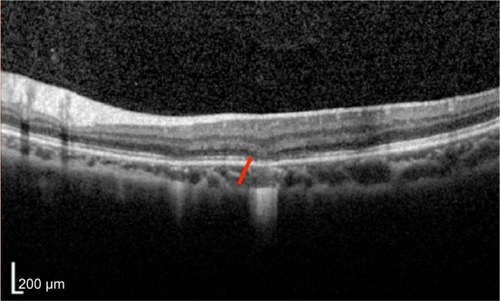
In the foveal region, SD-OCT imaging of the macula has conventionally been resolved into four bands (). The innermost and faintest band (first band) is believed to originate from the external limiting membrane (ELM),Citation8 which is an assembly of junctional complexes between Muller cells and photoreceptors. The second band is often referred to as the IS/OS boundary of the photoreceptors.Citation9,Citation10 The third band has been referred to as cone outer segment tips (COST),Citation11 intermediate line,Citation12 or as the Verhoeff membrane.Citation13,Citation14 The fourth band is supposed to represent the retinal pigment epithelium (RPE). Spaide and Curcio suggested that part of the fourth band is contributed by the Bruch’s membrane and the choriocapillaris.Citation15
Figure 1 Normal Heidelberg spectral domain optical coherence tomography (SD-OCT) with intact external limiting membrane (ELM), photoreceptor inner segment ellipsoid band (ISel), and retinal pigment epithelium (RPE).
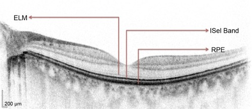
However, some questions were raised regarding authentication of the existence of these bands. As shown in several studies,Citation8,Citation16–Citation19 the length of the inner segment and outer segment was found to be approximately the same for both foveal rods and cones. From this, it can be deduced that IS/OS junction should lie at the midpoint between the ELM and the RPE. However, in normal SD-OCT, the second reflective band lies much closer to the ELM than the RPE. These doubts about the second band observed on SD-OCT were subsequently clarified. Fernandez et al demonstrated the anatomy of human foveal cone photoreceptors on SD-OCT and suggested that they visualized the inner segment ellipsoid band.Citation20 Spaide and Curcio constructed a scale model drawing of the outer retina based on published histology to enable visual comparisons.Citation15 Using this model for analysis, their conclusion on the origins of the first and fourth bands was the same as previously suggested. However, their analysis about the origin of the second band suggested that it originated from the inner segment ellipsoids. The third band seemed to have originated from the posterior tip of the cone outer segment ie, the contact cylinder. Lu et al used line scan OCT on excised living leopard frog retina to establish the origin of the existence of the IS/OS junction.Citation21 They speculated that the inner segment ellipsoid may contribute to the IS/OS junction in OCT images.
In this review, considering the evidence put forward by the above landmark studies, IS/OS junction will be referred to as inner segment ellipsoid (ISel), and various studies that assessed the role of the ISel band in SD-OCT in the diagnosis and prognosis evaluation of various surgical and medical retinal diseases will be discussed. Developments in several areas of OCT research promise exciting advances in performance and clinical applications. Adaptive optics uses deformable elements that corrects ocular aberrations and improves transverse resolution by decreasing spot size of the sample beam on the retina.Citation22 Coupled with OCT, isotropic cellular-resolution imaging may be achieved, which may be useful for studying photoreceptor morphology including ISel band in greater detail. This will further enhance our understanding of the role of ISel band in various retinal diseases.Citation23
Inner segment ellipsoid in surgical retinal diseases
Idiopathic macular hole
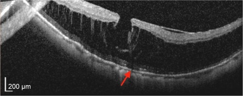
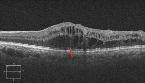
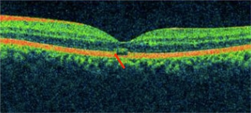
Ultra high resolution OCT has enabled imaging of previously undetectable changes in photoreceptor ISel band associated with macular hole pathology and its post-operative repair (). Macular hole reparative process was studied using SD-OCT in various studies and correlation analysis with best corrected visual acuity (BCVA) was performed. Vitrectomy, internal limiting membrane (ILM) peeling and internal gas tamponade is the standard treatment for achieving closure of macular hole. The reparative process of macular hole closure using SD-OCT was evaluated retrospectively and better visual outcome was observed in eyes with a continuous photoreceptor ISel band compared with those with a disrupted ISel band.Citation24 In another study, Oh et al evaluated photoreceptor ISel band defect parameters in linear and composite SD-OCT formats, assessing diameter and area of the ISel band defect and macular density.Citation4 They concluded that BCVA correlated better with the area of the photoreceptor ISel band defect than with linear measurements after macular hole closure.
Figure 2 Macular hole with disrupted ISel and ELM.
Visualization of the ISel band was demonstrated to be an important indicator of photoreceptor integrity for both standard resolution and ultra high resolution OCT images.Citation9 Thereafter, many studies were conducted to establish a relationship between the photoreceptor ISel band and BCVA in patients undergoing macular hole surgery. Presence of a normal photoreceptor ISel band was associated with better BCVA after surgery.Citation25 The external limiting membrane was the first structure to recover after macular hole closure.Citation26,Citation27 During follow-up, foveal cysts may develop, which may later fill, leading to formation of an intact photoreceptor ISel band.Citation27 Integrity of the ELM is directly correlated with restoration of the photoreceptor ISel band.Citation28
In further studies, even more detailed analysis was performed. The SD-OCT characteristics evaluated included macular hole diameter, ELM defect diameter, photoreceptor ISel band defect diameter, the presence or absence of subretinal fluid (SRF), central foveal thickness (CFT), and outer foveal thickness (defined as the distance between the ELM and the inner border of the RPE). Parameters that best correlated with BCVA postoperatively were outer foveal thickness and the photoreceptor ISel band respectively. Disrupted ELM was associated with poor BCVA. Outer foveal thickness gradually increased during the follow-up period. The results suggested that increase in outer foveal thickness indicates restoration of photoreceptor ISel band integrity, which is associated with better postoperative visual acuity.Citation29,Citation30
A recent retrospective study demonstrated that distinct a COST line was first seen at 6 months after the surgery. Moreover, in eyes with an intact photoreceptor ISel band and ELM, a distinct or irregular COST line had significantly better BCVA at 12 months compared with those with a disrupted COST line. It was concluded that appearance of a distinct or irregular COST line indicates recovery of foveal photoreceptor microstructure.Citation31
A meta-analysis on 1,654 eyes with macular holes treated by pars plana vitrectomy revealed that 87.5% of eyes achieved anatomical closure and 12.5% failed to do so.Citation32 Therefore, persistence of macular holes is still the major concern after macular hole surgery and further refinement of the surgical technique to enhance closure of macular holes is needed. Nonetheless, we should not forget to also focus our efforts on the analysis of predictive factors in determining not only surgical but also functional success in order to better advise our patients concerning their visual prognosis.
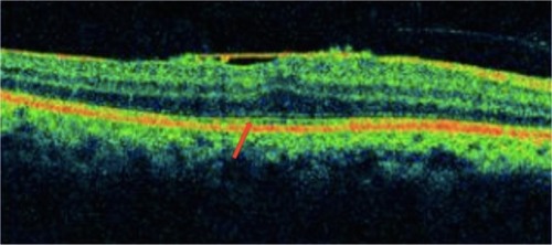
Idiopathic epiretinal membrane
Epiretinal membranes (ERMs) are collections of collagenous cells that occur on the inner surface of the central retina. These membranes have contractile properties and can lead to visual changes and metamorphopsia because of their effects on the underlying retina. ERMs can be treated with microincisional vitrectomy surgery and membrane peeling. However, visual acuity might not fully recover even after complete removal of an ERM. OCT is particularly useful in assessing the thickness and points of adhesions of the ERM in the macula (). Various studies were conducted to assess the predictive factors of good BCVA postoperatively. An intact photoreceptor ISel band on SD-OCT represents morphological and functional recovery of photoreceptors and was associated with good BCVA after surgery.Citation33 A disrupted photoreceptor ISel band was associated with poorer BCVA, and this disruption was found to be irreversible. Therefore, in patients with a disrupted photoreceptor ISel band, early removal of the ERM was warranted.Citation34 Other studies also suggested that the presence or absence of a photoreceptor ISel band together with central retinal thickness, ILM profile, and foveal contour on SD-OCT are good predictors of BCVA postoperatively.Citation35,Citation36 Inoue et al suggested that with a preoperative intact photoreceptor ISel band, recovery of this band can occur over a follow-up period of 1 year after surgery.Citation5 Preoperative integrity of the photoreceptor ISel band may be an important prognostic factor for better visual recovery after surgery. In another retrospective case series study on patients with secondary ERM after retinal detachment surgery, it was found that integrity of the photoreceptor ISel band and ELM were the main factors that predict final BCVA after ERM peeling.Citation37 Disrupted photoreceptor ISel bands found preoperatively with normal autofluorescence can be associated with recovery of photoreceptor ISel bands following ERM peeling. It can be explained by the presence of a functional retinal pigment epithelium-photoreceptor complex, supporting normal photoreceptor activity.Citation38
Figure 3 Epiretinal membrane showing photoreceptor ISel disruption (red arrow).
Based on several retrospective observational and interventional studies,Citation39–Citation42 it was established that integrity of the photoreceptor ISel band is correlated with BCVA. A number of studies also suggested that in addition to the photoreceptor ISel band and the COST line, photoreceptor outer segment length can also be considered to be an important prognostic factor for predicting BCVA postoperatively.Citation40,Citation41,Citation43 Degree of metamorphopsia correlated with inner nuclear layer thickness in patients with ERM.Citation39 Significant predictors for post-operative metamorphopsia outcome included the degree of preoperative metamorphopsia, central foveal thickness, and the photoreceptor ISel band integrity at baseline.Citation44 Recently, it was concluded that photoreceptor ISel band restoration in the parafoveal quadrants also contributed significantly to the recovery of BCVA following ERM surgery in patients with metamorphopsia. Therefore, functional and morphological tests of the macular area should not be limited to the fovea but should be extended to the parafoveal region.
Retinal detachment
A retrospective observational study on patients with poor visual acuity after retinal detachment (RD) surgery demonstrated disruption of photoreceptor ISel bands on ultrahigh resolution OCT and confirmed the prior histological finding of damage to photoreceptor outer segments occurring after RD.Citation45 Preoperative macula off RDs, the integrity of photoreceptor ISel bands and ELM signals on SD-OCT appeared to correlate with postoperative visual recovery.Citation46–Citation48 Preservation of the ELM postoperatively may predict the subsequent restoration of the photoreceptor layer.Citation46 Preoperative intraretinal separation and outer retinal undulation was associated with higher incidence of disrupted photoreceptor ISel bands postoperatively.Citation49 After correlating the OCT results with anatomic and functional outcome after scleral buckling surgery, it was concluded that postoperative visual outcome was associated with the integrity of the photoreceptor ISel band.Citation50
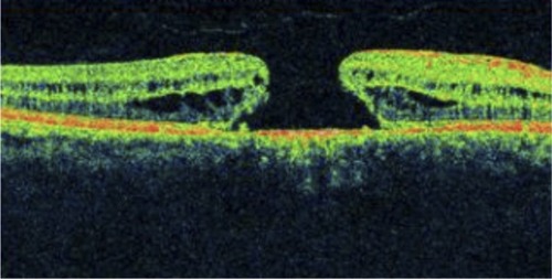
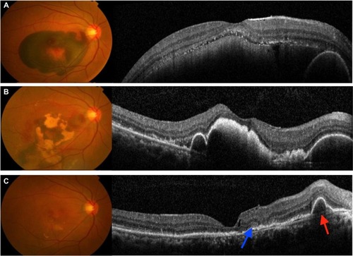
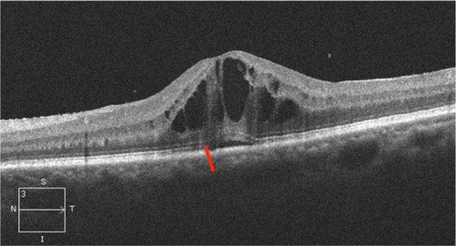
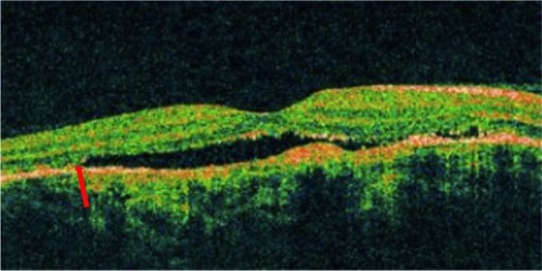
Myopic foveoschisis and foveal retinoschisis
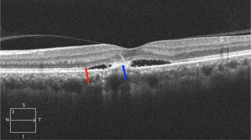
Myopic foveoschisis affects highly myopic patients with posterior staphyloma, and patients may later develop reduced vision due to foveal detachment and macular hole. SD-OCT imaging can demonstrate the characteristic splitting of retina with intraretinal bridges in myopic foveoschisis (). In a study conducted to correlate the pathologic features of eyes with myopic foveoschisis with findings of SD-OCT, defects in photoreceptor ISel bands were found.Citation51 In our study of two cases of bilateral X-linked foveal retinoschisis of different age groups, SD-OCT findings suggested disruption of the photoreceptor ISel band along with the outer nuclear layer and ELM disruption in the subfoveal region.Citation52 Several surgical options have been proposed, which include pars plana vitrectomy with or without ILM peeling (traditional or fovea sparing), gas tamponade as well as scleral and suprachoroidal buckling techniques. The SD-OCT findings of an observational case study on patients with myopic foveoschisis undergoing vitrectomy suggested that postoperative visual acuity depended on recovery of the photoreceptor ISel band.Citation53
Figure 4 Myopic foveoschisis showing disruption of ISel band (red arrow) and ELM.
Inner segment ellipsoid in medical retinal diseases
Age-related macular degeneration
SD-OCT is currently the modality of choice for diagnosis and monitoring of neovascular age-related macular degeneration (AMD), as it is noninvasive and provides high resolution imaging ( and ). Depending on the layer of choroidal neovascularization (CNV) involvement, neovascular AMD can be divided into several subtypes. Type I lesions have CNV beneath the RPE corresponding to occult lesions in fluorescein angiography (FA). Type II lesions are located beneath the retina but above the RPE corresponding to classic lesions in FA. SD-OCT helps in differentiating between the two types of lesions. Type III CNV includes retinal angiomatous proliferation and polypoidal choroidal vasculopathy (PCV).Citation54 Microperimetry, SD-OCT, and fundus autofluorescence (FAF) have been used for evaluation of patients with progressive geographical atrophy secondary to AMD, and it was concluded that retinal sensitivity changes are related to integrity of the photoreceptor ISel band.Citation55
Figure 5 Choroidal neovascular membrane showing disruption of the photoreceptor ISel band, ELM, and RPE (red arrow).
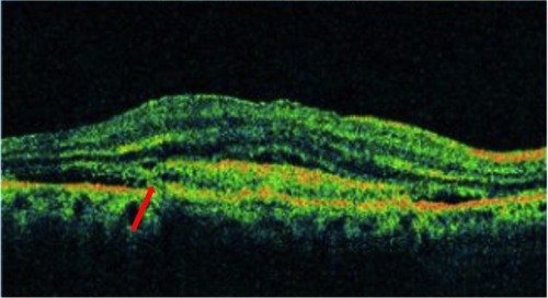
Figure 6 Submacular hemorrhage in neovascular AMD.
Abbreviations: AMD, age-related macular degeneration; ISel, inner segment ellipsoid.
Several retrospective studies have evaluated the SD-OCT changes in patients who were treated for neovascular AMD.Citation56–Citation58 After being successfully treated with photodynamic therapy (PDT), it was found that eyes with a continuous or discontinuous photoreceptor ISel band beneath the fovea were associated with better final BCVA than eyes without an ISel band. Visual outcome was also found to be significantly better in eyes with PCV. At final visit, it was found that 86.7% of eyes with typical AMD had no photoreceptor ISel band, whereas 36.1% of eyes with PCV had no photoreceptor ISel band beneath the fovea.Citation56 Other studies have evaluated the correlation between the outer retinal structural changes on SD-OCT and treatment outcome in patients treated with intravitreal antivascular endothelium growth factor (anti-VEGF) therapy.Citation57,Citation58 In a study on patients with neovascular AMD treated with intravitreal bevacizumab, univariate analysis showed that pretreatment damage to ELM was associated with poor visual outcome. On multivariate analysis, pretreatment BCVA and integrity of the ELM and photoreceptor ISel band were found to be important predictors of visual outcome in 37% of cases.Citation55 Prognostic factors for good visual outcome following 3 monthly injections of intravitreal bevacizumab in patients with neovascular AMD included integrity of the photoreceptor ISel band and ELM, but baseline visual acuity had the best predictive value.Citation58
In nonexudative AMD, disruption of the photoreceptor ISel band may be associated with progression of drusen.Citation59 Defects in the ISel band on SD-OCT and reduction in cone density using adaptive optics imaging have been described in eyes with subretinal drusenoid deposits.Citation60 As these subretinal drusenoid deposits regress, loss of the ISel band can be observed, and this was shown to be associated with significant visual loss.Citation60,Citation61
Myopic choroidal neovascularization hemorrhage
The development of CNV is one of the most vision-threatening complications associated with pathologic myopia. Progression of myopic maculopathy occurs in a substantial proportion of patients resulting in significant visual loss. In the past decade, PDT with verteporfin has been used for treating myopic CNV. However, the long-term outcome of PDT is not favorable, as patients generally had no improvement in visual acuity following treatment. The short-term efficacy of intravitreal bevacizumab and ranibizumab has been demonstrated by various studies in treating myopic CNV.Citation62–Citation79 Most of the studies have demonstrated significant mean visual improvement after anti-VEGF therapy, and the beneficial effects were maintained at 12 months. Several recent studies have also reported the long-term visual outcomes up to 2 years following intravitreal bevacizumab for myopic CNV.Citation68,Citation80,Citation81 As compared with the short-term results, these longer term results were more variable, as studies have reported that the initial visual gain might no longer be significant at 2 years.Citation68,Citation81 The microstructural changes and prognostic factors of final visual outcome in myopic subretinal hemorrhages without CNV were studied. The parameters investigated included the maximal hemorrhagic height, intraretinal hyperscattering signal (intraretinal hyperreflective sign) across the retina at 6 months and the integrity of the photoreceptor ISel band and the ELM. It was found that significant visual improvement and resolution of hemorrhages occurred in 92.3% of eyes by 6 months. The intraretinal hyperreflective sign in all eyes extended into the outer nuclear layer in five (38.5%) eyes, to the ILM in four (30.8%) eyes, and between the two layers in the remaining four (30.8%) eyes. The location of the hyperreflective signs at 6 months coincided with the ruptured retinal layers at baseline in all eyes. Moreover, the photoreceptor ISel band and the ELM were found to be intact in 46.2% of the eyes. Significant association was found between BCVA and integrity of the photoreceptor ISel band and ELM. It was concluded that correlation exists between the photoreceptor function and the intraretinal hyperreflective sign, which was presumed to be due to scarring through the disrupted outer retina.Citation82
Diabetic macular edema
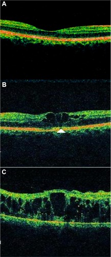
Diabetic retinopathy can result in structural changes in retina and the extent of the structural changes have been shown to correlate with the severity of retinopathy. OCT has been extremely useful to demonstrate various structural changes in patients with diabetic macular edema (DME) (). Currently, there is still no universally accepted classification of DME based on OCT. However, five common patterns have been described, including diffuse retinal edema, cystoid macular edema, serous retinal detachment, posterior hyaloidal traction, and tractional retinal detachment.Citation83 Photoreceptor dysfunction may be a significant predictor of visual acuity in patients with diabetic retinopathy. Several retrospective observational studies were conducted to establish the correlation between photoreceptor ISel band and visual acuity in patients with DME. Maheshwary et alCitation84 graded ISel disruption from grade 0–2, with grade 0 being intact ISel, grade 1 being focal ISel disruption of 200 μm or less, and grade 2 being ISel disruption of more than 200 μm. Grades from each patient’s horizontal and vertical scan were added to yield a global disruption scale. They demonstrated that the percentage of photoreceptor ISel band disruption would be an important predictor of visual acuity among DME patients.Citation84 Previous study has shown that the transverse length of the disrupted or absent ISel band has been related to visual impairment.Citation85 Our recent study demonstrated that increased levels of serum vascular endothelial growth factor and intercellular adhesion molecule-1 levels in diabetic retinopathy are associated with an increase in the severity of diabetic retinopathy and the grade of subfoveal ELM and ISel band disruption. Disruption of the photoreceptor ISel band and ELM was graded by SD-OCT as follows: grade 0 being no disruption of ELM and ISel band (), grade 1 being ELM disrupted with intact ISel band (), and grade 2 having both ELM and ISel band disrupted ().Citation86 Disrupted ELM and photoreceptor ISel band on SD-OCT is associated with hyperreflective foci in the outer retina and visual outcomes in DME.Citation87 However, only a few interventional studies have been conducted in this area. A statistically significant association of intact ELM and ISel band in the fovea with BCVA was found in patients with resolved macular edema following pars plana vitrectomy with ILM removal for DME.Citation88 In a retrospective interventional study, patients with DME who were successfully treated with intravitreal triamcinolone were included. Based on the restoration of the photoreceptor ISel band and ELM on SD-OCT at the final visit, eyes were divided into three groups: 1) group A with a completely visible ISel band and ELM, 2) group B with a disrupted ISel band and intact ELM, and 3) group C with a disrupted or loss of the ISel band and ELM. It was concluded that integrity of the photoreceptor ISel band correlated with final BCVA. Final BCVA of group A or group B was found to be better than group C. They also concluded that shorter length of a disrupted photoreceptor ISel band and ELM was associated with better BCVA. However, on multivariate analysis, it was found that mean disrupted photoreceptor ISel band and ELM length showed greatest correlation with final BCVA compared with disrupted photoreceptor ISel band and ELM. They also found that pretreatment visual acuity and photoreceptor status helped in predicting the posttreatment recovery of the photoreceptor ISel band and subsequent visual recovery.Citation89 Another study was performed on patients with DME who received intravitreal bevacizumab (IVB) injections. It was concluded that correlation between FAF with SD-OCT and visual acuity can help to predict restoration of photoreceptor ISel band integrity after treatment.Citation90 SD-OCT findings in patients with DME who underwent vitrectomy demonstrated that median axial length was longer in those with a visible photoreceptor ISel band at 12 months after surgery, and longer axial length predicted better visual outcome after vitrectomy in DME.Citation91
Figure 7 Diabetic macular edema showing disruption of both photoreceptor ISel band and ELM (red arrow), large cystic spaces, and serous detachment.
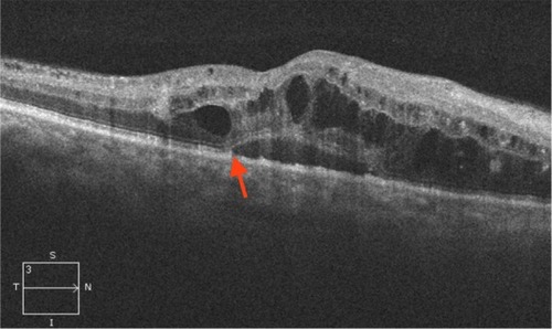
Figure 8 Spectral domain optical coherence tomography (SD-OCT) showing various grades of ELM and ISel band disruption.
Abbreviations: ISel, inner segment ellipsoid; ELM, external limiting membrane.
Central retinal vein occlusion
Several studies have been carried out to establish a correlation between integrity of the photoreceptor ISel band and final visual outcomes after resolution of macular edema associated with central retinal vein occlusion (CRVO) ().Citation92,Citation93 The authors concluded that loss of foveal photoreceptor ISel band and inner retinal layers on SD-OCT significantly correlated with poorer visual acuity in CRVO patients. In another study, it was noted that factors indicating better visual acuity were shorter length of the photoreceptor ISel band and good visual acuity at initial presentation.Citation94
Figure 9 Central retinal vein occlusion associated with macular edema showing large cystic spaces associated with photoreceptor ISel band disruption (red arrow).
Macular telangiectasia
Macular telangiectasia (MacTel) type 2 or juxtafoveal retinal telangiectasia is a bilateral disease of unknown cause with characteristic biomicroscopic and angiographic characteristics. Advances in imaging techniques, particularly using FAF imaging and SD-OCT, have led to remarkable progress in the understanding of MacTel (). Reduction in macular pigment has been demonstrated in this condition, which is supported by the finding of loss of normal central attenuation of FAF. SD-OCT features in MacTel include loss or disruption of the photoreceptor ISel band and hyporeflective cavities at the level of the inner or outer retina and retinal atrophy in later stages. Integrity of the photoreceptor ISel band was found, which correlated with visual acuity.Citation95 Studies have demonstrated that segmentation and en face imaging of the photoreceptor ISel band were found to correlate with visual function and aid in assessing disease severity in MacTel type 2.Citation96–Citation98
Figure 10 Macular telangiectasia showing juxtafoveal disruption of ISel band and ELM (red arrow).

Central serous chorioretinopathy
Studies using SD-OCT have enhanced our understanding of the pathogenesis of central serous chorioretinopathy (CSC). A number of studies have been performed using SD-OCT to identify the retinal morphological changes in CSC ( and ). Visual loss in CSC is now believed to result from photoreceptor loss. SD-OCT has demonstrated disruption of the ISel band and loss of the COST line, presence of subretinal and intraretinal bright spots, decreased outer nuclear layer thickness and in chronic cases, posterior cystoid retinal degeneration.Citation99–Citation102 A prospective observational study noted that photoreceptor ISel band was missing before neurosensory retinal reattachment but later became present after reattachment.Citation102 Visualization of the 3-D relationship between the ELM and each photoreceptor layer before and after macular reattachment have enhanced our understanding of the anatomical and visual changes due to CSC. In a similar study,Citation103 correlation of the photoreceptor ISel band with prediction of visual recovery after macular reattachment was established. In another study, the photoreceptor ISel band was not visible in eyes with serous retinal detachment, but became visible after its resolution.Citation104 Several studies have also evaluated the OCT changes in patients with chronic CSC treated by PDT. Significant increase in macular sensitivity that correlated with recovery of photoreceptor ISel bands and COST lines was seen 6 and 12 months after half-dose verteporfin PDT.Citation105 In another retrospective study on patients treated with standard PDT, it was concluded that morphological and functional changes correlated neither with the treatment nor with the progression of disease.Citation106 Outer nuclear layer thickness reduction and discontinuity of the photoreceptor ISel band correlated with poor visual recovery in resolved CSC eyes following treatment with intravitreal ranibizumab.Citation107 Various OCT parameters were evaluated to assess the visual function of eyes with resolved CSC. It was concluded that a disrupted photoreceptor ISel band was associated with worse BCVA.Citation108 For accurate assessment of visual function, various parameters showing retinal status should be combined and interpreted together.Citation109 Integrity of the ISel band has also correlated with functional assessment evaluated by microperimetry.Citation110,Citation111
Figure 11 Acute central serous chorioretinopathy showing serous detachment with pigment epithelium detachment.
Abbreviations: ISel, inner segment ellipsoid; ELM, external limiting membrane.
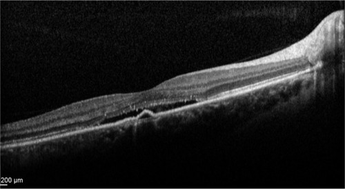
Figure 12 Chronic central serous chorioretinopathy showing disruption of ISel band and ELM (red arrow), and serous detachment and fibrin collection (blue arrow).
Abbreviations: ISel, inner segment ellipsoid; ELM, external limiting membrane; S, superior; N, nasal; I, inferior; T, temporal.
Retinitis pigmentosa
A significant correlation between photoreceptor ISel band and visual acuity has been identified in patients with retinitis pigmentosa (RP).Citation112 Assessment of the integrity of the photoreceptor ISel band was essential while monitoring cystoid macular edema associated with RP.Citation113 Although the macular volume and length of the photoreceptor ISel band were shown to correlate weakly with the amplitude of focal macular electroretinography, a preserved macular morphology did not necessarily guarantee normal-amplitude focal macular electroretinography in RP patients.Citation114 A few studies have attempted to correlate the FAF findings with the retinal structural changes in RP. According to one such study, FAF appeared to depict the integrity of the photoreceptor ISel band.Citation115 Residual photoreceptor ISel band could be found inside the hyperautofluorescent ring, which correlated with visual outcome.Citation116 During RP progression, the progressive constriction of the FAF ring correlated with the length of the photoreceptor ISel band.Citation117 Presence of a photoreceptor ISel band can be considered to be an objective sign in predicting visual recovery after retinal prosthesis implantation.Citation118 Eyes with cone-rod dystrophy (CRD) have significantly different features on SD-OCT compared with eyes with RP. Loss of the ISel band has been demonstrated in a significant proportion of patients with CRD or RP. However absence of an interdigitation zone and foveal cavitation have been shown to be more consistently seen in CRD.Citation119,Citation120 Similarly in eyes with achromatopsia, foveal ellipsoid zone disruption and foveal hypoplasia have been described on SD-OCT.Citation121 An SD-OCT based staging system has been proposed recently to facilitate classification of this condition.Citation121 In stage 1, intact outer retina is seen; stage 2 had ISel band disruption; stage 3 showed presence of an optically empty space; stage 4 showed optically empty space with partial retinal pigment epithelium disruption; and stage 5 had complete retinal pigment epithelium disruption and/or loss of the outer nuclear layer.Citation122
Stargardt’s disease
SD-OCT of patients with reduced vision due to Stargardt’s disease typically demonstrate disruption of the ISel band (). Gomes et al concluded that there was a correlation between findings of SD-OCT and FAF in Stargardt’s disease. They also suggested that structural changes affecting integrity of photoreceptors as seen in SD-OCT may occur earlier than changes in retinal pigment epithelium as detected on FAF.Citation123 The extent of the photoreceptor ISel band preservation showed a correlation with the extension of foveal abnormalities assessed by FAF imaging.Citation124 After mutation analysis, association of G1961E mutation of the ABCA4 gene with milder Stargardt’s disease phenotype was established, as patients with this mutation did not have extensive loss of photoreceptor ISel band.Citation125
Figure 13 Stargardt’s disease showing disruption of ISel band, increased foveal pit with hyperreflectivities at the level of retinal pigment epithelium, suggestive of flecks.
Occult macular dystrophy
Occult macular dystrophy is a rare disorder which presents as bilaterally decreased visual acuity and occurs in the absence of any significant ophthalmoscopic findings. A study was conducted on five patients with markedly decreased visual acuity, and no abnormality was found on full field electroretinogram, fundus photography and FA. However, multifocal electroretinogram was abnormal. SD-OCT was conducted, and all patients showed decreased bowing, and few patients showed disruption of the photoreceptor ISel band.Citation126 The findings of SD OCT were helpful in determining the pathophysiology and diagnostic criteria of occult macular dystrophy.Citation126,Citation127
Acute zonal occult outer retinopathy complex
Optical coherence tomography has demonstrated disruption of ISel in various chorioretinal inflammatory diseases including acute zonal occult outer retinopathy (AZOOR), multiple evanescent white dot syndrome (MEWDS), multifocal choroiditis and panuveitis, punctate inner choroidopathy (), acute macular choroidopathy, and acute idiopathic blind spot enlargement syndrome. AZOOR is typified by acute loss of one or more zones of outer retinal function associated with photopsia, visual field defects, and minimal changes on fundus. Multifocal electroretinogram showed decreased response from the blind spots and other visual field defects with corresponding loss or disruption of the photoreceptor ISel band on SD-OCT. Hangai reported reflective focal lesions within the outer photoreceptor layer and disrupted ISel band, which corresponded to the hypofluorescent areas in the late phase of indocyanine green angiography in MEWDS.Citation128 In addition, thinning of the outer nuclear layer has also been described.Citation129 In several studies conducted to establish correlation between visual field defects and SD-OCT findings, it was found that disruption of photoreceptor ISel band correlated with blind spot enlargement.Citation130 Photoreceptor ISel band disruption was found on SD-OCT in all patients with MEWDS as evidenced by a few studies.Citation131–Citation133
Figure 14 Punctate inner choroidopathy showing disruption of photoreceptor ISel band and ELM (red arrow).
Solar retinopathy
Solar retinopathy can develop following exposure to arc welding, viewing of a solar eclipse, or direct sun gazing in religious ritual participants, military personnel, people with mental disturbances, and sunbathers (). Damage to the inner and outer segment of the photoreceptors in patients with both acute and chronic presentations without involvement of the RPE was demonstrated in patients with solar retinopathy on SD-OCT.Citation134 RPE defects were more prominent in the acute exposure group, and photoreceptor defects were found more often in the chronic exposure group. While this was significant, RPE defects were still observed in the chronic exposure group, and photoreceptor defects were also seen in the acute exposure group. It was concluded that solar retinopathy mainly involved the inner photoreceptor segment. The authors of this study further added that visual loss in solar retinopathy can occur from chronic photoreceptor changes in the inner photoreceptor layer or at the junction of the inner and outer photoreceptor layers. A large fragment of hyporeflectance at the level of the outer and inner photoreceptor layer with an abrupt disruption of the photoreceptor ISel band was found to be correlated with decreased visual acuity.Citation135 A retrospective observational study demonstrated defects in photoreceptor ISel bands with foveal atrophy in all patients on SD-OCT, whereas FA identified classic window defects in only a few eyes.Citation136 In addition to distinct defects in the photoreceptor ISel band and photoreceptor outer segments, bilateral foveal thinning was demonstrated on high-speed ultra high definition OCT in a 34-year-old man with chronic solar retinopathy.Citation137 An observational case series demonstrated abnormally high reflectivity in the outer foveal retina as disruption of the photoreceptor ISel band and full thickness involvement of the photoreceptor layer was associated with poor visual acuity.Citation138
Figure 15 Solar retinopathy showing disruption of photoreceptor ISel band with intact ELM (red arrow).
Uveitis
Various retinal complications can occur in patients with uveitis and OCT has documented changes such as CNV, macular edema, and ISel band disruption in patients with uveitis (). In a study, two morphological patterns were observed on SD-OCT performed on patients with uveitis macular edema: cystoid macular edema in 69% of eyes and diffuse macular edema in 31%. It was concluded that BCVA negatively correlated with cystoid pattern and foveal thickening. In addition, a disrupted photoreceptor ISel band was also associated with poor visual acuity.Citation139 A disrupted photoreceptor ISel band was associated with poor visual outcome in another study of patients with ERM secondary to uveitis.Citation140
Figure 16 Uveitis associated with macular edema showing discontinuous photoreceptor ISel band and ELM (red arrow), and cystic spaces with serous detachment.
Sympathetic ophthalmia
Gupta et al conducted a study to investigate the photoreceptor changes occurring in the acute phase of sympathetic ophthalmia.Citation141 Six consecutive patients with sympathetic ophthalmia underwent FA and SD-OCT assessments at presentation. Patients received intravenous methylprednisolone followed by oral corticosteroids and serial OCT examinations were performed 48 hours after the initiation of treatment and 1, 2, and 12 weeks later. Outer retinal segment showed serous retinal detachment and disruption to the continuity of the two inner hyperreflective bands in all the eyes and elongation of photoreceptors could be seen in four eyes. However, after 4 weeks, there was resolution of serous detachment and restoration of the photoreceptor ISel band. This study supported the hypothesis that prompt and aggressive anti-inflammatory therapy is capable of reversing the photoreceptor changes in sympathetic ophthalmia.
Vogt-Koyanagi-Harada disease
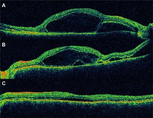
Vogt-Koyanagi-Harada (VKH) disease commonly occurs in pigmented individuals and can result in multifocal exudative retinal detachment during the acute phase. OCT is useful in documenting the changes in exudative retinal detachment following treatment for VKH disease (). Ishihara et al conducted a study to evaluate membranous structures seen on SD-OCT in patients with acute VKH disease.Citation142 It was noted that the membranous structure appeared to include a highly reflective line that seemed to be in conjunction with the photoreceptor ISel band in attached retina. Intraretinal split was seen to overlie the abnormal photoreceptor ISel band in the vicinity of cystoid spaces involving the fovea in nine (45%) of the 20 eyes. During the course of corticosteroid therapy, the membranous structure changed to a granular structure. The authors hypothesized that inflammatory products like fibrin bound with the membranous form of the outer segment and corticosteroid therapy causes its dissolution, thereby changing the membranous structure into a granular structure.
Figure 17 VKH disease after treatment with intravenous methylprednisolone showing (A) subretinal fluid, (B) resolving subretinal fluid, and (C) residual subretinal fluid.
Abbreviations: VKH, Vogt-Koyanagi-Harada; ISel, inner segment ellipsoid; ELM, external limiting membrane.
Behcet’s disease
A study by Unoki et al evaluated patients with remitting ocular Behcet’s disease, and the correlation between SD-OCT findings and BCVA was evaluated. Foveal thickness and integrity of the ELM and the photoreceptor ISel band on SD-OCT were analyzed, and it was found that visual acuity correlated with integrity of the photoreceptor ISel band and not with the ELM.Citation143
Choroidal hemangioma
Choroidal hemangioma is a benign vascular tumor arising in the choroidal and can result in visual loss due to exudation (). A retrospective review of 14 eyes with symptomatic circumscribed choroidal hemangioma that was treated with PDT was evaluated using time-domain OCT.Citation144 It was found that a preserved photoreceptor ISel band was associated with good visual acuity after PDT.
Figure 18 Choroidal hemangioma showing disruption of photoreceptor ISel band, ELM (red arrow) and serous detachment.
Drug-related maculopathies
Several studies were conducted to correlate visual field defects with SD-OCT findings in patients who developed retinal toxicity after treatment with hydroxychloroquine or chloroquine.Citation145–Citation147 It was found that structural changes in the photoreceptor ISel band correlated topographically with field defects seen on Humphrey visual field 10-2. Moreover, SD-OCT may be helpful in determining subclinical abnormalities.Citation145 It has been shown that photoreceptor ISel band changes correlated with changes on multifocal electroretinography.Citation146,Citation147 Findings of FAF imaging were consistent with disruptions in photoreceptor ISel band in patients who developed hydroxychloroquine maculopathy.Citation148 To this effect, SD-OCT was listed as one of the recommended screening procedures for early detection of hydroxychloroquine toxicity according to the revised American Academy of Ophthalmology guideline.Citation149
Conclusion
Based on this review, we conclude that photoreceptor dysfunction may be a significant predictor of visual acuity in a wide spectrum of surgical and medical retinal diseases. Both preoperative and postoperative disruption of ISel band has been found to correlate with BCVA. In addition to the ISel band, various other OCT parameters including ELM and COST line can be evaluated to determine the prognosis of various retinal diseases.
Disclosure
The authors have no conflicts of interest in this work.
References
- ChangLKKoizumiHSpaideRFDisruption of the photoreceptor inner segment-outer segment junction in eyes with macular holesRetina200828796997518698299
- SpaideRFKoizumiHFreundKBPhotoreceptor outer segment abnormalities as a cause of blind spot enlargement in acute zonal occult outer retinopathy-complex diseasesAm J Ophthalmol2008146111112018439564
- WangNKChouCLLimaLHFundus autofluorescence in cone dystrophyDoc Ophthalmol2009119214114419340470
- OhJSmiddyWEFlynnHWJrGregoriGLujanBPhotoreceptor inner/outer segment defect imaging by spectral domain OCT and visual prognosis after macular hole surgeryInvest Ophthalmol Vis Sci20105131651165819850825
- InoueMMoritaSWatanabeYInner segment/outer segment junction assessed by spectral-domain optical coherence tomography in patients with idiopathic epiretinal membraneAm J Ophthalmol2010150683483920719295
- HoodDCLazowMALockeKGGreensteinVCBirchDGThe transition zone between healthy and diseased retina in patients with retinitis pigmentosaInvest Ophthalmol Vis Sci201152110110820720228
- KitaguchiYKusakaSYamaguchiTMihashiTFujikadoTDetection of photoreceptor disruption by adaptive optics fundus imaging and Fourier-domain optical coherence tomography in eyes with occult macular dystrophyClin Ophthalmol2011534535121468344
- DrexlerWSattmannHHermannBEnhanced visualization of macular pathology with the use of ultrahigh-resolution optical coherence tomographyArch Ophthalmol2003121569570612742848
- KoTHFujimotoJGDukerJSComparison of ultrahigh- and standard-resolution optical coherence tomography for imaging macular hole pathology and repairOphthalmology2004111112033204315522369
- SrinivasanVJKoTHWojtkowskiMNoninvasive volumetric imaging and morphometry of the rodent retina with high-speed, ultrahigh-resolution optical coherence tomographyInvest Ophthalmol Vis Sci200647125522552817122144
- SrinivasanVJMonsonBKWojtkowskiMCharacterization of outer retinal morphology with high-speed, ultrahigh-resolution optical coherence tomographyInvest Ophthalmol Vis Sci20084941571157918385077
- OishiAHataMShimozonoMMandaiMNishidaAKurimotoYThe significance of external limiting membrane status for visual acuity in age-related macular degenerationAm J Ophthalmol201015012732.e120609705
- ZawadzkiRJJonesSMOlivierSSAdaptive-optics optical coherence tomography for high-resolution and high-speed 3D retinal in vivo imagingOpt Express200513218532854619096728
- PucheNQuerquesGBenhamouNHigh-resolution spectral domain optical coherence tomography features in adult onset foveomacular vitelliform dystrophyBr J Ophthalmol20109491190119620576764
- SpaideRFCurcioCAAnatomical correlates to the bands seen in the outer retina by optical coherence tomography: literature review and modelRetina20113181609161921844839
- AngerEMUnterhuberAHermannBUltrahigh resolution optical coherence tomography of the monkey fovea. Identification of retinal sublayers by correlation with semithin histology sectionsExp Eye Res20047861117112515109918
- SpaideRFQuestioning optical coherence tomographyOphthalmology20121191122032204.e123122463
- KrebsWKrebsIPrimate Retina and Choroid: Atlas of Fine Structure in Man and MonkeyNew York, NYSpringer-Verlag1991
- HoganJAAlvaradoMJWeddellJEHistology of the Human Eye: An Atlas and TextbookPhiladelphia, PAWB Saunders1971
- FernandezEJHermannBPovazayBUltrahigh resolution optical coherence tomography and pancorrection for cellular imaging of the living human retinaOpt Express20081615110831109418648422
- LuRWCurcioCAZhangYInvestigation of the hyper-reflective inner/outer segment band in optical coherence tomography of living frog retinaJ Biomed Opt201217606050422734727
- FernandezEJPovazayBHermannBThree-dimensional adaptive optics ultrahigh-resolution optical coherence tomography using a liquid crystal spatial light modulatorVision Res200545283432344416249013
- ZhangYCenseBRhaJHigh-speed volumetric imaging of cone photoreceptors with adaptive optics spectral-domain optical coherence tomographyOpt Express200614104380439419096730
- SanoMShimodaYHashimotoHKishiSRestored photoreceptor outer segment and visual recovery after macular hole closureAm J Ophthalmol20091472313318.e118835472
- BabaTYamamotoSAraiMCorrelation of visual recovery and presence of photoreceptor inner/outer segment junction in optical coherence images after successful macular hole repairRetina200828345345818327138
- WakabayashiTFujiwaraMSakaguchiHKusakaSOshimaYFoveal microstructure and visual acuity in surgically closed macular holes: spectral-domain optical coherence tomographic analysisOphthalmology201011791815182420472291
- BottoniFDe AngelisSLuccarelliSCigadaMStaurenghiGThe dynamic healing process of idiopathic macular holes after surgical repair: a spectral-domain optical coherence tomography studyInvest Ophthalmol Vis Sci20115274439444621345999
- TheodossiadisPGGrigoropoulosVGTheodossiadisGPThe significance of the external limiting membrane in the recovery of photoreceptor layer after successful macular hole closure: a study by spectral domain optical coherence tomographyOphthalmologica2011225317618421293159
- ShimozonoMOishiAHataMKurimotoYRestoration of the photoreceptor outer segment and visual outcomes after macular hole closure: spectral-domain optical coherence tomography analysisGraefes Arch Clin Exp Ophthalmol2011249101469147621499768
- Ruiz-MorenoJMLugoFMonteroJAPineroDPRestoration of macular structure as the determining factor for macular hole surgery outcomeGraefes Arch Clin Exp Ophthalmol2012250101409141422354370
- ItohYInoueMRiiTHiraokaTHirakataASignificant correlation between visual acuity and recovery of foveal cone microstructures after macular hole surgeryAm J Ophthalmol20121531111119.e121880295
- MesterVKuhnFInternal limiting membrane removal in the management of full-thickness macular holesAm J Ophthalmol2000129676977710926987
- MitamuraYHiranoKBabaTYamamotoSCorrelation of visual recovery with presence of photoreceptor inner/outer segment junction in optical coherence images after epiretinal membrane surgeryBr J Ophthalmol200993217117518971234
- SuhMHSeoJMParkKHYuHGAssociations between macular findings by optical coherence tomography and visual outcomes after epiretinal membrane removalAm J Ophthalmol20091473473480.e319054492
- OsterSFMojanaFBrarMYusonRMChengLFreemanWRDisruption of the photoreceptor inner segment/outer segment layer on spectral domain-optical coherence tomography is a predictor of poor visual acuity in patients with epiretinal membranesRetina201030571371820038861
- Falkner-RadlerCIGlittenbergCHagenSBeneschTBinderSSpectral-domain optical coherence tomography for monitoring epiretinal membrane surgeryOphthalmology2010117479880520045567
- TheodossiadisPGTheodossiadisGPCharonisAEmfietzoglouIGrigoropoulosVGLiarakosVSThe photoreceptor layer as a prognostic factor for visual acuity in the secondary epiretinal membrane after retinal detachment surgery: imaging analysis by spectral-domain optical coherence tomographyAm J Ophthalmol2011151697398021457925
- BritoPNGomesNLVieiraMPPossible role for fundus autofluorescence as a predictive factor for visual acuity recovery after epiretinal membrane surgeryRetina201434227328023881227
- OkamotoFSugiuraYOkamotoYHiraokaTOshikaTAssociations between metamorphopsia and foveal microstructure in patients with epiretinal membraneInvest Ophthalmol Vis Sci201253116770677522969078
- ShimozonoMOishiAHataMThe significance of cone outer segment tips as a prognostic factor in epiretinal membrane surgeryAm J Ophthalmol20121534698704704.e122245463
- WatanabeKTsunodaKMizunoYAkiyamaKNodaTOuter retinal morphology and visual function in patients with idiopathic epiretinal membraneJAMA Ophthalmol2013131217217723411882
- CobosEAriasLRuiz-MorenoJPreoperative study of the inner segment/outer segment junction of photoreceptors by spectral-domain optical coherence tomography as a prognostic factor in patients with epiretinal membranesClin Ophthalmol201371467147023901254
- ShionoAKogoJKloseGPhotoreceptor outer segment length: a prognostic factor for idiopathic epiretinal membrane surgeryOphthalmology2013120478879423290984
- BaeSHKimDParkTKHanJRKimHNamWPreferential hyperacuity perimeter and prognostic factors for metamorphopsia after idiopathic epiretinal membrane surgeryAm J Ophthalmol20131551109117.e323022166
- SchocketLSWitkinAJFujimotoJGUltrahigh-resolution optical coherence tomography in patients with decreased visual acuity after retinal detachment repairOphthalmology2006113466667216581427
- WakabayashiTOshimaYFujimotoHFoveal microstructure and visual acuity after retinal detachment repair: imaging analysis by Fourier-domain optical coherence tomographyOphthalmology2009116351952819147231
- ChoMWitmerMTFavaroneGChanRPD’AmicoDJKissSOptical coherence tomography predicts visual outcome in macula-involving rhegmatogenous retinal detachmentClin Ophthalmol20126919622275812
- DelolmeMPDugasBNicotFMuselierABronAMCreuzot-GarcherCAnatomical and functional macular changes after rhegmatogenous retinal detachment with macula offAm J Ophthalmol2012153112813621937016
- JoeSGKimYJChaeJBStructural recovery of the detached macula after retinal detachment repair as assessed by optical coherence tomographyKorean J Ophthalmol201327317818523730110
- AkkoyunIYilmazGOptical coherence tomography: anatomic and functional outcome after scleral buckling surgery in macula-off rhegmatogenous retinal detachmentKlin Monbl Augenheilkd20132308814819 German23670523
- FujimotoSIkunoYNishidaKPostoperative optical coherence tomographic appearance and relation to visual acuity after vitrectomy for myopic foveoschisisAm J Ophthalmol20131565968973.e123938124
- SaxenaSManishaMeyerCHThree-dimensional spectral domain optical coherence tomography in X linked foveal retinoschisisBMJ Case Rep20132013
- SayanagiKIkunoYSogaKTanoYPhotoreceptor inner and outer segment defects in myopic foveoschisisAm J Ophthalmol2008145590290818342829
- GerendasBSKroisamerJSSulzbacherFSchmidt-ErfurthUNeovascular age-related macular degenerationMidenaEMicroperimetry and Multimodal Retinal ImagingBerlin, HeidelbergSpringer-Verlag Berlin Heidelberg2014
- PilottoEBenettiEConventoEMicroperimetry, fundus autofluorescence, and retinal layer changes in progressing geographic atrophyCan J Ophthalmol201348538639324093185
- HayashiHYamashiroKTsujikawaAOtaMOtaniAYoshimuraNAssociation between foveal photoreceptor integrity and visual outcome in neovascular age-related macular degenerationAm J Ophthalmol200914818389.e119327745
- ChhablaniJKimJSFreemanWRKozakIWangHYChengLPredictors of visual outcome in eyes with choroidal neovascularization secondary to age related macular degeneration treated with intravitreal bevacizumab monotherapyInt J Ophthalmol201361626623549041
- OishiAShimozonoMMandaiMHataMNishidaAKurimotoYRecovery of photoreceptor outer segments after anti-VEGF therapy for age-related macular degenerationGraefes Arch Clin Exp Ophthalmol2013251243544022576370
- HartmannKIGomezMLBartschDUSchusterAKFreemanWREffect of change in drusen evolution on photoreceptor inner segment/outer segment junctionRetina20123281492149922481478
- MrejenSSatoTCurcioCASpaideRFAssessing the cone photoreceptor mosaic in eyes with pseudodrusen and soft Drusen in vivo using adaptive optics imagingOphthalmology2014121254555124183341
- CurcioCAMessingerJDSloanKRMcGwinGMedeirosNESpaideRFSubretinal drusenoid deposits in non-neovascular age-related macular degeneration: morphology, prevalence, topography, and biogenesis modelRetina201333226527623266879
- YamamotoIRogersAHReichelEYatesPADukerJSIntravitreal bevacizumab (Avastin) as treatment for subfoveal choroidal neovascularisation secondary to pathological myopiaBr J Ophthalmol200791215716016870653
- SakaguchiHIkunoYGomiFIntravitreal injection of bevacizumab for choroidal neovascularisation associated with pathological myopiaBr J Ophthalmol200791216116516914470
- ChanWMLaiTYLiuDTLamDSIntravitreal bevacizumab (Avastin) for myopic choroidal neovascularization: six-month results of a prospective pilot studyOphthalmology2007114122190219617599414
- Hernandez-RojasMLQuiroz-MercadoHDalma-WeiszhauszJShort-term effects of intravitreal bevacizumab for subfoveal choroidal neovascularization in pathologic myopiaRetina200727670771217621179
- ChanWMLaiTYChanKPChanges in aqueous vascular endothelial growth factor and pigment epithelial-derived factor levels following intravitreal bevacizumab injections for choroidal neovascularization secondary to age-related macular degeneration or pathologic myopiaRetina20082891308131318728623
- ChanWMLaiTYLiuDTLamDSIntravitreal bevacizumab (Avastin) for myopic choroidal neovascularisation: 1-year results of a prospective pilot studyBr J Ophthalmol200993215015418801766
- IkunoYNagaiYMatsudaSTwo-year visual results for older Asian women treated with photodynamic therapy or bevacizumab for myopic choroidal neovascularizationAm J Ophthalmol2010149114014619846061
- Ruiz-MorenoJMMonteroJAAriasLTwelve-month outcome after one intravitreal injection of bevacizumab to treat myopic choroidal neovascularizationRetina201030101609161520856171
- ScupolaATibertiACSassoPMacular functional changes evaluated with MP-1 microperimetry after intravitreal bevacizumab for subfoveal myopic choroidal neovascularization: one-year resultsRetina201030573974720038860
- WakabayashiTIkunoYGomiFDifferent dosing of intravitreal bevacizumab for choroidal neovascularization because of pathologic myopiaRetina201131588088621242860
- GharbiyaMGiustolisiRAllieviFChoroidal neovascularization in pathologic myopia: intravitreal ranibizumab versus bevacizumab – a randomized controlled trialAm J Ophthalmol20101493458464.e120172072
- SilvaRMRuiz-MorenoJMNascimentoJShort-term efficacy and safety of intravitreal ranibizumab for myopic choroidal neovascularizationRetina20082881117112318788102
- LaiTYChanWMLiuDTLamDSIntravitreal ranibizumab for the primary treatment of choroidal neovascularization secondary to pathologic myopiaRetina200929675075619357555
- MonesJMAmselemLSerranoAGarciaMHijanoMIntravitreal ranibizumab for choroidal neovascularization secondary to pathologic myopia: 12-month resultsEye (Lond)200923612751280 quiz 128119478826
- LalloumFSouiedEHBastuji-GarinSIntravitreal ranibizumab for choroidal neovascularization complicating pathologic myopiaRetina201030339940620038864
- SilvaRMRuiz-MorenoJMRosaPIntravitreal ranibizumab for myopic choroidal neovascularization: 12-month resultsRetina201030340741220094007
- VaranoMTedeschiMOddoneFPerilloLCoppeAMParravanoMMicroperimetric retinal changes in myopic choroidal neovascularization treated with intravitreal ranibizumabRetina201030341341720010453
- HeierJSBrownDCiullaTRanibizumab for choroidal neovascularization secondary to causes other than age-related macular degeneration: a phase I clinical trialOphthalmology2011118111111820678799
- GharbiyaMAllieviFConflittiSIntravitreal bevacizumab for treatment of myopic choroidal neovascularization: the second year of a prospective studyClin Ter20101613e87e9320589348
- Ruiz-MorenoJMMonteroJAIntravitreal bevacizumab to treat myopic choroidal neovascularization: 2-year outcomeGraefes Arch Clin Exp Ophthalmol2010248793794120221624
- AsaiTIkunoYNishidaKMacular microstructures and prognostic factors in myopic subretinal hemorrhagesInvest Ophthalmol Vis Sci201455122623224327618
- KimBYSmithSDKaiserPKOptical coherence tomographic patterns of diabetic macular edemaAm J Ophthalmol2006142340541216935584
- MaheshwaryASOsterSFYusonRMChengLMojanaFFreemanWRThe association between percent disruption of the photoreceptor inner segment-outer segment junction and visual acuity in diabetic macular edemaAm J Ophthalmol201015016367.e120451897
- SaxenaSJainAAlterations in “in vivo” histology of retina in bilateral chronic central serous chorioretinopathy after intravitreal bevacizumabJ Ocul Biol Dis Infor20114413714023476721
- JainASaxenaSKhannaVKShuklaRKMeyerCHStatus of serum VEGF and ICAM-1 and its association with external limiting membrane and inner segment-outer segment junction disruption in type 2 diabetes mellitusMol Vis2013191760176823922493
- UjiAMurakamiTNishijimaKAssociation between hyperreflective foci in the outer retina, status of photoreceptor layer, and visual acuity in diabetic macular edemaAm J Ophthalmol20121534710717717.e122137207
- YanyaliABozkurtKTMacinAHorozogluFNohutcuAFQuantitative assessment of photoreceptor layer in eyes with resolved edema after pars plana vitrectomy with internal limiting membrane removal for diabetic macular edemaOphthalmologica20112262576321555906
- ShinHJLeeSHChungHKimHCAssociation between photoreceptor integrity and visual outcome in diabetic macular edemaGraefes Arch Clin Exp Ophthalmol20122501617021874345
- ChungHParkBShinHJKimHCCorrelation of fundus autofluorescence with spectral-domain optical coherence tomography and vision in diabetic macular edemaOphthalmology201211951056106522342014
- WakabayashiYKimuraKMuramatsuDAxial length as a factor associated with visual outcome after vitrectomy for diabetic macular edemaInvest Ophthalmol Vis Sci201354106834684024052638
- OtaMTsujikawaAKitaMIntegrity of foveal photoreceptor layer in central retinal vein occlusionRetina200828101502150818997611
- LimaVCYeungLCastroLCLandaGRosenRBCorrelation between spectral domain optical coherence tomography findings and visual outcomes in central retinal vein occlusionClin Ophthalmol2011529930521468337
- ShinHJChungHKimHCAssociation between integrity of foveal photoreceptor layer and visual outcome in retinal vein occlusionActa Ophthalmol2011891e35e4021155986
- PaunescuLAKoTHDukerJSIdiopathic juxtafoveal retinal telangiectasis: new findings by ultrahigh-resolution optical coherence tomographyOphthalmology20061131485716343625
- SalloFBPetoTEganC“En face” OCT imaging of the IS/OS junction line in type 2 idiopathic macular telangiectasiaInvest Ophthalmol Vis Sci201253106145615222899757
- YannuzziLABardalAMFreundKBChenKJEandiCMBlodiBIdiopathic macular telangiectasiaArch Ophthalmol2006124445046016606869
- Charbel IssaPGilliesMCChewEYMacular telangiectasia type 2Prog Retin Eye Res201334497723219692
- MatsumotoHKishiSOtaniTSatoTElongation of photoreceptor outer segment in central serous chorioretinopathyAm J Ophthalmol2008145116216818028861
- KonYIidaTMarukoISaitoMThe optical coherence tomography-ophthalmoscope for examination of central serous chorioretinopathy with precipitatesRetina200828686486918536604
- PiccolinoFCDe La LongraisRRManeaMCicinelliSPosterior cystoid retinal degeneration in central serous chorioretinopathyRetina20082871008101218698305
- OjimaYHangaiMSasaharaMThree-dimensional imaging of the foveal photoreceptor layer in central serous chorioretinopathy using high-speed optical coherence tomographyOphthalmology2007114122197220717507096
- PiccolinoFCde la LongraisRRRaveraGThe foveal photoreceptor layer and visual acuity loss in central serous chorioretinopathyAm J Ophthalmol20051391879915652832
- OjimaAIidaTSekiryuTMarukoISuganoYPhotopigments in central serous chorioretinopathyAm J Ophthalmol20111516940952.e121457927
- FujitaKShinodaKImamuraYCorrelation of integrity of cone outer segment tips line with retinal sensitivity after half-dose photodynamic therapy for chronic central serous chorioretinopathyAm J Ophthalmol2012154357958522818904
- VasconcelosHMarquesISantosARLong-term chorioretinal changes after photodynamic therapy for chronic central serous chorioretinopathyGraefes Arch Clin Exp Ophthalmol201325171697170523389551
- OzdemirOErolMKMorphologic changes and visual outcomes in resolved central serous chorioretinopathy treated with ranibizumabCutan Ocul Toxicol201433212212623848591
- MatsumotoHSatoTKishiSOuter nuclear layer thickness at the fovea determines visual outcomes in resolved central serous chorioretinopathyAm J Ophthalmol20091481105110.e119327740
- KimSWOhJHuhKCorrelations among various functional and morphological tests in resolved central serous chorioretinopathyBr J Ophthalmol201296335035521617156
- OjimaYTsujikawaAHangaiMRetinal sensitivity measured with the micro perimeter 1 after resolution of central serous chorioretinopathyAm J Ophthalmol20081461778418405876
- OzdemirHKaracorluSASenturkFKaracorluMUysalOAssessment of macular function by microperimetry in unilateral resolved central serous chorioretinopathyEye (Lond)200822220420816936642
- AizawaSMitamuraYBabaTHagiwaraAOgataKYamamotoSCorrelation between visual function and photoreceptor inner/outer segment junction in patients with retinitis pigmentosaEye (Lond)200923230430818188175
- OishiAOtaniASasaharaMPhotoreceptor integrity and visual acuity in cystoid macular oedema associated with retinitis pigmentosaEye (Lond)20092361411141618724276
- SugitaTKondoMPiaoCHItoYTerasakiHCorrelation between macular volume and focal macular electroretinogram in patients with retinitis pigmentosaInvest Ophthalmol Vis Sci20084983551355818441311
- WakabayashiTSawaMGomiFTsujikawaMCorrelation of fundus autofluorescence with photoreceptor morphology and functional changes in eyes with retinitis pigmentosaActa Ophthalmol2010885e177e18320491687
- IriyamaAYanagiYFundus autofluorescence and retinal structure as determined by spectral domain optical coherence tomography, and retinal function in retinitis pigmentosaGraefes Arch Clin Exp Ophthalmol2012250333333921947266
- AizawaSMitamuraYHagiwaraASugawaraTYamamotoSChanges of fundus autofluorescence, photoreceptor inner and outer segment junction line, and visual function in patients with retinitis pigmentosaClin Experiment Ophthalmol201038659760420456441
- TamakiMMatsuoTOptical coherence tomographic parameters as objective signs for visual acuity in patients with retinitis pigmentosa, future candidates for retinal prosthesesJ Artif Organs201114214015021505820
- LimaLHSallumJMSpaideRFOuter retina analysis by optical coherence tomography in cone-rod dystrophy patientsRetina20133391877188023648999
- InuiEOishiAOishiMTomographic comparison of cone-rod and rod-cone retinal dystrophiesGraefes Arch Clin Exp Ophthalmol20141524318530
- YangPMichaelsKVCourtneyRJRetinal Morphology of Patients With Achromatopsia During Early Childhood: Implications for Gene TherapyJAMA Ophthalmol2014
- GreenbergJPShermanJZweifelSASpectral-domain optical coherence tomography staging and autofluorescence imaging in achromatopsiaJAMA Ophthalmol2014132443744524504161
- GomesNLGreensteinVCCarlsonJNA comparison of fundus autofluorescence and retinal structure in patients with Stargardt diseaseInvest Ophthalmol Vis Sci20095083953395919324865
- TestaFRossiSSodiACorrelation between photoreceptor layer integrity and visual function in patients with Stargardt disease: implications for gene therapyInvest Ophthalmol Vis Sci20125384409441522661472
- RencovaEStudnickaJMarakJDvorakovaHLangrovaHThe coincidence of the junction layer of inner and outer photoreceptors segments (IS/OS) defects localization on SD OCT and functional defects in case of Stargardt disease – a case reportCesk Slov Oftalmol20126828486 88. Czech22913873
- KoizumiHMaguireJISpaideRFSpectral domain optical coherence tomographic findings of occult macular dystrophyOphthalmic Surg Lasers Imaging200940217417619320307
- KimYGBaekSHMoonSWLeeHKKimUSAnalysis of spectral domain optical coherence tomography findings in occult macular dystrophyActa Ophthalmol2011891e52e5620560888
- HangaiMFujimotoMYoshimuraNFeatures and function of multiple evanescent white dot syndromeArch Ophthalmol2009127101307131319822847
- NguyenMHWitkinAJReichelEMicrostructural abnormalities in MEWDS demonstrated by ultrahigh resolution optical coherence tomographyRetina200727441441817420691
- TsunodaKFujinamiKMiyakeYSelective abnormality of cone outer segment tip line in acute zonal occult outer retinopathy as observed by spectral-domain optical coherence tomographyArch Ophthalmol201112981099110121825201
- WolffEThe external limiting membrane of the retina and its relation to Verhoeff’s membraneTrans Ophthalmol Soc U K1950606167
- JoussenFSpitznasMThe fine structure of the human retina at the ora serrataAlbrecht Von Graefes Arch Klin Exp Ophthalmol197218531771884538828
- FeeneyLEditorial: Intercellular junctions: sites of permeability barriers and cellular communicationInvest Ophthalmol197413118118144215036
- ChenKCJungJJAlzmanAHigh definition spectral domain optical coherence tomography findings in three patients with solar retinopathy and review of the literatureOpen Ophthalmol J20126293522798967
- KlemencicSMcMahonJUpadhyaySMessnerLSpectral domain optical coherence tomography as a predictor of visual function in chronic solar maculopathyOptom Vis Sci20118881014101921552175
- JainADesaiRUCharalelRAQuiramPYannuzziLSarrafDSolar retinopathy: comparison of optical coherence tomography (OCT) and fluorescein angiography (FA)Retina20092991340134519934824
- ChenRWGorczynskaISrinivasanVJFujimotoJGDukerJSReichelEHigh-speed ultrahigh-resolution optical coherence tomography findings in chronic solar retinopathyRetin Cases Brief Rep20082210310519756263
- JorgeRCostaRAQuirinoLSOptical coherence tomography findings in patients with late solar retinopathyAm J Ophthalmol200413761139114315183809
- IannettiLSpinucciGAbboudaADe GeronimoDTortorellaPAccorintiMSpectral-domain optical coherence tomography in uveitic macular edema: morphological features and prognostic factorsOphthalmologica20122281131822508138
- IannettiLTortorellaPD’AmbrosioESpenaRZitoRGharbiyaMEpiretinal membranes in patients with uveitis: morphological and functional analysis with spectral domain optical coherence tomographyBiomed Res Int2013201328482124294602
- GuptaVGuptaADograMRSinghIReversible retinal changes in the acute stage of sympathetic ophthalmia seen on spectral domain optical coherence tomographyInt Ophthalmol201131210511021331811
- IshiharaKHangaiMKitaMYoshimuraNAcute Vogt-Koyanagi-Harada disease in enhanced spectral-domain optical coherence tomographyOphthalmology200911691799180719643489
- UnokiNNishijimaKKitaMHayashiRYoshimuraNStructural changes of fovea during remission of Behcet’s disease as imaged by spectral domain optical coherence tomographyEye (Lond)201024696997519786959
- LiuWZhangYXuGQianJJiangCLiLOptical coherence tomography for evaluation of photodynamic therapy in symptomatic circumscribed choroidal hemangiomaRetina201131233634321102373
- StepienKEHanDPSchellJGodaraPRhaJCarrollJSpectral-domain optical coherence tomography and adaptive optics may detect hydroxychloroquine retinal toxicity before symptomatic vision lossTrans Am Ophthalmol Soc2009107283320126479
- Rodriguez-PadillaJAHedgesTR3rdMonsonBHigh-speed ultra-high-resolution optical coherence tomography findings in hydroxychloroquine retinopathyArch Ophthalmol2007125677578017562988
- KellnerSWeinitzSKellnerUSpectral domain optical coherence tomography detects early stages of chloroquine retinopathy similar to multifocal electroretinography, fundus autofluorescence and near-infrared autofluorescenceBr J Ophthalmol200993111444144719692385
- KelmensonATBrarVSMurthyRKChalamKVFundus autofluorescence and spectral domain optical coherence tomography in early detection of Plaquenil maculopathyEur J Ophthalmol201020478578820099229
- MarmorMFKellnerULaiTYLyonsJSMielerWFRevised recommendations on screening for chloroquine and hydroxychloroquine retinopathyOphthalmology2011118241542221292109