
Abstract
Herpes simplex virus is responsible for numerous ocular diseases, the most common of which is herpetic stromal keratitis. This is a recurrent infection of the cornea that typically begins with a subclinical infection of the cornea that establishes a latent infection of sensory ganglia, most often the trigeminal ganglia. Recurring infections occur when the virus is reactivated from latency and travels back to the cornea, where it restimulates an inflammatory response. This inflammatory response can lead to decreased corneal sensation, scarring, and blindness. The diagnosis of these lesions as the result of a recurrent herpes simplex virus infection can at times be problematic. Currently, herpetic stromal keratitis is diagnosed by its clinical presentation on the slit-lamp examination, but the literature does not always support the accuracy of these clinical findings. Other diagnostic tests such as polymerase chain reaction assay, enzyme-linked immunosorbent assay, immunofluorescent antibody, and viral cultures have provided more definitive diagnosis, but also have some limitations. That said, accurate diagnosis is necessary for proper treatment, in order to prevent serious consequences. Current treatment reduces the severity of lesions and controls further viral spread, but does not provide a cure.
Introduction
Herpes simplex virus (HSV) is a ubiquitous DNA virus that can infect virtually anywhere in the body, particularly when newborns are infected.Citation1–Citation3 However, in an individual with a normal immune system, the most common sites of infection are the mouth, genitalia, and eyes. In very young children, and in rare instances adults, the brain may also become infected. HSV infections of the eye are the leading cause of infectious corneal blindness in developed countries.Citation4 Approximately 500,000 people in the US are currently infected with ocular HSV.Citation5,Citation6 The costs of treatment for this disease are in the tens of millions spent annually in the US alone.Citation6 While most infections are unilateral, around 1.3%–12% of affected individuals have bilateral ocular infections. Bilateral infections are seen mostly in immunocompromised patients.Citation7–Citation9 Infections can occur in both anterior and posterior segments of the eye, but it most commonly infects the corneal epithelia.Citation4,Citation7,Citation8 It is primarily diagnosed by its clinical presentation, but atypical presentation of the infection can impede accurate diagnoses and thus proper treatment.Citation10
Pathophysiology
HSV is a linear double-stranded DNA virus that is classified as an α-member of the Herpesviridae family.Citation5,Citation11 Primary infection results after HSV spread via direct contact with mucous membrane of the host.Citation5,Citation11 In the case of ocular infections, the virus is transported following primary retrograde infection via sensory neurons to establish latency in trigeminal ganglia; here, it remains asymptomatic until reactivation of the virus leads to secondary or recurrent infections.Citation4,Citation8,Citation12,Citation13 The host cell’s DNA polymerase, located in the nucleus of the cell, is required for HSV to transcribe and replicate.Citation8,Citation14
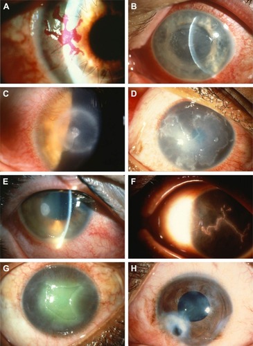
Herpetic stromal keratitis (HSK) comprises three major subtypes: epithelial, stromal, and endothelial ().Citation15 Clinical findings in epithelial keratitis include geographic corneal ulcers with a dendritic tail or dendritic keratitis. This occurs after direct invasion by the virus and is the most common subtype.Citation13,Citation15 SK develops as a result of immune response to the virus. The stromal subtype can be further divided into disciform keratitis, immune SK, and necrotizing keratitis. Endothelial keratitis manifests as rejection line-like keratic precipitates and stromal edema. Variation in presentation between different subtypes has posed a challenge in accurately diagnosing this condition.Citation15
Figure 1 Representative images of various corneal damages due to HSV1 infection.
Abbreviations: HSV, herpes simplex virus; WBC, white blood cell.
Diagnosis
HSK is primarily diagnosed by its clinical presentation on the slit-lamp examination.Citation4 Common symptoms include redness, discharge, watery eyes, irritation, itching, pain, and photophobia. In most patients, symptoms begin to subside after the first 2 weeks.Citation4 The most common subtype, epithelial keratitis, appears as coarse granular spots that form punctuate lesions, but these quickly coalesce to form dendritic lesions.Citation16–Citation18 A physical examination may reveal whitened area.Citation19 On the slit-lamp examination, epithelial keratitis presents as a dendritic lesion with a terminal bulb, swollen borders, and intraepithelial cell infiltration.Citation15 It is visualized by staining the lesion with either lissamine green or rose bengal dye.Citation20 For atypical epithelial lesions, polymerase chain reaction (PCR) has been used to confirm HSK.Citation21 Newer methods, such as tear collection and immunofluorescence antibody assay (IFA), have also been employed to aid identification of epithelial lesions.Citation22,Citation23 SK on physical examination appears opaque or whitened, due to stromal infiltration.Citation20,Citation24 Similarly, the necrotizing form of SK appears as gray-white or opaque, but there is accompanying necrosis and ulceration on slit-lamp examination.Citation20,Citation24 Edema and abscess may be apparent as well.Citation4,Citation25 In immunomediated SK, necrosis or ulceration is lacking, but there is stromal infiltration.Citation26 Unfortunately, PCR has been less helpful for identification of immunomediated SK. Another form of HSK, disciform lesion, has a ground-glass appearance and is disk shaped with stromal edema on slit-lamp examination.Citation27 Lastly, the endothelial form, keratic precipitates, and iritis may be visible. Stromal edema is also present.Citation27 Enzyme-linked immunosorbent assay (ELISA) and viral cultures have been used as diagnostic tools for all subgroups of lesions.Citation15,Citation28 Dendritiform epithelial lesions on slit-lamp examination are pathognomonic for keratitis. However, atypical lesions can make diagnosis difficult.Citation15 Factors affecting diagnosis include duration of illness, systemic diseases, previous medication use, and corneal transplantation, as these can change the appearances of lesions on slit-lamp examination.Citation21
Misdiagnosis in clinical settings is not uncommon, as other pathogens can present with similar lesions.Citation10 Amebic and fungal infections have been mistakenly identified as HSV keratitis.Citation13 In a study conducted by Rübben et al, 8% of the clinically diagnosed HSV lesions were identified on PCR assay as lesions caused by another member of the Herpesviridae family – varicella zoster virus.Citation29 Similarly, another study uncovered 5% of clinically diagnosed HSK lesions as lesions caused by adenovirus, 3.2% as lesions caused by cytomegalovirus, and 2.7% by enterovirus.Citation30 When PCR was used to confirm clinical diagnosis of epithelial dendritiform lesions, there was only moderate correlation (K=0.485, P<0.0001) between diagnosis made by an ophthalmologist and diagnosis made using PCR.Citation10 In a study designed to address the diagnosis of atypical HSK lesions, Koizumi et al defined atypical lesions as those where “dendritic or geographical ulcers with terminal bulbs and epithelial infiltrations are not evident”. They found very little agreement (P=0.22) between PCR results and clinical diagnosis.Citation21
While most diagnoses of HSK are based on clinical presentation, PCR provides better sensitivity. In a study conducted by El-Aal et al, PCR detected 29.2% more cases than cell culture.Citation22 In another study, while viral culture identified 12% of suspected patients with HSK, PCR identified 88% of suspected patients.Citation22 PCR is highly sensitive, but large variation has been observed in various studies between the rate of HSV detection by PCR when compared to clinical diagnosis.Citation10 PCR is more likely to identify patients that present with typical lesions or patients who have not used antiviral medications (P=0.022). It is less responsive in patients with atypical lesions or in patients who previously used or currently use antiviral medications (P=0.968).Citation28 It has been hypothesized by McGilligan et al that because SK is a result of immune response to the virus, rather than the viral infection itself, it may explain the negative PCR results. Variation was also seen (4%–6% variability) when the same samples were reanalyzed using different amplification regions of the gene.Citation10 In another study, there was an 80% decrease of detectable virus in patients who had been taking 400 mg of acyclovir twice daily.Citation31 This can lead to reduction in PCR sensitivity.Citation13 PCR-based tests can also result in false negatives.Citation31,Citation32
Similarly, ophthalmologists use oxybuprocaine, a local anesthetic that is used in conjunction with fluorescein-based dye to visualize possible HSV corneal lesions better, but can decrease the effectiveness of PCR by interfering with the PCR reaction, resulting in a decrease of more than 2 logs (DNA copies/sample).Citation33 Other dyes, such as rose bengal and lissamine green, also inhibit the detection of HSV DNA by PCR assay.Citation34
PCR assays require corneal scrapings. Unfortunately, patients with recurrent infections may have decreased corneal thickness, thus restricting the ophthalmologist from performing corneal scraping to obtain a specimen.Citation23 Alternatively, viral load can be determined from patient tears.Citation23 Satpathy et al compared viruses collected by this less invasive technique with that collected by corneal scraping using immunofluorescence assay, PCR, and viral titering.Citation23 The results indicated that immunofluorescence assay detected viral antigen in 12.53% of tear specimens and 22.87% of corneal scrapings, PCR detected the virus in 13.97% of tear specimens and 36.66% of corneal scrapings. Lastly, infectious virus was detected from 5.2% of tears and 11.11% of corneal scrapings. While PCR of virus collected from tears was more sensitive than both viral isolation and immunofluorescence from tears, it was significantly less sensitive than corneal scrapings (P<0.0005).Citation23 Once again, the timing of testing tear film is a concern, as the virus rarely persists as long as the corneal lesions do.Citation23
Along with PCR, ELISA has also been used to detect virus in tear collections. Shoji et al measured HSV DNA using real-time PCR and measured HSV-specific secretory immunoglobulin A (IgA) antibody using ELISA in tears of clinically suspected HSK patients.Citation15 Overall, sensitivity of ELISA was 49.2% and specificity 82.6%. On the other hand, sensitivity and specificity for real-time PCR were higher at 55.8% and 100%, respectively, in tears collected. However, when divided into subgroups, real-time PCR detected HSV DNA levels in the disciform keratitis subgroup (median 3.1×10² copies/sample), significantly less than it detected HSV DNA in the dendritic/geographic keratitis subgroup (median 2.3×10⁴ copies/sample) (P<0.05, Mann–Whitney test). Detection of HSV DNA was also low in the atypical subgroup.Citation15 Furthermore, viral load in tears decreased after day 11 of illness which could have further increased false negatives in all subgroups.Citation15
Viral culture is considered the gold standard for identifying HSV.Citation28 When compared to viral culture, clinical diagnosis is only 55%–65% accurate.Citation28 Unfortunately, culturing HSV is time-consuming and can take a week or longer when few infectious viruses are in the sample, and typically underestimates the number of patients whose disease is due to HSV.Citation23,Citation35 IFA has also been used to diagnose HSV and detects 33.3% more positive cases than viral culture. It also had sensitivity of 80%, specificity of 71.4%, positive predictive value of 63.6%, and negative predictive value of 81.8%. In other studies, the sensitivity of IFA in diagnosing HSV ranged from 77% to 86%.Citation22 However, sample size and false-positive and false-negative results can unfavorably influence IFA.Citation23
HSK is a leading cause of corneal blindness.Citation4 Accurate and prompt diagnosis is necessary to start the proper treatment and prevent further complications. Variation in different subtypes has posed a challenge to accurately diagnose HSK.Citation15 This has also prevented a unified or specific test from properly diagnosing all of the different subtypes in a quick and effective manner.
Challenges to clinical management
Most infections of HSK are self-limiting, even without treatment. However, healing is prolonged without the use of proper medication, and inappropriate treatment can worsen corneal inflammation and lead to recurring lesions and vision loss. On the other hand, other subtypes, such as geographic epithelial keratitis, are difficult to treat and require prolonged therapy.Citation13 Although episodes can be self-limiting, it is essential to treat the infection at the earliest onset to reduce viral replication, shorten disease course, and maintain latency, in order to prevent further complications.
Current treatment for HSK includes acyclovir, ganciclovir, triflurothymidine, penciclovir, and valacyclovir.Citation13,Citation36 Acyclovir and its derivatives are nucleoside analogs that are selectively phosphorylated by the virally encoded thymidine kinase to be used as a substrate by DNA polymerase; this drug is not a substrate for the host thymidine kinase, and thus has a reduced side-effect profile. Once the analog is phosphorylated, it is incorporated into the viral DNA as it is being synthesized. Since this analog does not possess the chemical structure for subsequent nucleosides to be added, this results in chain termination and prevents viral replication by inhibiting DNA elongation.Citation8,Citation37,Citation38 Because it affects only newly synthesized viral DNA, it does not cure infected cells of the virus, but it does prevent new viruses from being produced. Since acyclovir has poor bioavailability, high doses and increased frequency of administration are required.Citation8,Citation36,Citation39,Citation40 Valacyclovir, another nucleoside analog, has improved bioavailability, and thus it has reduced frequency of administration and can lead to better patient adherence.Citation41 Both acyclovir and valacyclovir can cause nausea, vomiting, diarrhea, and other gastrointestinal side effects.Citation41 Ganciclovir works in a similar manner as acyclovir. Though it has fewer side effects, it can cause blurred vision, punctuate keratitis, and eye irritation.Citation8,Citation42,Citation43 Nonetheless, long-term treatment with these nucleoside analogs has resulted in resistance, especially in immunocompromised hosts, due to mutations in thymidine kinase or in DNA polymerase, which are selected for when the immune system does not efficiently remove newly made infectious virus.Citation5,Citation8,Citation36,Citation44–Citation46 Second-line treatment includes foscarnet and cidofovir, but they have less specificity for viral DNA and are more likely to have significant toxicity in patients. Early generation drugs, such as idoxuridine, iododeoxyuridine, vidarabine, and trifluridine, are no longer used, because of increased side-effect profile and low bioavailability.Citation8,Citation47
As alluded to earlier, current treatment for HSV does not provide a cure, but rather decreases duration of symptoms and helps maintain the virus in latency. Recurrence can still occur, despite treatment with antiviral drugs. In the HEDS study, oral administration of 400 mg of acyclovir decreased ocular HSV1 diseases by 45% (32% placebo vs 19% acyclovir).Citation48 In another study, recurrence of ocular HSV diseases was 23.1% in patients taking acyclovir.Citation49 Risk of recurrence is 20% by 2 years, 40% by 5 years, and 67% by 7 years, and the risk increases with subsequent episodes. It should be stressed that acyclovir does not prevent SK, as the pathogenesis of SK is immunomediated, though by reducing viral load it can reduce the magnitude of the inflammatory response. Topical steroids help reduce progression of stromal inflammation, but they do not decrease epithelial disease.Citation7 A quick summary of current treatment for different subtypes can be found in .
Table 1 Summary of current treatment for different subtypes
Relapsing and recurring stromal and endothelial diseases significantly increase the risk of corneal scarring from fibrosis and neovascularisation.Citation4,Citation7,Citation8,Citation11 Reactivation can be attributed to stress, trauma, and ultraviolet radiation.Citation51,Citation52 Endothelial keratitis due to recurrent infections can cause cell loss, permanent swelling, corneal scarring, opacities, tissue damage, and irregular astigmatism.Citation53,Citation54 As the number of episodes increases, corneal sensitivity to mechanical stimulation decreases.Citation53
Stromal infections are immunomediated and are the leading cause of corneal blindness in developed countries. They occur as a result of chronic viral reactivation, and lead to neurotrophic keratitis, a degenerative condition. A normal cornea is densely innervated, but lacks blood vessels. Subsequent episodes not only damage nerves, leading to decreased corneal sensation (corneal hypoesthesia), but also lead to angiogenesis, and neovascularization.Citation53,Citation54 Decreased corneal sensation leads to loss of the corneal blink reflex.Citation54 This immunomediated reaction occurs as a result of cytokines released by CD4+ T cells.Citation55–Citation57 While recurrent episodes of HSK can lead to stromal opacification, long-term use of antiviral drugs to prevent future episodes can increase the risk of resistance and toxicity.Citation5,Citation6,Citation11 Since stromal response is immunomediated, steroids can help decrease recurrence, but they do not eliminate the virus.Citation8,Citation58
Additional changes due to HSK include changes in corneal thickness. In a study conducted by Wilhelmus et al, corneal thickness of disciform SK decreased 15% (95% confidence interval 10%–20%).Citation59 Other complications include necrotizing SK, where ulceration and necrosis of the cornea are visible on slit-lamp examination.Citation56,Citation60 Keratitis has also been shown to cause dryness in patients with stromal infection.Citation53
Each subsequent episode increases the patient’s risk of developing corneal scaring and blindness. As current treatment helps maintain latency and only shortens the course of the disease, all infected patients are at risk of reactivation. Corneal scarring that leads to blindness is an indication for corneal transplantation. However, transplantation is complicated by increased risk of graft rejection in patients with HSK.Citation10
Conclusion
HSK is an infection of the cornea caused by HSV. Primary infection is the result of direct exposure of the host’s mucous membranes to infectious HSV. Following primary infection and the establishment of latency in the sensory ganglia, the virus can be stimulated to enter an infectious cycle, from which it returns to the cornea. Once there, this recurrent infection can cause various complications, in particular an inflammatory response, which if strong enough can compromise the integrity of the cornea, leading to corneal scarring and in severe cases blindness.
HSK is primarily a clinical diagnosis based on the findings of the split-lamp examination. Dendritiform epithelial lesions on slit-lamp examination are pathognomonic for keratitis. However, previous studies have shown that ocular lesions caused by cytomegalovirus, herpes zoster, adenovirus, and fungal infections have been misdiagnosed as HSK lesions.Citation7,Citation10,Citation11 Other diagnostic tests, such as PCR assay, ELISA, IFA, and viral cultures, have provided a more definitive diagnosis, but have their own limitations. Additionally, variation in different subtypes of keratitis has made diagnosis of atypical lesions more difficult.
Accurate and prompt diagnosis is necessary to aid the physician to know which treatment will have the best outcomes and thus prevent further complications. Latency of HSV has prevented pharmacotherapy from eliminating the virus. Current pharmacotherapy treatments have helped decrease recurrence and maintain latency, but secondary infections can still occur.Citation23,Citation24 Recurring lesions increase a patient’s risk of developing fibrosis, scarring, and neovascularization of the cornea. SK, an immunomodulated response, is the major cause of decreased corneal sensation and blindness. Acyclovir is not effective against the inflammatory stage of SK, as there is little virus to be found during peak inflammation. It should be noted that each episode of recurrent infection increases the risk of subsequent episodes and further complicates clinical management.Citation4
Acknowledgments
This work was supported by National Institutes of Health grants EY16352 (PMS) and EY21247 (PMS) and an unrestricted grant from Research to Prevent Blindness to the Department of Ophthalmology, Saint Louis University.
Disclosure
The authors report no conflicts of interest in this work.
References
- PinnintiSGKimberlinDWNeonatal herpes simplex virus infectionsPediatr Clin North Am201360235136523481105
- KimberlinDWLinCYJacobsRFNatural history of neonatal herpes simplex virus infections in the acyclovir eraPediatrics2001108222322911483781
- LangenbergAGCoreyLAshleyRLLeongWPStrausSEChiron HSV Vaccine Study Group A prospective study of new infections with herpes simplex virus type 1 and type 2N Engl J Med1999341191432143810547406
- DarougarSWishartMSViswalingamNDEpidemiological and clinical features of primary herpes simplex virus ocular infectionBr J Ophthalmol1985691263965025
- FarooqAVShuklaDHerpes simplex epithelial and stromal keratitis: an epidemiologic updateSurv Ophthalmol201257544846222542912
- LairsonDRBegleyCEReynoldsTFWilhelmusKRPrevention of herpes simplex virus eye disease: a cost-effectiveness analysisArch Ophthalmol2003121110811212523894
- LiesegangTJHerpes simplex virus epidemiology and ocular importanceCornea200120111311188989
- TsatsosMMacGregorCAthanasiadisIMoschosMMHossainPAndersonDHerpes simplex virus keratitis: an update of the pathogenesis and current treatment with oral and topical antiviral agentsClin Exp Ophthalmol Epub201668
- SouzaPMHollandEJHuangAJBilateral herpetic keratoconjunctivitisOphthalmology2003110349349612623810
- McGilliganVEMooreJETallouziMA comparison of the clinical and molecular diagnosis of herpes simplex keratitisOpen J Ophthalmol2014436574
- LiesegangTJMeltonLJDalyPJDalyPJIlstrupDMEpidemiology of ocular herpes simplex: incidence in Rochester, Minn, 1950 through 1982Arch Ophthalmol19891078115511592787981
- LaVailJHTauscherANHicksJWHarrabiOMelroeGTKnipeDMGenetic and molecular in vivo analysis of herpes simplex virus assembly in murine visual system neuronsJ Virol20057917111421115016103165
- WilhelmusKRAntiviral treatment and other therapeutic interventions for herpes simplex virus epithelial keratitisCochrane Database Syst Rev20151CD00289825879115
- SearsAERoizmanBAmplification by host cell factors of a sequence contained within the herpes simplex virus 1 genomeProc Natl Acad Sci U S A19908723944194442174562
- ShojiJSakimotoTInadaNA diagnostic method for herpes simplex keratitis by simultaneous measurement of viral DNA and virus-specific secretory IgA in tears: an evaluationJpn J Opthalmol2016604294301
- ChangEJDreyerEBHerpesvirus infections of the anterior segmentInt Ophthalmol Clin199636317288989597
- GreenLKPavan-LangstonDHerpes simplex ocular inflammatory diseaseInt Ophthalmol Clin20064622737
- EdelhauserHFSchultzROVan HornDLExperimental herpes simplex keratitis: corneal hydration, electrolyte content and structural changesAm J Ophthalmol19696834584665807676
- Van HomeDLEdelhauserHFSchultzROExperimental herpes simplex keratitis: early alteration of corneal epithelium and stromaArch Ophthalmol197084167754316410
- ReidyJJ2011–2012 Basic and Clinical Science Course – Section 8: External Disease and CorneaSan FranciscoAmerican Academy of Ophthalmology2011
- KoizumiNNishidaKAdachiWDetection of herpes simplex virus DNA in atypical epithelial keratitis using polymerase chain reactionBr J Ophthalmol199983895796010413702
- El-AalAMEl SayedMMohammedEAhmedMFathyMEvaluation of herpes simplex detection in corneal scrapings by three molecular methodsCurr Microbiol200652537938216586022
- SatpathyGMishraAKTandonREvaluation of tear samples for herpes simplex virus 1 (HSV) detection in suspected cases of viral keratitis using PCR assay and conventional laboratory diagnostic toolsBr J Ophthalmol201195341541820852317
- KayeSChoudharyAHerpes simplex keratitisProg Retin Eye Res200625435538016807055
- Mayers-ElliotRPettitTMaxwellWViral antigens in the immune ring of herpes simplex stromal keratitisArch Ophthalmol19809858979046246864
- OlsenTWHardtenDRMeiusiRSHollandEJLinear endotheliitisAm J Opthalmol19941174468474
- KnickelbeinJEHendricksRLCharukamnoetkanokPManagement of herpes simplex virus stromal keratitis: an evidence-based reviewSurv Ophthalmol200954222623419298901
- KowalskiRPGordonYJRomanowskiEGAraullo-CruzTKinchingtonPRA comparison of enzyme immunoassay and polymerase chain reaction with the clinical examination for diagnosing ocular herpetic diseaseOphthalmology199310045305338386821
- RübbenABaronJMGrussendorf-ConenEIRoutine detection of herpes simplex virus and varicella zoster virus by polymerase chain reaction reveals that initial herpes zoster is frequently misdiagnosed as herpes simplexBr J Dermatol199713722592619292077
- MarangonFBMillerDAlfonsoELaboratory results in ocular viral diseases: implications in clinical-laboratory correlationArq Bras Oftalmol200770218919417589685
- WaldACoreyLConeRHobsonADavisGZehJFrequent genital herpes simplex virus 2 shedding in immunocompetent women: effect of acyclovir treatmentJ Clin Invest1997995109210979062368
- SharmaSDasDAnandRDasTKannabiranCReliability of nested PCR in the diagnosis of bacterial endophthalmitisAm J Ophthalmol2002133114214411755853
- GoldschmidtPRostaneHSaint-JeanCEffects of topical anaesthetics and fluorescein on the real-time PCR used for the diagnosis of herpesviruses and Acanthamoeba keratitisBr J Ophthalmol200690111354135616899529
- SeitzmanGDCevallosVMargolisTPRose bengal and lissamine green inhibit detection of herpes simplex virus by PCRAm J Ophthalmol2006141475675816564821
- MadhavanHNPriyaKAnandARThereseKLDetection of herpes simplex (HSV) genome using polymerase chain reaction (PCR) in clinical samples comparison of PCR with standard laboratory methods for detection of HSVJ Clin Virol199914214515110588457
- VadlapudiADVadlapatlaRKMitraAKUpdate on emerging antivirals for the management of herpes simplex virus infections: a patenting perspectiveRecent Pat Antiinfect Drug Discov201381556723331181
- TyringSKBakerDSnowdenWValacyclovir for herpes simplex virus infection: long-term safety and sustained efficacy after 20 years’ experience with acyclovirJ Infect Dis2002186Suppl 1S40S4612353186
- LiscoAVanpouilleCTchesnokovEPAcyclovir is activated into a HIV-1 reverse transcriptase inhibitor in herpesvirus-infected human tissuesCell Host Microbe20084326027018779052
- De MirandaPBlumMRPharmacokinetics of acyclovir after intravenous and oral administrationJ Antimicrob Chemother198312Suppl B2937
- WagstaffAJFauldsDGoaKLAciclovir: a reappraisal of its antiviral activity, pharmacokinetic properties and therapeutic efficacyDrugs19944711532057510619
- PerryCMFauldsDValaciclovir: a review of its antiviral activity, pharmacokinetic properties, and therapeutic efficacy in herpesvirus infectionsDrugs19965257547719118821
- KaufmanHEHawWHGanciclovir ophthalmic gel 0.15%: safety and efficacy of a new treatment for herpes simplex keratitisCurr Eye Res201237765466022607463
- ColinJHohHBEastyDLGanciclovir ophthalmic gel (Virgan; 0.15%) in the treatment of herpes simplex keratitisCornea19971643933999220235
- KudoEShiotaHNaitoTSatakeKItakuraMPolymorphisms of thymidine kinase gene in herpes simplex virus type 1: analysis of clinical isolates from herpetic keratitis patients and laboratory strainsJ Med Virol19985621511589746072
- PiretJBoivinGResistance of herpes simplex viruses to nucleoside analogues: mechanisms, prevalence, and managementAntimicrob Agents Chemother201155245947221078929
- SauerbreiADeinhardtSZellRWutzlerPTesting of herpes simplex virus for resistance to antiviral drugsVirulence20101655555721178503
- MorfinFThouvenotDHerpes simplex virus resistance to antiviral drugsJ Clin Virol2003261293712589832
- WilhelmusKRBeckRWMokePSAcyclovir for the prevention of recurrent herpes simplex virus eye diseaseN Engl J Med199833953003069696640
- MiserocchiEModoratiGGalliLRamaPEfficacy of valacyclovir vs acyclovir for the prevention of recurrent herpes simplex virus eye disease: a pilot studyAm J Ophthalmol2007144454755117692271
- HillGMKuESDwarakanathanSHerpes simplex keratitisDis Mon201460623924624906668
- No authors listedPsychological stress and other potential triggers for recurrences of herpes simplex virus eye infectionsArch Ophthalmol20011181216171625
- No authors listedPredictors of recurrent herpes simplex virus keratitisCornea200120212312811248812
- GallarJTervoTMNeiraWSelective changes in human corneal sensation associated with herpes simplex virus keratitisInvest Ophthalmol Vis Sci20105194516452220375335
- HamrahPCruzatADastjerdiMHCorneal sensation and subbasal nerve alterations in patients with herpes simplex keratitis: an in vivo confocal microscopy studyOphthalmology2010117101930193620810171
- Al-DujailiLJClerkinPPClementCOcular herpes simplex virus: how are latency, reactivation, recurrent disease and therapy interrelated?Future Microbiol20116887790721861620
- TangQChenWHendricksRLProinflammatory functions of IL-2 in herpes simplex virus corneal infectionJ Immunol19971583127512839013970
- TangQHendricksRLInterferon γ regulates platelet endothelial cell adhesion molecule 1 expression and neutrophil infiltration into herpes simplex virus-infected mouse corneasJ Exp Med19961844143514478879215
- WilhelmusKRGeeLHauckWWHerpetic Eye Disease Study: a controlled trial of topical corticosteroids for herpes simplex stromal keratitisOphthalmology199410112188318967997324
- WilhelmusKRSugarJHyndiukRAStultingRDCorneal thickness changes during herpes simplex virus disciform keratitisCornea200423215415715075884
- HollandEJSchwartzGSClassification of herpes simplex virus keratitisCornea199918214415410090359