
 ?Mathematical formulae have been encoded as MathML and are displayed in this HTML version using MathJax in order to improve their display. Uncheck the box to turn MathJax off. This feature requires Javascript. Click on a formula to zoom.
Dear editor
In a recent edition of Clinical Ophthalmology, Zlotcavitch et al presented a case of progressive diabetic traction retinal detachment in the fellow eye 1 week after vitrectomy with intravitreal bevacizumab.Citation1 This interesting observation extends previous original work by the same authors in which proliferative diabetic retinopathy was noted to regress following a bevacizumab injection into the fellow eye.Citation2 Several points pertaining to this thought-provoking report deserve further discussion.
Bevacizumab exits the eye through the trabecular meshwork and choroidal circulation, and enters the bloodstream unchanged. Since the intravitreal half-life of bevacizumab in human eyes is considerably shorter than the intravascular half-life (9.8 daysCitation3 vs 20 daysCitation4), the drug accumulates in the circulation. Concentrations increase initially, peak at approximately 2 weeks, and then decrease exponentially as intraocular concentrations fall further. Bevacizumab circulates to the fellow eye and enters both the vitreous and anterior chamber, although it remains unclear whether intravitreal or intravascular drug is primarily responsible for vascular inhibition. Since intravascular bevacizumab contacts neovascular endothelium directly, the blood concentration of bevacizumab, and not the intravitreal concentration, may be the primary determinant of contralateral effects.
RabbitCitation5 and monkeyCitation6 models, along with a small human study,Citation7 show that bevacizumab exits the eye more rapidly following vitrectomy. The magnitude of the intravitreal half-life reduction varies between reports, but the 46% decrease contended by Zlotcavitch et al resulting in a human half-life of 5.3 days, is a reasonable assumption. With these rates in mind, we mathematically modeled the time-dependent intravitreal and intravascular bevacizumab concentrations in patients before and after vitrectomy. Using the half-lives mentioned above, the concentrations of bevacizumab following a 1.25 mg intravitreal injection are as follows:
where [B]V is the intravitreal concentration of bevacizumab and [B]S is the serum concentration of bevacizumab. The time-dependent concentrations of bevacizumab in both vitreous and serum can be seen in .
Figure 1 Time-dependent vitreous and serum bevacizumab concentrations in patients before and after vitrectomy.
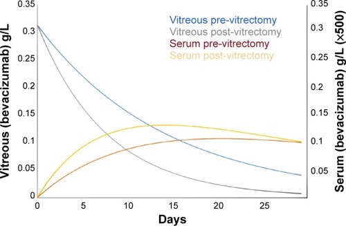
Several important observations regarding serum concentrations and the resultant exposure of the fellow eye to bevacizumab can be made from the graph. In a post-vitrectomy patient, the serum concentration rises faster and peaks earlier than in a pre-vitrectomy patient, with maximum concentrations at 14 days and 20 days, respectively. More importantly, the serum concentration at 7 days in a post-vitrectomy patient is 1.53 times that in a pre-vitrectomy patient and the area under the curve ratio through 7 days is 1.6 times. Therefore, a vitrectomy significantly increases the exposure of the fellow eye to bevacizumab during the first week, which helps to explain the observation made by Zlotcavitch et al.
As a monoclonal antibody against vascular endothelial growth factor (VEGF), bevacizumab works by decreasing the concentration of unbound (metabolically active) VEGF. In eyes with proliferative diabetic retinopathy, the degree of fibrosis depends upon the relative amounts of connective tissue growth factor and VEGF. The introduction of bevacizumab alters the ratio of connective tissue growth factor to VEGF in favor of fibrosis,Citation8 as occurred in this case.
Disclosure
The authors report no conflicts of interest in this communication.
Dear editor
We read with interest the comments of Stewart et al regarding our case of progressive diabetic traction retinal detachment in the fellow eye 1 week after vitrectomy with the use of intravitreal bevacizumab.Citation1 The letter to the editor estimates the vitreous and serum time-dependent concentrations of bevacizumab after an intravitreal injection in vitrectomized and non-vitrectomized eyes. These calculations support the clinical course observed in our patient. Further studies are necessary to quantitatively assess the possible bilateral effect of intravitreal medications and the effects of vitrectomy on the pharmacodynamics of anti-vascular growth factor agents. Although the bilateral response to a unilaterally injected medication is usually beneficial, one should be cognizant of potential progression of diabetic traction retinal detachment.
Disclosure
The authors report no conflicts of interest in this communication.
Reference
- ZlotcavitchLFlynnHWJrAveryRLRachitskayaAProgression to macula-off tractional retinal detachment after a contralateral intraoperative intravitreal bevacizumab injection for proliferative diabetic retinopathyClin Ophthalmol20159697125609907