
Abstract
The treatment of presbyopia has been the focus of much scientific and clinical research over recent years, not least due to an increasingly aging population but also the desire for spectacle independence. Many lens and nonlens-based approaches have been investigated, and with advances in biomaterials and improved surgical methods, removable corneal inlays have been developed. One such development is the KAMRA™ inlay where a small entrance pupil is exploited to create a pinhole-type effect that increases the depth of focus and enables improvement in near visual acuity. Short- and long-term clinical studies have all reported significant improvement in near and intermediate vision compared to preoperative measures following monocular implantation (nondominant eye), with a large proportion of patients achieving Jaeger (J) 2 to J1 (~0.00 logMAR to ~0.10 logMAR) at the final follow-up. Although distance acuity is reduced slightly in the treated eye, binocular visual acuity and function remain very good (mean 0.10 logMAR or better). The safety of the inlay is well established and easily removable, and although some patients have developed corneal changes, these are clinically insignificant and the incidence appears to reduce markedly with advancements in KAMRA design, implantation technique, and femtosecond laser technology. This review aims to summarize the currently published peer-reviewed studies on the safety and efficacy of the KAMRA inlay and discusses the surgical and clinical outcomes with respect to the patient’s visual function.
Introduction
Treatment for the correction of presbyopia has continued to be the focus of considerable research. Typically affecting people from 40 years of age, the loss of near visual acuity is often attributed to increased lens nucleus hardness and subsequent inability of the lens capsule to compress the lens to a more convex state over time.Citation1–Citation3 However, as lens thickness increases with age, the space between the lens and ciliary body reduces, and the angle of zonule insertion may change and therefore render ciliary body contraction ineffective.Citation4–Citation6 Presbyopia can significantly impact the quality of life and combined with an increasingly aging global population it poses a greater demand for spectacle independence.Citation7
Approaches to treat presbyopia have included the use of intracorneal inlays to either change the refractive power of the cornea based on corneal multifocalityCitation8 or increase the refractive power of the central cornea by changing its curvature.Citation9,Citation10 Another inlay method which has been studied in great detail is the use of small-aperture optics to increase the depth of focus based on the pinhole effect.Citation11,Citation12 This commercially available inlay is known as the KAMRA™ inlay (AcuFocus Inc., Irvine, CA, USA), and this review aims to summarize the efficacy and safety of currently published clinical studies of this procedure.
Methodology
Clinical trials of the KAMRA inlay used in this literature review were searched in PubMed using the following keywords alone and in combination (where appropriate): KAMRA, corneal inlay, safety, efficacy, and visual outcomes. In total, 14 clinical trials were identified and used for analysis.
The KAMRA inlay
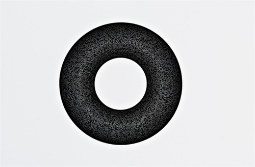
The KAMRA design (ACI7000PDT) consists of a 3.8 mm diameter microperforated (8,400 holes 5–11 µm in diameter) tinted disc with 1.6 mm central aperture at 6 µm thick and is made of polyvinylidene fluoride and carbon nanoparticles. shows the size of the KAMRA inlay compared to a 14 mm soft contact lens. The inlay is designed to be inserted in the line if sight of the nondominant eye and implanted in a femtolaser created corneal lamellar pocket at least 220 µm deep. shows a schematic of the inlay design.
Figure 1 The size of the KAMRA inlay compared to a 14 mm diameter soft contact lens.

Figure 2 A schematic of the KAMRA inlay design.
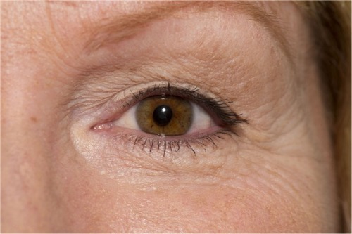
The inlay is designed to allow light to enter through the central aperture, thus reducing retinal image blur and increasing depth of focus to allow increased near and intermediate visual acuity. As the inlay does not split light between different focal points, this allows the patient to maintain binocular summation.Citation13 shows the inlay in situ in a patient’s cornea.
Figure 3 The KAMRA inlay inserted in a patient’s cornea.
Given that this is an additive procedure (ie, no corneal tissue is removed), it can be combined with refractive laser vision correction procedures where the eyes are made emmetropic – here the inlay is situated in a lamellar pocket at least 100 µm beneath the initial laser in situ keratomileusis (LASIK) flap.Citation14 Further, it can be implanted in previously pseudophakic eyes, which has been shown, albeit in a few cases, to produce a significant improvement in near acuity without affecting distance acuity.Citation15 Based on an eye model, it has been suggested that the best depth of focus is achieved where the dominant eye is made plano and the nondominant eye is made myopic (−0.75 to −1.00 D).Citation16
Clinical performance
The efficacy of the KAMRA inlay has been investigated in several studies, albeit in case series where pre- and postoperative measures were compared rather than case-control clinical studies. Nonetheless, all have reported significant improvements in near visual acuity following implantation. However, it should be borne in mind by the reader that all currently published studies are company sponsored (AcuFocus).
In a study comprising hyperopic, myopic, and emmetropic patients (180 patients), the KAMRA inlay (model ACI7000PDT) was implanted in the nondominant eye together with a bilateral LASIK for the ametropic patients. Although only 64 patients were available for follow-up, the KAMRA-treated eye resulted in a seven-line improvement in logMAR uncorrected near visual acuity (UNVA) in hyperopic eyes (to mean of 0.18 logMAR), two lines in myopic eyes (0.12 logMAR), and six lines in emmetropic eyes (0.10 logMAR) after 6 months.Citation14 The smaller improvement in myopic eyes was not unexpected due to preoperative good UNVA, and this was reflected in the patient satisfaction scores for this myopic group where the improvement in overall vision was not statistically significant.Citation14 Uncorrected distance visual acuity (UDVA [logMAR]) also improved in the treated eye, by three lines in hyperopic eyes (to mean of −0.04 logMAR), ten lines in myopic eyes (−0.01 logMAR), and one line in emmetropic eyes (−0.07 logMAR) – again the smaller improvements were not unexpected in the emmetropic and hyperopic eyes.Citation14 Although there were significant differences in UNVA and UDVA between each group preoperatively, no significant differences were observed 6 months after implantation; thus, the KAMRA inlay can be implanted after a LASIK procedure and the postoperative results appear similarly successful despite preoperative ametropia.Citation14
Another case series by the same study group investigated the visual outcomes of the KAMRA inlay (model ACI7000PDT; again implanted in the nondominant eye) in 223 presbyopic patients who had previously undergone LASIK refractive surgery for emmetropia (mean spherical equivalent of −0.18 D in treated eye). After 6 months, the mean UNVA improved from Jaeger (J) 8 (~0.50 logMAR) to J2 (~0.10 logMAR) in the treated eye, but unfortunately binocular UNVA (BUNVA) was not reported.Citation17 However, despite mean UDVA reducing slightly by one line from −0.10 logMAR to 0.00 logMAR in the treated eye, the mean binocular UDVA (BUDVA) remained very good (−0.20 logMAR).Citation17 Although 29% of patients had >0.50 D change, with a slight myopic shift compared to baseline, mean spherical equivalent refraction remained stable.Citation17 Patient satisfaction of their visual status (1= least, 7= most satisfied) without reading glasses under bright light conditions improved significantly compared to baseline for all near (reading newspaper: 3.3±2.1 to 5.0±1.4; reading stock price on medicine bottle: 1.5±1.1 to 4.1±1.8) and intermediate tasks (reading the computer screen: 2.8±1.7 to 5.6±1.2) examined.Citation17
Two-year follow-up of the efficacy of this inlay has also been investigated in 24 emmetropic presbyopes who underwent monocular implantation in the nondominant eye. In this study, the mean UNVA improved from 0.40 logMAR to 0.10 logMAR in the treated eye, with 83% achieving 0.10 logMAR or better.Citation18 Mean unaided intermediate visual acuity (UIVA) improved from 0.20 logMAR to 0.10 logMAR, but UDVA decreased by one line compared to baseline in the treated eye (−0.10 logMAR to 0.00 logMAR). However, this is considered very good acuity and BUDVA remained stable (−0.10 logMAR over the 2-year period).Citation18
Longer-term studies have also been reported, but mainly with the previous (original) version of the KAMRA implant (model ACI7000). This implant is slightly thicker (10 µm) than the current design and has fewer porosity holes (1,600 holes 25 µm in diameter). In a prospective cohort study, 32 naturally emmetropic patients who underwent implantation in the nondominant eye achieved mean UNVA of J2 (~0.10 logMAR) after 2 years in the treated eye compared to J7/J8 (~0.48/0.50 logMAR) preoperatively, with 96.9% of patients reading J3 (~0.18 logMAR) or better.Citation19 Mean BUNVA also improved significantly from J6 (~0.40 logMAR) preoperatively to J1 (~0.00 logMAR). Mean UIVA improved from 0.30 logMAR to 0.10 logMAR in the treated eye and from 0.20 logMAR to 0.00 logMAR binocularly, with 71.9% of patients achieving 0.00 logMAR or better.Citation19 Although there was no significant difference between preoperative (−0.10 logMAR) and postoperative (−0.10 logMAR) BUDVA, six patients experienced a reduction to 0.10 logMAR and two patients to 0.20 logMAR.Citation19 However, mean UDVA in the treated eye remained 0.00 logMAR over the 2-year follow-up period.Citation19 The same study group also reported at 3 years postoperatively on the same patient cohort. Mean UNVA was J1 (~0.00 logMAR), UIVA was 0.10 logMAR, and UDVA was 0.00 logMAR in the treated eye.Citation20
Yılmaz et al investigated the efficacy of the original inlay design up to 4 years postoperatively (n=22 patients) in the natural and post-LASIK (to correct hyperopia) emmetropic presbyopes.Citation21 Here, UNVA improved significantly from J7 (0.40 logMAR) preoperatively to J1 (0.00 logMAR) in the treated eye (mean improvement of 3.8±1.5 lines; 96% reading J3 [~0.18 logMAR] or better) at the last study visit. Compared to baseline, UDVA decreased, albeit statistically insignificantly, by one line (0.00 logMAR to 0.10 logMAR) in the treated eye over the 4-year period.Citation21 The longest follow-up with the KAMRA inlay (ACI7000) was recently reported by Dexl et al, where it was implanted in the nondominant eye of 32 natural emmetropic presbyopes.Citation22 Mean UNVA improved significantly from J7/J8 (~0.50 logMAR) preoperatively to J1 (~0.00 logMAR) at 1 year and remained stable over the next 3 years before tapering slightly to J3 (~0.18 logMAR) after 5 years, with 74.2% of patients reading J3 (~0.18 logMAR) or better in the treated eye. The BUNVA demonstrated the same pattern, but maintained consistently better acuity compared to monocular status, achieving a mean of J2 (~0.10 logMAR) after 5 years, with 45.2% reading at J1 (~0.00 logMAR) or better.Citation22 This pattern was also observed for UIVA in both monocular (0.20 logMAR; remaining similar to preoperative after 5 years) and binocular (0.10 logMAR after 5 years compared to 0.20 logMAR preoperatively) states, with over 50% reading 0.10 logMAR or better.Citation22 As observed in previous studies, mean UDVA decreased slightly from −0.10 logMAR preoperatively before tapering over the next 5 years to 0.10 logMAR in the treated eye; however, mean BUDVA remained very good (−0.10 logMAR) with over 90% achieving 0.00 logMAR or better.Citation22 In this study, acuity in the fellow, untreated eye was also measured preoperatively and at 5 years postoperatively. A similar decrease in UDVA was also observed. As a result, the authors attributed the loss of UDVA in both eyes to natural age-related hyperopic shift previously identified in the Beaver Dam and Liwan Eye Studies.Citation23,Citation24
In addition to measures of acuity, reading performance has also been assessed with the original KAMRA inlay. Dexl et al reported significant improvements in reading distance (reduced working distance), reading acuity at best working distance, and smallest print size in over a 2-year period in 24 natural emmetropic presbyopes.Citation25 However, although an increase in reading speed was also observed, this was not statistically significant.Citation25
More recently, Tomita and Waring divided their patient cohort (n=277) into three age groups (40–49, 50–59, and 60–65 years) and performed simultaneous LASIK (to correct hyperopia) and KAMRA (ACI7000PDT) implantation to investigate the effect of age on safety and clinical outcomes over a 1-year period. The mean UNVA and UDVA were similar between groups, but the 60–65 years age group exhibited the largest gain in both outcomes at the final follow-up visit.Citation26 Although this result was not unexpected, this group had lowest reduction in spectacle independence. The authors concluded that age should be taken into consideration during consultation in order to manage patient expectations postoperatively.Citation26
Safety and adverse events
From the longer-term studies previously mentioned, it is apparent that UDVA in the treated eye and under binocular conditions becomes slightly compromised with the KAMRA inlay. However, in order to establish whether this is due to uncorrected residual ametropia or otherwise, measures of best corrected distance visual acuity (CDVA) have been evaluated. In the 3-year follow-up study by Seyeddain et al, although CDVA remained stable over time, 28.3% of patients lost one line and 3.1% lost two lines of acuity in the treated eye. Binocular CDVA was, however, stable and no patient lost a line of acuity during the follow-up period.Citation20 No inlays had to be explanted, but two had to be recentered after 6 months due to misalignment and no observable improvement in the near and intermediate acuity; once recentered, both patients subsequently achieved a significant improvement in these outcomes.Citation20 One patient developed flap striae at 1 month and epithelial ingrowth at the flap interface, but were successfully resolved following surgical intervention. Of note, however, was the development of iron deposits in 56.2% of patients within a median interval of 18±9 months after implantation. Although these deposits were not associated with visual or refractive outcomes, corneal topography revealed very small areas of flattening overlying the deposits.Citation20 Corneal endothelial cell density decreased slightly (5.73%) after 6 months, but further significant loss was not observed thereafter.Citation20 The most common patient-reported symptoms at the final study visit (3 years) were night vision problems (40.6% mild, 6.3% moderate, and 15.6% severe cases) and halos (34.4%, 25.0%, and 3.1%). Although dryness and glare were also reported, most cases were mild or moderate in nature.Citation20
In the 4-year follow-up study, 27% of patients lost more than one line of CDVA, but mean CDVA did not change significantly from baseline (0.00 logMAR) to the final study visit (0.00 logMAR) in the treated eye.Citation21 Four patients had the inlay explanted: one at 6 weeks postimplantation due to the detection of a buttonhole flap, two at 3 months due to large refractive shifts (−2.00 D and +3.00 D), and one at 17 months due to shallow implantation. All four of these patients were, however, successfully treated and no loss of monocular or binocular CDVA was observed.Citation21 Complications reported included dry eye (n=4 treated eyes) and epithelial ingrowth (n=5) related to LASIK, but it is not clear how the authors differentiated the cause between previous LASIK procedure and that of KAMRA implantation. In the 5-year follow-up study by Dexl et al, mean CDVA remained stable at −0.10 logMAR for the first 3 years before reducing to 0.00 logMAR after 5 years in the treated eye, with 45.2% of patients losing one line and 22.6% losing two or more lines.Citation22 A similar pattern emerged under binocular conditions, where mean CDVA reduced slightly from −0.20 logMAR preoperatively to −0.10 logMAR after 5 years, with 51.6% losing one line and 16.1% losing two or more lines.Citation22 As observed in the study by Seyeddain et al (old inlay design ACI7000), iron deposits developed in 56.3% of treated eyes at the 3-year follow-up and were associated with overlying corneal flattening; however, no further cases were observed for the remaining study period.Citation20,Citation22 The inlay was explanted from only one eye at the 36-month follow-up due to a hyperopic shift causing dissatisfaction with near and distance vision.
With the current inlay, Tomita et al reported no significant change in CDVA from baseline to 6 months postoperatively, although 14% of eyes lost one line of acuity in the treated eye. Despite this, all patients had monocular CDVA of 0.00 logMAR or better.Citation17 Visual symptoms were also evaluated, albeit using a nonvalidated scale (0= no symptoms, 7= very heavy symptoms); here, dryness, glare, halo, and night vision disturbances increased significantly, but were considered mild by the study authors.Citation17 All patients were post-LASIK and therefore may be predisposed to such symptoms, which are typically associated with laser refractive procedures, but these symptoms should not be discounted, particularly when gaining consent for surgery.Citation17,Citation27 Similar results were also found by Seyeddain et al, where CDVA reduced by a mean of 2.5 letters from −0.10 logMAR to 0.00 logMAR in the treated eye, with 16.7% of patients losing one line of acuity at the last follow-up (2 years); however, all of these patients achieved 0.00 logMAR.Citation18 No implants had to be recentered or explanted, and no ocular inflammation was observed during the study period.Citation18 Adverse events included epithelial ingrowth at the pocket entrance at 1 month in one patient, epithelial iron deposits near the inlay margin at 18 months in one patient, while several others (number not reported) developed a thin hazy appearance at the outer and or inner rim of the inlay; however, they did not require treatment and were not associated with any visual or refractive outcomes.Citation18 Further, endothelial cell count (ECC) and central corneal thickness (CCT) were not affected over the 2-year follow-up period.Citation18 In another study of 24 emmetropic presbyopes who underwent monocular (nondominant) implantation, despite 16.7% losing one line and 4.1% losing two lines of CDVA in the treated eye at the final (2-year) follow-up, over 95% achieved CDVA of 0.00 logMAR and binocular CDVA (−0.10 logMAR) remained stable over the entire study period.Citation28 No deposits were observed in or on the cornea, ECC and CCT were unaffected, and no patient required the inlay to be recentered or explanted.Citation28 Only one patient experienced epithelial ingrowth at the pocket entrance after 1 month but remained stable and required no intervention.Citation28
Confocal microscopy studies describing the corneal appearance with the implant in situ and postexplantation have also been performed. Abboud et al found that the implant changed the normal structure of the cornea (decreased keratocyte density in the anterior stroma, loss of subbasal nerve plexus), but this did not result in visual complications.Citation29 However, keratocyte activation, also observed in typical laser refractive surgery procedures, was observed and this was significantly correlated with reduced UNVA, corrected near visual acuity, and CDVA.Citation29 Thus, using lower laser energy, creating a thicker flap, and applying intensive steroid therapy postoperatively are suggested as key for good visual outcomes and healing response.Citation29
Discussion
With both the original and current designs of the KAMRA implant, the clinical studies have clearly demonstrated significant improvements in both near and intermediate visual acuity with a minimal impact on distance vision following monocular implantation in the nondominant eye of presbyopes. Although longer-term studies for the current design are not yet reported, it is likely that the reduced incidence of loss of UDVA and CDVA is a result of improved surgical technique and implant design.Citation27 The ACI7000 inlay was implanted under a corneal flap 170–180 µm deep using a microkeratome or femtosecond laser,Citation20–Citation22 whereas the thinner ACI7000PDT inlay is implanted at least 220 µm deep in a pocket created with a femtosecond laser, thus the latter is less likely to affect corneal topography and subsequent visual acuity.Citation18 Not only does the pocket technique allow for better centration, it also requires a smaller incision such that fewer corneal nerves are cut and therefore reduces the likelihood of postoperative dry eye typically associated with laser refractive procedures.Citation13,Citation30 Using femtosecond laser over mechanical microkeratome to create corneal flaps or pockets has been shown to provide lower incidence of postoperative dry eye, faster visual recovery, better UDVA, and more predictable incision depths.Citation18,Citation30,Citation31 Although both inlay designs are microperforated to allow water and nutritional flow, ACI7000PDT is thinner (6 µm vs 10 µm) and has more holes (8,400 vs 1,600), so is less likely to induce corneal thinning and epithelial decompensation.Citation13,Citation32 Indeed, only one in 20 patients developed epithelial iron deposits with the new design compared to over 56% with the ACI7000 inlay.Citation18,Citation20 However, in either case, these deposits neither interfered with vision nor were they associated with refractive outcomes.Citation18
In cases where the inlays were explanted, all resolved without sequelae and without significant impact on distance vision;Citation2 indeed, Alio et al reported that the KAMRA inlay is safe to remove and removal has minimal impact on corneal topography and aberrometry during and after recovery if explanted before 6 months.Citation33 Thereafter, the changes in corneal topography may remain permanent.Citation33 Despite decentration as little as 0.5 mm significantly affecting retinal image quality, recentration can be performed easily with subsequent improvements in near and distance acuity.Citation14,Citation20 Another major advantage of this surgery is that it is additive such that where the KAMRA inlays are removed, future options for presbyopia correction, including corneal approaches, are still available to the patient.Citation32
Given that the inlay relies upon the pinhole effect to achieve improvement in near vision, the effect of pupil size on clinical outcome has been investigated, as pupil size is well known to influence optical image quality based on the amount of visual aberrations that pass in the eye after refractive surgery.Citation34–Citation36 An optical simulation of the KAMRA implant has shown that for combinations of pupil sizes and field angles, image brightness on the retina may be attenuated up to 60% due to its central position in front of the pupil and opaque nature, and it is predicted that this vignetting effect may lead to a clinically relevant reduction in contrast sensitivity.Citation37 However, in a study of 584 actual KAMRA inlay treated eyes (584 patients), Tomita et al report that pupil size had no impact on visual acuity after implantation. There was no statistically significant difference between uncorrected and distance-corrected near visual acuity for both mesopic and photopic size groups.Citation38 One study has reported a statistically significant reduction in contrast sensitivity in eyes with KAMRA inlays compared to preoperative measurements after 24 months in photopic and mesopic conditions, but these were found at higher spatial frequencies and the measures were within the range of the normal population.Citation18 Another paper published by Vilupuru et al reported no loss of binocular contrast in either photopic or mesopic conditions for a series of 507 patients implanted monocularly with the KAMRA inlay. This same study also compared contrast sensitivity results for KAMRA inlay subjects to subjects treated with bilateral multifocal or accommodating intraocular lenses. Under all conditions, the KAMRA inlay patients demonstrated better contrast sensitivity than patients with the tested lenses.Citation39
Conclusion
The KAMRA inlay is a safe and effective clinical procedure for the treatment of presbyopia, where significant improvement in near and intermediate visual acuity and function has been reported in several large and long-term follow-up studies. Although distance visual acuity has been compromised in some patients, the reductions were not clinically significant. Iron deposits within the corneal epithelium have been observed, but these are not considered to affect vision, and the incidence has reduced with improvements in surgical methods and the inlay design itself. Further studies with the latest KAMRA inlay are required to establish the longer-term safety and clinical stability of visual acuity.
Disclosure
The authors report no conflicts of interest in this work. The figures were supplied courtesy of AcuFocus Inc. (Irvine, CA, USA).
References
- PauHKranzJThe increasing sclerosis of the human lens with age and its relevance to accommodation and presbyopiaGraefes Arch Clin Exp Ophthalmol199122932942961869070
- GlasserACampbellMCPresbyopia and the optical changes in the human crystalline lens with ageVision Res19983822092299536350
- HeysKRTruscottRJThe stiffness of human cataract lenses is a function of both age and the type of cataractExp Eye Res200886470170318289531
- StrenkSASemmlowJLStrenkLMMunozPGronlund-JacobJDeMarcoJKAge-related changes in human ciliary muscle and lens: a magnetic resonance imaging studyInvest Ophthalmol Vis Sci19994061162116910235549
- StrenkSAStrenkLMSemmlowJLHigh resolution MRI study of circumlental space in the aging eyeJ Ref Surg2000165659660
- StrenkSAStrenkLMKoretzJFThe mechanism of presbyopiaProg Retin Eye Res200524337939315708834
- PatelIWestSKPresbyopia: prevalence, impact, and interventionsCommunity Eye Health20072063404117971909
- LimnopoulouANBouzoukisDIKymionisGDVisual outcomes and safety of a refractive corneal inlay for presbyopia using femtosecond laserJ Refract Surg2013291121823311737
- ChayetAGarzaEBCombined hydrogel inlay and laser in situ keratomileusis to compensate for presbyopia in hyperopic patients: one-year safety and efficacyJ Cataract Refract Surg201339111713172124021565
- GarzaEBGomezSChayetADishlerJOne-year safety and efficacy results of a hydrogel inlay to improve near vision in patients with emmetropic presbyopiaJ Refract Surg201329316617223446012
- TaberneroJSchwarzCFernándezEJArtalPBinocular visual simulation of a corneal inlay to increase depth of focusInvest Ophthalmol Vis Sci20115285273527721436279
- LindstromRLMacRaeSMPeposeJSHoopesPCSrCorneal inlays for presbyopia correctionCurr Opin Ophthalmol201324428128723703461
- DexlAKSeyeddainORihaWOne-year visual outcomes and patient satisfaction after surgical correction of presbyopia with an intracorneal inlay of a new designJ Cataract Refract Surg201238226226922138501
- TomitaMKanamoriTWaringGOSimultaneous corneal inlay implantation and laser in situ keratomileusis for presbyopia in patients with hyperopia, myopia, or emmetropia: six-month resultsJ Cataract Refract Surg201238349550622340607
- HuseynovaTKanamoriTWaringGOIVTomitaMSmall-aperture corneal inlay in presbyopic patients with prior phakic intraocular lens implantation surgery: 3-month resultsClin Ophthalmol201371683168624003304
- TaberneroJArtalPOptical modelling of a corneal inlay in real eyes to increase depth of focus: optimum centration and residual defocusJ Cataract Refract Surg201238227027722177883
- TomitaMKanamoriTWaringGONakamuraTYukawaSSmall-aperture corneal inlay implantation to treat presbyopia after laser in situ keratomileusisJ Cataract Refract Surg201339689890523688876
- SeyeddainOBacherneggARihaWFemtosecond laser-assisted small-aperture corneal inlay implantation for corneal compensation of presbyopia: two-year follow-upJ Cataract Refract Surg201339223424123245507
- SeyeddainORihaWHohensinnMNixGDexlAKGrabnerGRefractive surgical correction of presbyopia with the AcuFocus small aperture corneal inlay: two-year follow-upJ Refract Surg2010261070771520438021
- SeyeddainOHohensinnMRihaWSmall-aperture corneal inlay for the correction of presbyopia: 3-year follow-upJ Cataract Refract Surg2012381354522018596
- YılmazÖFAlagözNPekelGIntracorneal inlay to correct presbyopia: long-term resultsJ Cataract Refract Surg20113771275128121570249
- DexlAKJellGStrohmaierCLong-term outcomes after monocular corneal inlay implantation for the surgical compensation of presbyopiaJ Cataract Refract Surg201541356657525726504
- LeeKEKleinBEKleinRWongTYChanges in refraction over 10 years in an adult population: the Beaver Dam Eye studyInvest Ophthalmol Vis Sci20024382566257112147586
- HeMHuangWLiYZhengYYinQFosterPJRefractive error and biometry in older Chinese adults: the Liwan eye studyInvest Ophthalmol Vis Sci200950115130513619553618
- DexlAKSeyeddainORihaWHohensinnMHitzlWGrabnerGReading performance after implantation of a small-aperture corneal inlay for the surgical correction of presbyopia: two-year follow-upJ Cataract Refract Surg201137352553121262559
- TomitaMWaringGOOne-year results of simultaneous laser in situ keratomileusis and small-aperture corneal inlay implantation for hyperopic presbyopia: comparison by ageJ Cataract Refract Surg201541115216125532642
- MelkiSAAzarDTLASIK complications: etiology, management, and preventionSurv Ophthalmol20114629511611578645
- DexlAKSeyeddainORihaWReading performance and patient satisfaction after corneal inlay implantation for presbyopia correction: two-year follow-upJ Cataract Refract Surg201238101808181622921231
- AbboudAJavaloyJAlióJLConfocal microscopy evaluation of the corneal response following AcuFocus KAMRA inlay implantationJ Refract Surg201430317217824763721
- SalomãoMQAmbrósioRWilsonSEDry eye associated with laser in situ keratomileusis: mechanical microkeratome versus femtosecond laserJ Cataract Refract Surg200935101756176019781472
- KezirianGMStonecipherKGComparison of the IntraLase femtosecond laser and mechanical keratomes for laser in situ keratomileusisJ Cataract Refract Surg200430480481115093642
- ArltEMKrallEMMoussaSGrabnerGDexlAKImplantable inlay devices for presbyopia: the evidence to dateClin Ophthalmol2015912913725609913
- AlióJLAbboudaAHuseynliSKnorzMCDurrieDSRemovability of a small aperture intracorneal inlay for presbyopia correctionJ Refract Surg201329855055623909782
- MartinezCEApplegateRAKlyceSDMcDonaldMBMedinaJPHowlandHCEffect of pupillary dilation on corneal optical aberrations after photorefractive keratectomyArch Ophthalmol19981168105310629715686
- MillerJMAnwaruddinRStraubJSchwiegerlingJHigher order aberrations in normal, dilated, intraocular lens, and laser in situ keratomileusis corneasJ Refract Surg2002185S579S58312361161
- WangBCiuffredaKJDepth-of-focus of the human eye: theory and clinical implicationsSurv Ophthalmol2006511758516414364
- LangenbucherAGoebelsSSzentmáryNSeitzBEppigTVignetting and field of view with the KAMRA corneal inlayBiomed Res Int2013201315459324324954
- TomitaMKanamoriTWaringGOHuseynovaTRetrospective evaluation of the influence of pupil size on visual acuity after KAMRA inlay imp lantationJ Refract Surg201430744845324983830
- VilupuruSLinLPeposeJSComparison of contrast sensitivity and through focus in small aperture inlay, accommodating intraocular lens, or multifocal intraocular lens subjectsAm J Ophthalmol2015160115016225896457