
Abstract
Purpose
To describe the use of a ruler to increase the accuracy and customization of Lester Jones tube placement.
Materials and methods
This was a review of 92 patients who underwent minimally invasive conjunctivodacryocystorhinostomy using this instrument.
Results
The success rate was 100%, with an average operating time of 15 minutes. In nine patients, late migration of the Jones tube into the nasal cavity required a secondary intervention, with successful Jones tube repositioning. In five cases, obstruction of the tube opening was caused by conjunctival overgrowth.
Conclusion
The millimetric ruler engraved in the sleeve of the trocar allows measuring and placing a personalized tube for each patient.
Introduction
Conjunctivodacryocystorhinostomy (CDCR) with a Jones tube is the chosen technique to treat higher lacrimal duct obstructions.Citation1,Citation2 However, the complications associated with it and the difficulty of the technique may turn out to be frustrating.Citation3,Citation4 In this study, we present a technique that facilitates the insertion of the Jones tube with excellent results.
Materials and methods
This study adhered to the principles outlined in the Declaration of Helsinki, and was approved by the Institutional Review Board – IRB 00003580. In this retrospective study, the charts of 92 patients who underwent CDCR as a primary or revision surgery at Hospital Italiano de Buenos Aires between April 2008 and April 2014 and had follow-up for more than 6 months were reviewed.
Patients comprised 60% males (n=56) and 40% females (n=36). The mean age was 51 years, and the mean follow-up period was 30 months (range: 18–60 months). A variety of Jones tubes ranging from 19 to 23 mm in length were used. The exclusion criteria were lacrimal duct obstruction due to tumor, sinusitis, and bone diseases.
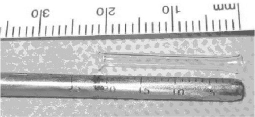
A video endoscope (Tricam SL; KARL STORZ GmbH and Co KG, Tuttlingen, Germany) was introduced into the middle nasal cavity for visualization and electrocauterization. The inferior third of the caruncle was excised. The trocar () was inserted from the semilunar directed to the middle meatus through the thin lacrimal bone (). Once the trocar was in a satisfactory position, the central wire was removed, and the sleeve allowed the direct insertion of a guide rod. Before removing it, the millimetric ruler (), which was engraved in the sleeve, was checked. In this way, we were able to find a personalized tube for each patient. After the trocar sleeve was removed, a Pyrex Lester Jones tube was inserted down the guide rod. The proximal end of the tube was secured in position at the medial canthus with a 6.0 nylon suture, which was removed 20 days after the surgery. See the full video of the surgery using the millimetric ruler.
Figure 1 Trocar for Jones tube placement, with a millimetric ruler engraved.
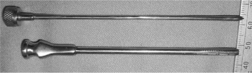
Figure 2 Trocar inserted through the lacrimal bone.
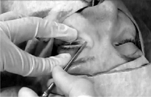
Figure 3 Measurement by Jones tube for a personalized placement.
Results
The success rate was 85%. The other 15% presented mild complications, which were solved with a second intervention. The surgeries were performed in an average time of 15 minutes.
Migration of the tube was the most frequent complication. In nine cases, the tube migrated into the nose, with successful repositioning. Obstruction of the Jones tube by the conjunctiva was the second-most frequent complication, and five cases needed excision of the conjunctiva.
Discussion
Lacrimal canalicular bypass tract surgery, first described by Jones, is indicated for epiphora secondary to obstruction at the level of the canaliculi, failed DCR, and lacrimal pump dysfunction.Citation1–Citation3,Citation5 Unfortunately, this procedure is associated with multiple complications; therefore, it requires long-term follow-up.Citation6–Citation8
A minimally invasive approach with no skin incision has been described.Citation9,Citation10 However, placement of a Jones glass tube can be difficult with this technique. The distal end is wider than the tube’s outer diameter, so soft tissue within the tunnel is often drawn in front of the tube, complicating placement. Moreover, the length of the tube cannot be calculated beforehand, and its choice respresent a challenge for the surgeon. Some authors identify the latter as a major reason of failure.Citation11
The use of a steel double-component trocar guide by endoscopic view maintains the desired tract for positioning the tube; this reduces the operating time and the risk of malpositioning. We add an advantage to the trocar with a millimetric ruler, which is engraved in the sleeve to measure the tube for every patient.
With this technique, the placement of the tube is easier and quicker, and not many changes are necessary to choose the most appropriate one. The limitations of our study are as follows: it is retrospective, there was no control group, and longer follow-up may be needed to determine long-term complications.
Conclusion
This millimetric ruler engraved on the trocar allows measuring and placing a personalized tube for each patient. It also eases the Jones tube placement with endoscopic control, maintaining the desired tract for positioning the tube. This reduces the operating time and the risk of malposition.
Disclosure
The authors report no conflicts of interest in this work.
References
- JonesLTConjunctivodacryocystorhinostomyAm J Ophthalmol19655977378314288913
- DoucetTWHurwitzJJCanaliculodacryocystorhinostomy in the management of unsuccessful lacrimal surgeryArch Ophthalmol19821006196217073579
- WelhamRAGuthoffRThe Lester-Jones tube: a 15-year follow-upGraefes Arch Clin Exp Ophthalmol19852231061084007506
- VicinanzoMGAllamneniCComptonCJLongJANabaviCBThe prevalence of air regurgitation and its consequences after conjunctivodacryocystorhinostomy and dacryocystorhinostomy in continuous positive airway pressure patientsOphthal Plast Reconstr Surg201531269271
- RoseGEWelhamRAJones’ lacrimal canalicular bypass tubes: twenty-five years’ experienceEye (Lond)1991513192060661
- SendulSYCagatayHHDirimBComparison of Medpor coated tear drainage tube versus silicon tear drainage tube in conjunctivodacryocystorhinostomy: problems and solutionsScientificWorldJournal2014201416483425379518
- SekharGCDortzbachRKGonneringRSLemkeBNProblems associated with conjunctivodacryocystorhinostomyAm J Ophthalmol19911125025061951585
- SteinsapirKDGlattHJPuttermanAMA 16-year study of conjunctival dacryocystorhinostomyAm J Ophthalmol19901093873932330941
- DevotoMHBernardiniFPde ConciliisCMinimally invasive conjunctivodacryocystorhinostomy with Jones tubeOphthal Plast Reconstr Surg200622253255
- KomínekPCervenkaSConjunctivodacryocystorhinostomy tube placement with a urologic catheterOphthal Plast Reconstr Surg200521235236
- ChangMLeeHParkMBaekSLong-term outcomes of endoscopic endonasal conjunctivodacryocystorhinostomy with Jones tube placement: a thirteen-year experienceJ Craniomaxillofac Surg20154371025459376