
Abstract
Purpose
The aim of the study was to detect the expression of Sox10 in human nasopharyngeal carcinoma (NPC) and investigate the relationship between its expression and the clinicopathological characteristics of NPC patients.
Patients and methods
Tumor specimens (n=105) were retrospectively collected from patients with NPC diagnosed between 2004 and 2005 who presented at Hunan Cancer Hospital. Immunohistochemistry analyses were performed to characterize the expression of Sox10 in NPC. Kaplan–Meier survival and Cox regression analyses were employed to evaluate the prognosis of 105 NPC patients.
Results
The results showed that Sox10 was markedly overexpressed in human NPC tissues. Analysis of clinicopathological parameters showed that high Sox10 expression was significantly correlated with the clinical stage (P=0.032), T classification (P=0.034), and lymph node metastasis (P=0.03). Cox regression analyses further showed that Sox10 expression was an independent prognostic factor for overall survival (P=0.005). This is the first time Sox10 has shown its importance in predicting NPC progressiveness and survival outcomes.
Conclusion
Sox10 serves as a potential biomarker for NPC patients. It may hopefully become a novel therapeutic target for NPC patients.
Introduction
Nasopharyngeal carcinoma (NPC) is a highly invasive malignancy that rises from the epithelial lining of the nasopharynx. With an estimated 86,700 new cases and 50,800 deaths in 2012, NPC is viewed as a relatively rarer form of cancer globally. However, in endemic regions, including Southern China and Southeast Asia, the annual incidence rate reached 15–50/100,000.Citation1 Although early stage NPC can be managed with high cure rate under radiotherapy, 70%–90% of NPC patients, at the time of initial diagnosis, are characterized by cervical nodal metastasis.Citation2,Citation3 The prognosis of NPC has improved a lot with the use of radiotherapy and chemotherapy, but for locally advanced and metastatic NPC, the result remains unsatisfactory due to local relapse and distant metastasis.Citation4 An urgent need exists to identify novel and specific biomarkers that have clinicopathologic and prognostic values in NPC, and hopefully, help to guide the development of new therapeutic regimens.
Transcription factors of the Sox (sex-determining region Y-box protein) protein family, characterized by a conserved high mobility group DNA binding domain, regulate many diverse developmental processes as well as control homeostasis in adult tissues.Citation5 There were 20 Sox genes identified. Among these, Sox10 gene was the first Sox gene discovered to be involved in neural crest development. It functions by directing the activity of other genes that signal differentiation of neural crest into other cells, so it is essential for the survival of neural crest precursor cells and for proper differentiation of neural crest-derived melanocytes and glia.Citation6 The association between Sox10 mutation and myelination deficiency in human being’s Waardenburg–Hirschsprung syndrome and disruptions of neural crest development has been established.Citation7
In the last few years, an increasing amount of evidence has revealed that dysfunction and mutations of Sox factors are implicated in several human diseases, including a variety of cancers. These cancer cells are mostly originated in tissues overlapping with their expression pattern during embryonic development. The involvement of Sox10 in human cancer has been almost exclusively in melanomas and some brain tumors.Citation8 Sox10 activates the expression of microphthalmia-associated transcription factor, a well-established oncogene in melanoma, and high expression of Sox10 in melanoma has been found to be involved in metastatic potential of melanoma.Citation9–Citation11 However, currently, little information has been addressed concerning the association between Sox10 and carcinogenesis and progression of NPC.
In the present study, for the first time, we investigated Sox10 expression in NPC tissue specimens by immunohistochemistry (IHC) analyses. Besides this, through further analyses, we studied the relationship between Sox10 expression and NPC clinical parameters, as well as the value of Sox10 in predicting NPC prognosis.
Patients and methods
Study patients and treatments
Between May 2004 and July 2005, 105 consecutive patients with undifferentiated nonkeratinizing nasopharyngeal carcinoma NPC in the Department of Radiotherapy, the Affiliated Cancer Hospital of Xiangya School of Medicine, Central South University, Changsha, People’s Republic of China, were enrolled retrospectively in this study. The medical ethics committee of Hunan Cancer Hospital approved the present retrieval method of cancer specimens. The criteria are listed as follows: histologically confirmed NPC, age ranging from 17 years to 77 years, managed in accordance with a uniform protocol, enough available tissue, and clinical follow-up. All patients underwent restaging according to the 2007 International Union against Cancer tumor–node–metastasis (TNM) classification. All patients received conventional two-dimensional radiotherapy. Radiotherapy was administered five times a week at a dose of 2 Gy/day. The accumulated dose of radiation ranged between 66 Gy and 78 Gy. In all, 34 patients were treated with boost dose to skull base. Patients did not receive chemotherapy unless metastasis or recurrence occurred. The primary end point was overall survival. Overall survival was defined as the time (in months) from the date of admission to the date of death from any causes or the last follow-up. The follow-up period ended on December 31, 2010, with a median follow-up of 61 months (range 9–80 months).
IHC and evaluation of immunohistochemical staining
Detailed steps for IHC were following the manufacturer’s protocol. Briefly, antigen retrieval was performed in citrate buffer (pH 6.0) for 20 minutes using a microwave oven. After blocking in 10% normal blocking serum at room temperature for 10 minutes, slides were incubated with Sox10 antibody (1:200; Santa Cruz Biotechnology Inc., Dallas, TX, USA) at 4°C overnight. Negative (omission of the primary antibody) and positive controls were included according to manufacturer’s datasheet of each antibody.
Semiquantitative assessment of the staining, including analysis of both the intensity and percentage of the stained cells, was used. The staining intensity was scored as 0, 1+, 2+, or 3+ corresponding to colorless, buff, brownish yellow, and dark brown, respectively. The percentage of positive stained cells at each intensity was estimated. Final scores ranging from 0 to 300 was calculated by multiplying the intensity and the percentage scores. The cutoff value for Sox10 was chosen based on a measure of heterogeneity using the log-rank test statistical analysis with respect to overall survival. An optimal cutoff value was identified: a staining index of ≥14 was used to define tumors of high expression and ≤13, tumors of low expression.
Statistical analysis
SPSS 18.0 software (SPSS Inc., Chicago, IL, USA) was used for statistical analysis. The Pearson’s chi-squared test was used to determine the correlation between Sox10 expression and clinical factors. Survival curves were plotted using the Kaplan–Meier method and compared with the log-rank test. Statistically significant P-value was set as <0.05.
Results
Sox10 is highly expressed in NPC patients
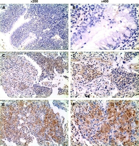
To determine Sox10 expression in clinical samples, we performed IHC analysis in 105 NPC specimens and adjacent tissues. It was revealed that Sox10 was expressed in 84.8% of tumor samples (89 of 105). In contrast, the adjacent tissues exhibited undetectable or low Sox10 staining ().
Figure 1 Sox10 expressed in NPC tissue and adjacent nasopharyngeal tissue.
Abbreviation: NPC, nasopharyngeal carcinoma.
Sox10 is associated with clinical aggressiveness in NPCpatients
To elaborate the relationship between Sox10 expression levels and clinicopathological characteristics, 105 paraffin- embedded NPC tissue specimens were selected for Sox10 IHC staining. A summary of Sox10 expression and clinicopathological parameters is presented in . No statistically significant associations were observed between Sox10 expression and sex, age, or N classification at the initial diagnosis (P>0.05). However, high Sox10 expression was significantly correlated with the clinical stage (P=0.032), T classification (P=0.034), and advanced N stage (P=0.03).
Table 1 Correlation between Sox10 expression and clinicopathologic characteristics of NPC
Sox10 expression is closely correlated with adverse NPC overall survival time
To evaluate the prognostic importance of high Sox10 expression in patients with NPC, we used Cox proportional hazards regression model and Kaplan–Meier method. According to univariate analysis, the overall survival of the NPC patients was correlated with Sox10 expression (P=0.005) and age (P=0.027). Multivariate analysis showed that Sox10 may be viewed as an independent predictor for NPC overall survival (P=0.011; ). Kaplan–Meier method and the log-rank test also showed a significantly inverse correlation between high Sox10 expression level and patient overall survival (P=0.004), revealing that higher levels of Sox10 expression were associated with shorter survival rate (). These results, all together, revealed that Sox10 may be a potential biomarker for the prediction of prognosis of NPC patients receiving radiotherapy.
Figure 2 Survival analysis of 105 NPC patients by Kaplan–Meier method.
Abbreviation: NPC, nasopharyngeal carcinoma.
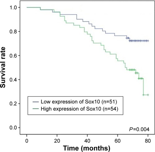
Table 2 Univariate and multivariate statistical analyses for various prognostic parameters in NPC patients
Discussion
Characterized by a conserved high mobility group DNA binding domain, the Sox family is a group of related transcription factors that have demonstrated their importance in chondrogenesis, hematopoiesis, neural crest development, and neurogenesis. Over the years, continuous studies have revealed that many genes first identified as key genes in cancer development play crucial roles in embryogenesis. Conversely, many of the proteins controlling embryonic development are also involved in carcinogenesis, indicating that both processes are closely related.Citation12 An increasing number of studies have discovered the association between Sox genes and tumorigenesis in various kinds of malignancies, including glioma, breast cancer, non-small cell lung cancer, gastric cancer, esophageal cancer, and colon rectal cancer.Citation13–Citation18 The most thoroughly studied member in this family is Sox2. Luo et al found that Sox2 was highly expressed in NPC compared with the nontumorous tissues. Furthermore, Sox2 correlated significantly with several clinical–pathological factors and epithelial–mesenchymal transition-associated indicators (E-cadherin/N-cadherin and Snail).Citation19
With regard to Sox10, it was found to exist in various tissues, including heart, lungs, brain, adrenal glands, bladder, pancreas, salivary glands, colon, prostate, and testis in adulthood. The existence of Sox10 in visceral tissues was believed to be attribute to the presence of glial components in the peripheral nervous system in these tissues.Citation20 Although many studies have discovered the role of Sox10 mainly in melanoma, in salivary gland tumors, and brain tumors, like anaplastic oligodendroglioma, studies focusing on Sox10 in NPC are limited.Citation8,Citation21
Our study discovered that Sox10 was highly expressed in NPC tissue, with staining found in both the nucleus and the cytoplasm. This is similar to the result of Sox10 staining in salivary adenoid cystic and breast basal-like carcinomas.Citation22
We discovered that high expression of Sox10 was correlated with advanced tumor stages and advanced N stage in NPC, which suggests that high expression of Sox10 correlates with locally invasive features. Many studies showed the involvement of Sox10 in tumor aggressiveness. In melanoma, Hoek et al described that high levels of Sox10 have been correlated with a proliferative gene expression signature and low levels are associated with the gene expression profile of a highly invasive phenotype.Citation23,Citation24 Hoang et al’sCitation25 research retrospectively studied 428 cases of invasive ductal carcinoma of the breast. A significant association between Sox10 expression and high-grade, estrogen receptor/progesterone receptor-negative, triple-negative, or basal-like invasive ductal carcinoma was discovered. These results may be explained by the mechanism in which Sox10 works. One of the genes that is crucial in regulating the growth, differentiation, and survival of cells of the melanocytic lineage is microphthalmia-associated transcription factor (MITF).Citation26 One way that Sox10 works is to act on MITF upstream, activate MITF expression, as well as synergize with MITF to activate downstream targets.Citation27 Cronin et al’sCitation28 study discovered previously unknown mutations in MITF and Sox10. These mutations added up to >9% in primary melanoma tumors and 22% in metastatic melanoma tumors, which implied the involvement of MITF and Sox10 in melanoma.
Using Kaplan–Meier analysis, we discovered that high expression of Sox10 is associated with adverse overall survival and could be used as an independent prognostic marker. Our finding may be explained by the involvement of Sox10 in stem cell marker potential. Sox10 has been viewed as a stem cell marker and is found to be expressed extensively in melanoma.Citation29 Studies also found that stable Sox10 knockdown in human melanoma cells arrested cell growth, altered cellular morphology and cell cycle, and induced senescence.Citation30 Apart from in melanoma,Citation31 Sox10 has also shown its stem cell marker potential in breast cancer. Sox10 was specifically expressed in mammary cells exhibiting the highest levels of stem/progenitor activity. This included fetal and adult mammary cells in vivo and mammary organoids in vitro. Sox10 was functionally relevant, as its deletion reduced stem/progenitor competence, whereas its overexpression increased stem/progenitor activity.Citation32 Also, as a key downstream effector of Sox9 in the mammary stem cell induction, Sox10 promotes the tumorigenic and metastasis-seeding abilities of human breast cancer cells and is associated with poor patient survival.Citation31 Tumor stem cells can lead to tumor cell proliferation and resistance to treatment. The mechanism on which Sox10 works on NPC remains to be explored by more work.
However, there are some limitations in our study. First, considering the favorable prognosis of patients diagnosed with NPC, we selected patients from 2004 to 2005 to better evaluate the prognosis. This resulted in incomplete data and inability to analyze progression-free survival because of lack of information in the Province Public Security Bureau and many people changed their phone numbers. Also, no patients included received concurrent chemoradiotherapy since it was not the standard treatment. From another viewpoint, this may better review the relationship between Sox10 expression and radiosensitivity. Second, the number of patients selected was 105. The results would be more convincing if more patients were included.
Next, we are planning to further validate the relationship between Sox10, radiosensitivity, and prognosis in NPC by downregulating (through knockdown) or upregulating Sox10 expression in NPC cell lines.
Conclusion
In summary, we found that Sox10 was highly expressed in NPC and was associated with tumor aggressiveness and prognosis. It may serve as a potential biomarker for NPC patients. Finally, Sox10 may hopefully become a novel therapeutic target for NPC patients.
Acknowledgments
This work was partially supported by the National Key Clinical Specialty, Oncology Department (National Health and Family Planning Commission of the PRC 2013/544); the National Natural Science Foundation of China (Nos 81472802, 81201982, 81572500); the Specialized Research Fund for the Doctoral Program of Higher Education (No 20120171120110); and the Research Project of Health and Family Planning Commission of Hunan Province (No B2014-112).
Disclosure
The authors report no conflicts of interest in this work.
References
- TorreLABrayFSiegelRLFerlayJLortet-TieulentJJemalAGlobal cancer statistics, 2012CA Cancer J Clin20156528710825651787
- AgaMBentzGLRaffaSExosomal HIF1alpha supports invasive potential of nasopharyngeal carcinoma-associated LMP1-positive exosomesOncogene201433374613462224662828
- LinDCMengXHazawaMThe genomic landscape of nasopharyngeal carcinomaNat Genet201446886687124952746
- ChanKCChanLSIpJCTherapeutic targeting of CBP/beta-catenin signaling reduces cancer stem-like population and synergistically suppresses growth of EBV-positive nasopharyngeal carcinoma cells with cisplatinSci Rep20155997925897700
- GubbayJCollignonJKoopmanPA gene mapping to the sex-determining region of the mouse Y chromosome is a member of a novel family of embryonically expressed genesNature199034662812452502374589
- MollaaghababaRPavanWJThe importance of having your SOX on: role of SOX10 in the development of neural crest-derived melanocytes and gliaOncogene200322203024303412789277
- NgJCelebreAMunozDGKeithJLKaramchandaniJRSox10 is superior to S100 in the diagnosis of meningiomaAppl Immunohistochem Mol Morphol201523321521925265429
- de la RochaAMSampronNAlonsoMMMatheuARole of SOX family of transcription factors in central nervous system tumorsAm J Cancer Res20144431232425057435
- BakosRMMaierTBeschRNestin and SOX9 and SOX10 transcription factors are coexpressed in melanomaExp Dermatol2010198e89e9419845757
- HoekKSEichhoffOMSchlegelNCIn vivo switching of human melanoma cells between proliferative and invasive statesCancer Res200868365065618245463
- HoekKSSchlegelNCBraffordPMetastatic potential of melanomas defined by specific gene expression profiles with no BRAF signaturePigment Cell Res200619429030216827748
- CastilloSDSanchez-CespedesMThe SOX family of genes in cancer development: biological relevance and opportunities for therapyExpert Opin Ther Targets201216990391922834733
- GarrawayLASellersWRLineage dependency and lineage-survival oncogenes in human cancerNat Rev Cancer20066859360216862190
- Ben-PorathIThomsonMWCareyVJAn embryonic stem cell-like gene expression signature in poorly differentiated aggressive human tumorsNat Genet200840549950718443585
- LengerkeCFehmTKurthRExpression of the embryonic stem cell marker SOX2 in early-stage breast carcinomaBMC Cancer2011114221276239
- JeonHMSohnYWOhSYID4 imparts chemoresistance and cancer stemness to glioma cells by derepressing miR-9*-mediated suppression of SOX2Cancer Res20117193410342121531766
- SashikawaKMMutohHSuganoKSOX9 is expressed in normal stomach, intestinal metaplasia, and gastric carcinoma in humansJ Gastroenterol201146111292129921861142
- AndersenCLChristensenLLThorsenKDysregulation of the transcription factors SOX4, CBFB and SMARCC1 correlates with outcome of colorectal cancerBr J Cancer2009100351152319156145
- LuoWLiSPengBYeYDengXYaoKEmbryonic stem cells markers SOX2, OCT4 and Nanog expression and their correlations with epithelial-mesenchymal transition in nasopharyngeal carcinomaPLoS One201382e5632423424657
- KhongHTRosenbergSAThe Waardenburg syndrome type 4 gene, SOX10, is a novel tumor-associated antigen identified in a patient with a dramatic response to immunotherapyCancer Res200262113020302312036907
- OhtomoRMoriTShibataSSOX10 is a novel marker of acinus and intercalated duct differentiation in salivary gland tumors: a clue to the histogenesis for tumor diagnosisMod Pathol20132681041105023558573
- IvanovSVPanaccioneANonakaDDiagnostic SOX10 gene signatures in salivary adenoid cystic and breast basal-like carcinomasBr J Cancer2013109244445123799842
- HoekKSEichhoffOMSchlegelNCIn vivo switching of human melanoma cells between proliferative and invasive statesCancer Res200868365065618245463
- HoekKSSchlegelNCBraffordPMetastatic potential of melanomas defined by specific gene expression profiles with no BRAF signaturePigment Cell Res200619429030216827748
- HoangLLWangJTachaDExpression of SOX10 in invasive ductal carcinoma of the breastThirty-Seventh Annual CTRC-AACR San Antonio Breast Cancer Symposium[C]December 9–13, 2014San Antonio, TX
- HouLPavanWJTranscriptional and signaling regulation in neural crest stem cell-derived melanocyte development: do all roads lead to MITF?Cell Res200818121163117619002157
- LudwigARehbergSWegnerMMelanocyte-specific expression of dopachrome tautomerase is dependent on synergistic gene activation by the Sox10 and MITF transcription factorsFEBS Lett20045561–323624414706856
- CroninJCWunderlichJLoftusSKFrequent mutations in the MITF pathway in melanomaPigment Cell Melanoma Res200922443544419422606
- MohamedAGonzalezRSLawsonDWangJCohenCTumor stem cells (CD271, c-kit, SOX10) in melanomas: prognostic and outcome implicationsAppl Immunohistochem Mol Morphol201422214214523958542
- CroninJCWatkins-ChowDEIncaoASOX10 ablation arrests cell cycle, induces senescence, and suppresses melanomagenesisCancer Res201373185709571823913827
- GuoWKeckesovaZDonaherJLSlug and Sox9 cooperatively determine the mammary stem cell stateCell201214851015102822385965
- DravisCSpikeBTHarrellJCSox10 regulates stem/progenitor and mesenchymal cell states in mammary epithelial cellsCell Rep201512122035204826365194