
Abstract
Background
Decoy receptor 3 (DcR3) has been reported to be involved in different cancers. However, few related researches have been accomplished on the role of DcR3 in lung cancer.
Objective
To explore the expression level and clinicopathological implication of DcR3 protein in lung cancer tissues.
Materials and methods
Immunohistochemistry was used to examine DcR3 protein expression in lung cancer (n=365) and normal lung tissues (n=26). The relationships between DcR3 expression and clinical parameters were further investigated. Furthermore, the diagnostic and clinicopathological value of DcR3 mRNA was analyzed based on The Cancer Genome Atlas database in lung cancer patients.
Results
Compared to normal lung tissues, DcR3 expression was significantly higher in lung cancer (P=0.007) tissues, including small-cell lung cancer (P=0.001) and non-small-cell lung cancer (P=0.008). In addition, DcR3 expression was related to tumor-node-metastasis (TNM) stage (P<0.001), tumor diameter (P=0.007), distant metastasis (P<0.001), and lymph node metastasis (P<0.001) in lung cancers. When concerning non-small-cell lung cancer, consistent correlations between DcR3 expression and TNM stage (P<0.001), tumor diameter (P=0.019), distant metastasis (P<0.001), and lymph node metastasis (P<0.001) were found. Simultaneously, in small-cell lung cancer, TNM stage (P=0.004) and lymph node metastasis (P=0.005) were also associated with DcR3 expression. Additionally, receiver operator characteristic curve revealed that the area under curve (AUC) of DcR3 was 0.637 (95% confidence interval [CI] 0.531–0.742) for lung cancer. Furthermore, DcR3 was overexpressed in both adenocarcinoma and squamous cell carcinoma tissues than in noncancerous lung tissues (all P<0.0001) based on the data from The Cancer Genome Atlas. AUC of DcR3 was 0.726 (95% CI 0.644–0.788) for lung adenocarcinoma patients and 0.647 (95% CI 0.566–0.728) for squamous cell carcinoma patients. DcR3 expression was also related to the overall survival (P<0.001) and disease-free survival (P<0.001) of lung adenocarcinoma according to the data from The Cancer Genome Atlas.
Conclusion
Our study confirms that DcR3 might be involved in the tumorigenesis and deterioration of lung cancer. Therefore, the detection of DcR3 gains the potential to be applied in the clinic for screening and progression prediction of lung cancer.
Introduction
Lung cancer is the most common cancer and the first leading cause of cancer-related death worldwide.Citation1,Citation2 Over 1.8 million lung cancer patients were diagnosed per year, accounting for ~13% of total new cancer cases.Citation3 Lung cancer is divided into two categories based on the histological type: small-cell lung cancer (SCLC) and non-small-cell lung cancer (NSCLC). Among these two types, NSCLC accounts for 80%–85% of the newly diagnosed lung cancer cases. More than 70% of the NSCLC cases are in the advanced stage of the disease, and the 5-year survival rate for NSCLC is only 16%.Citation4 In the management of locally advanced or stage I lung cancer, surgery is the best option. In stage II/IIIA lung cancer, surgery or the combination with chemoradiotherapy might be the standard adjuvant approach. For late-stage (IIIB/IV) lung cancer, platinum-based combined chemoradiotherapy is the current standard treatment, which still has a great developing space.Citation5,Citation6 Hence, the research for efficient molecular biomarkers is of great significance to the diagnosis and treatment of lung cancer.
Decoy receptor 3 (DcR3), also identified as TNF family receptor 6, tumor necrosis factor receptor super family member 6B, or M68, is located on chromosome 20q13.3.Citation7–Citation10 Some studies have provided evidence for the overexpression of DcR3 in malignant tumors, such as renal cell carcinoma, hepatocellular carcinoma, glioma, and colorectal carcinoma.Citation11–Citation14 Additionally, DcR3 was confirmed to be associated with some biological functions, for example, proliferation, invasion, metastasis, and apoptosis,Citation11,Citation12 which might be related to immune evasion by malignant tumors.Citation15
Till date, only three studies related to the role of DcR3 in lung cancer have been published. Pitti et alCitation14 first reported the discovery of a soluble decoy receptor and named it as DcR3. They found that DcR3 gene was highly expressed in six out of 15 lung tumors. Sung et alCitation16 explored the role of DcR3 gene expression by comparing two different lung cancer cell lines. They found that DcR3 could inhibit p53-dependent apoptosis by regulating cellular response to ionizing radiation. Wu et alCitation17 used enzyme-linked immunosorbent assay to measure DcR3 serum levels in several malignant cancers, including lung cancer. They disclosed that a 10% positive incidence of DcR3 was detected in lung adenocarcinomas. In our previous work, we had studied the role of DcR3 in glioma, gastric cancer, and hepatocellular carcinoma.Citation12,Citation18–Citation21 However, the expression and clinicopathological implication of DcR3 in lung cancer tissues was still unclarified; hence, we designed this study to further explore the correlation between DcR3 protein level and lung cancer.
Materials and methods
Study design on clinical samples
A total of 365 tumor and 26 normal lung tissues were included in this retrospective study. All these lung tissues were constructed into three microarrays. All lung cancer tissues were collected from the First Affiliated Hospital of Guangxi Medical University, People’s Republic of China (from January 2010 to December 2012). All these cases were ensured to be selected randomly from initial pneumonectomies without treatment. The study protocol was approved by the Ethical Committee of the First Affiliated Hospital of Guangxi Medical University. Written informed consent was obtained from the patients and clinicians for the usage of the samples for research. The lung cancer and normal lung patients were aged 19–84 and 19–73 years, with a mean age of 57.67 and 54.03 years, respectively. Of these, 26 cases were SCLC and 339 were NSCLC. Among the NSCLC cases, 127 were adenocarcinomas, 175 were squamous cell carcinomas, 28 were adenosquamous carcinomas, eight were undifferentiated carcinomas, and one was large cell carcinoma (). All these tissues were annotated for DcR3 expression by two pathological doctors independently without knowing patient information. All the clinicopathological information was provided by medical records and summarized in –.
Table 1 Expression of DcR3 protein in lung cancer and normal lung tissues
Table 4 The status of DcR3 with respect to other clinicopathological parameters in SCLC
Evaluation of immunostaining
Expression of DcR3 was detected by immunohistochemistry (IHC). The DcR3 antibody (C-15, 1:100 dilution) was supplied by Santa Cruz Biotechnology Inc. (Dallas, TX, USA). Immunohistochemical staining reagents were purchased from Shanghai Changdao Biotech Co., Ltd (Shanghai, People’s Republic of China). The IHC assay was performed according to the manufacturer’s instructions. Two pathologists (Ping Li and Zuyun Li) scored the average percentage of positive cells as zero (0%), one (1%–25%), two (26%–50%), three (51%–75%), and four (76%–100%). The intensity of staining was recorded as zero (negative), one (weak), two (moderate), and three (strong). Finally, the pathological results of each sample were calculated by multiplying the scores obtained for the percentage of staining area and intensity of staining. The results were confirmed as positive for staining when the scores were more than two.
Extra information regarding the effect of DcR3 on lung cancer from TCGA
The Cancer Genome Atlas (TCGA) is a collection of exome sequencing, DNA methylation, single nucleotide polymorphism (SNP) array, miRNA-Seq, and RNA-Seq data.Citation22 Also, TCGA could be used to analyze complicated clinical profiles and cancer genomics.Citation23,Citation24 The cBio Cancer Genomics Portal (http://cbioportal.org) is an important part of TCGA. The data in cBioPortal include 20 cancer studies with more than 5,000 tumor samples. In this paper, the data regarding DcR3 expression in lung cancer were extracted and analyzed from cBioPortal. In addition, the original data regarding cancerous and noncancerous lung tissues were downloaded and analyzed.
Statistical analysis
SPSS20.0 was utilized for the statistical analysis. DcR3 expression among different tumor histological subtypes, pathological grading, and classifications were evaluated by Kruskal–Wallis H-test. Mann–Whitney U-test was used to compare DcR3 expression in different clinical features (age, sex, TNM stage, tumor size, distant metastasis, and lymph node metastasis). The relationships between DcR3 expression levels and clinicopathological parameters were assessed by Spearman’s correlation. The receiver operator characteristic (ROC) curve was applied to determine the potential of DcR3 protein in lung cancer diagnosis. A P-value <0.05 was considered statistically significant (two sides).
Results
Differential expression of DcR3 in lung cancer and normal lung tissues
The positive signaling of DcR3, located in the cytoplasm of lung cancer cells or pulmonary epithelium of normal cells, is indicated by the formation of diffuse brown-yellow or dark brown color on immunohistochemical staining (). In this study, among the 365 cases of lung cancer, 196 cases were DcR3-positive (53.7%), while positive DcR3 expression was observed in 15.4% of normal lung tissues (seven in 26 cases), which was significantly lower than that in lung cancer tissues ().
Figure 1 Immunohistochemical staining of DcR3 expression in lung cancers.
Abbreviation: DcR3, decoy receptor 3.
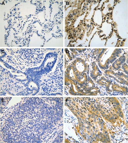
Then, DcR3 expression was assessed separately in SCLC and NSCLC. Higher levels of DcR3 expression were also found in both SCLC (P=0.001) and NSCLC (P=0.008), as compared to that in noncancerous lung tissues. However, only a slightly higher expression of DcR3 was observed in SCLC (73.1%) compared to that in NSCLC (53.1%, P=0.039) tissues. We also investigated the expression of DcR3 in different histologic types of NSCLC. We found that the expression of DcR3 was higher in adenocarcinoma (56.7%, P=0.005), squamous cell carcinoma (48.6%, P=0.031), adenosquamous carcinoma (60.7%, P=0.012), and undifferentiated carcinoma (75%, P=0.013) than in normal lung tissues (). In addition, ROC curve was applied to analyze the diagnostic value of DcR3 level in lung cancer. The area under curve (AUC) of DcR3 was 0.637 (95% confidence interval [CI] 0.531–0.742, P=0.02).
Relationships between DcR3 protein and other clinicopathological parameters in lung cancer
Relationships between DcR3 protein and other clinicopatho-logical parameters in lung cancer tissues were further investigated. In all 66 advanced-stage (III/IV) lung cancer cases, the positive expression of DcR3 (77.3%) was markedly higher compared to that in early stages (I and II, 48.2%, P<0.001). As for tumor diameter, the positive expression of DcR3 was found to be higher in tumors of larger size (>7 cm, 70.6%) than in those of smaller size (≤7 cm, 51.0%, P=0.007). Concerning distal metastasis, positive expression of DcR3 was found to be significantly higher in distant metastasis tumor group (15/16, 93.8%) than in group showing no metastasis (180/349, 51.6%, P<0.001). Additionally, the positive expression of DcR3 was observed to be significantly higher in patients with lymph node metastasis tumors (102/128, 79.7%) than in those with no lymph node metastasis (96/237, 40.5%, P<0.001, ). Spearman’s test was performed to assess the correlations between DcR3 and lung cancer, which showed that consistent correlations existed between DcR3 and TNM stage (r=0.225, P<0.001), tumor diameter (r=0.137, P=0.009), distant metastasis (r=0.173, P=0.001), and lymph node metastasis (r=0.375, P<0.001). We also calculated the correlation between the differential expression of DcR3 and age and sex, but no significant difference was observed ( and ).
Table 2 Differential expression of DcR3 protein in relation to other clinicopathological parameters in lung cancer
Table 5 The correlation between DcR3 expression and other clinicopathological parameters in lung cancer, SCLC, and NSCLC
Further, we studied the clinical contribution of DcR3 in the subtypes of SCLC and NSCLC. In NSCLC, a consistent correlation between TNM stage, tumor diameter, distal and lymph node metastasis, and DcR3 expression was found. Specifically, positive expression of DcR3 was found in 41 out of 53 NSCLC cases in advanced stages (III and IV, 77.4%), clearly higher than in early stages (I and II, 138/286, 48.3%, P<0.001). The positive expression of DcR3 was upregulated in larger tumors (>7 cm, 30/44, 68.2%) than in smaller ones (≤7 cm, 147/295, 48.3%, P=0.019). The positive expression of DcR3 was remarkably higher in distant metastasis tumor group (15/16, 93.8%) than in the group with no distant metastasis (164/323, 50.8%, P<0.001). Furthermore, when lymph node metastasis was concerned, the positive expression of DcR3 was found in 70 among 115 NSCLC cases with lymph node metastasis (60.9%), which was significantly higher than in those without (39.7%, 89/224, P<0.001, ). Spearman’s test showed that there were consistent correlations between DcR3 expression and TNM stage (r=0.212, P<0.001), tumor diameter (r=0.124, P=0.022), distant metastasis (r=0.183, P=0.001), and lymph node metastasis (r=0.201, P<0.001, ). No significant difference was found in groups with respect to age and sex ( and ).
Table 3 The status of DcR3 with respect to other clinicopathological parameters in NSCLC
Simultaneously, in SCLC, DcR3 expression was also found to be associated with TNM stage (r=0.580, P=0.004) and lymph node metastasis (r=0.564, P=0.005). The positive expression of DcR3 was found to be higher in advanced stages (III and IV, 100%) than in early stages (I and II, 6/13, 46.2%, P=0.003). With regard to tumor diameter, the positive expression of DcR3 was remarkably higher in larger tumors (>7 cm, 12/19, 63.2%) than in smaller ones (≤7 cm, 4/4, 100%, P=0.005). In addition, the positive expression of DcR3 was found in 12 out of 13 SCLC cases with lymph node metastasis (92.3%), clearly higher than in those with no lymph node metastasis (4/10, 40.0%, P=0.012, ). No significant difference was found with regard to age, sex, and distant metastasis ( and ).
Relationships between DcR3 expression and clinical parameters in lung cancer according to the data from TCGA
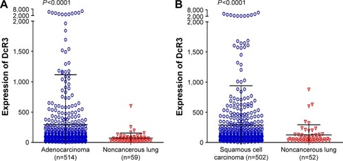
To further elucidate the clinical role of DcR3 in the survival of lung cancer patients, we searched the data of 521 adenocarcinoma and 504 squamous cell carcinoma cases in TCGA. Analysis of TCGA data by cBioPortal (http://www.cbioportal.org/public-portal/) demonstrated that overall, DcR3 was highly expressed in lung cancer, and the upregulated expression of DcR3 was observed in 18% of all adenocarcinoma cases (). We further confirmed this correlation at the mRNA level. We found that DcR3 mRNA was overexpressed in adenocarcinoma patients (). Similarly to adenocarcinoma, upregulated expression of DcR3 was also found in 21% of all squamous cell carcinoma samples (). Further, we confirmed this correlation at the mRNA level. A high expression of DcR3 mRNA was found in squamous cell carcinoma cases (). In order to further explore the clinical value of DcR3 in lung cancer, including adenocarcinoma and squamous cell carcinoma, the original data were downloaded and analyzed from TCGA datasets, and we found that DcR3 was overexpressed in both adenocarcinoma and squamous cell carcinoma tissues than in noncancerous lung tissues (all P<0.0001, ). We also compared the expression between adenocarcinoma and squamous cell carcinoma tissues, but no significant difference was noted between these two groups (294.5±36.23 vs 288.3±29.12, P=0.894).
Figure 2 The upregulated expression of DcR3 in all samples of adenocarcinoma.
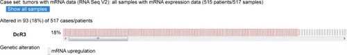
Figure 3 The expression of DcR3 mRNA in all samples of adenocarcinoma.
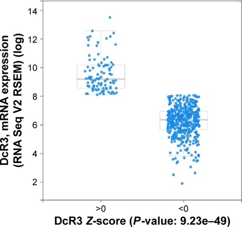
Figure 4 The upregulated expression of DcR3 in all samples of squamous cell carcinoma.
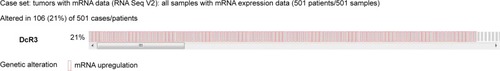
Figure 5 The expression of DcR3 mRNA in all samples of squamous cell carcinoma.
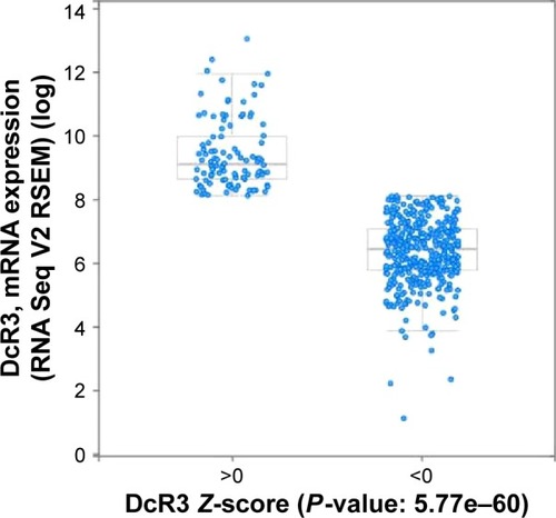
Figure 6 Clinical significance of DcR3 in lung adenocarcinoma and squamous cell carcinoma based on The Cancer Genome Atlas database.
Abbreviation: DcR3, decoy receptor 3.
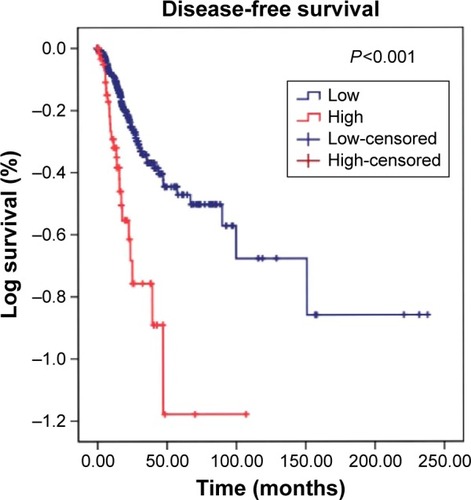
In addition, the ROC curve revealed that the AUC of DcR3 was 0.726 (95% CI 0.644–0.788) for lung adenocarcinoma patients and 0.647 (95% CI 0.566–0.728) for squamous cell carcinoma patients, which was applied to analyze the diagnostic value of DcR3 level in lung cancer. Interestingly, we also investigated the relationship between DcR3 level and patients’ survival. The upregulated expression of DcR3 was associated with overall survival (P<0.001, 98.8±9.34 vs 39.7±4.22) and disease-free survival of adeno-carcinoma patients (P<0.001, 133.0±12.92 vs 47.0±8.34, and ), which indicated that DcR3 could influence the prognosis.
Figure 7 The expression of DcR3 is associated with overall survival of adenocarcinoma patients.
Abbreviation: DcR3, decoy receptor 3.
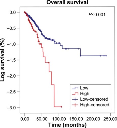
Figure 8 The expression of DcR3 is associated with disease-free survival of adenocarcinoma patients.
Abbreviation: DcR3, decoy receptor 3.
Discussion
Lung cancer is a general and frequently occurring disease that threatens human health seriously. With the acceleration of urbanization, the incidence and mortality of lung cancer has also been increasing. So far, a variety of studies related to genes involved in lung cancer have been carried out.Citation25,Citation26 For instance, Deben et alCitation25 explored the role of p53 in carcinogenesis and they found that the expression of p53 could be used to evaluate the prognostic and predictive significance in lung cancer patients. Garajova et alCitation26 found that c-Met could be a target for personalized therapy, but no report was available to explain the correlation between the expression of DcR3 and lung cancer. In this study, we attempted to evaluate the relationship between DcR3 and lung cancer and further investigate the prospective role of DcR3 expression in the prediction and diagnosis of lung cancer.
In our present study, we explored the DcR3 expression in lung cancer and normal lung samples by tissue microarray and IHC. We found that DcR3 expression in lung cancer, including SCLC and NSCLC, was clearly higher compared to that in the normal lung tissues. The ROC curve indicated that DcR3 protein might have a moderate diagnostic value for lung cancer (AUC =0.637). Also, in the study of the correlation between the expression of DcR3 and some clinicopathological parameters, we found that DcR3 expression was remarkably related to the deterioration of the disease, including both of NSCLC and SCLC. The higher expression of DcR3 might prompt an advanced stage or larger tumor size or metastasis. Also, the low survival rate showed that the overexpression of DcR3 might indicate a poor prognosis. We thus could draw a conclusion that DcR3 might act as a molecular biomarker in lung cancer, and our study confirms that DcR3 might be involved in the tumorigenesis and deterioration of lung cancer, although further verification is required. In other reports, Huang et al studied 125 glioma patients and 18 normal brain cases and found that the higher expression of DcR3 might be related to tumor cell differentiation and proliferation.Citation27 Zhou et alCitation28,Citation29 investigated the relationship between high expression of DcR3 and prognosis of pancreatic carcinoma. They found that high expression of DcR3 could predict the prognosis of pancreatic carcinoma and that the DcR3 level in sera could be used for the early diagnosis and prognostic judgment of pancreatic carcinoma. In addition, TCGA database was used to verify the DcR3 expression in lung cancer. Consistent with our results, DcR3 was found to be highly expressed in lung cancer patients according to the data from TCGA. All the aforementioned results demonstrated that DcR3 can be used for the early diagnosis and prognostic judgment of lung cancer.
The role of DcR3 in lung cancer has been preliminarily studied by several groups. In agreement with our results, Pitti et alCitation14 reported that the DcR3 gene was highly expressed in lung cancers. They studied 35 primary lung cancer cases and found that DcR3 played its role by binding specifically to FasL, thereby inhibiting its activity. DcR3 has also been reported to correlate with FasL in some malignant tumors, such as pancreatic cancer and hepatocellular carcinoma.Citation30,Citation31 FasL is an important factor involved in the regulation of the immune response. Also, the FasL-Fas receptor could act as a key physiological regulator in programmed cell death.Citation32 Zhang et alCitation30 found that DcR3 could suppress FasL-induced apoptosis via ERK1/2 pathway in pancreatic cancer cells, and reported that DcR3 could enhance ERK1/2 phosphorylation and oppose FasL signaling. In our previous study of DcR3 in glioma, we obtained results consistent with those reported by Zhang et al that DcR3 could cause an effect on cell growth suppression and apoptosis induction in glioma cells.Citation19 In our present study, we envision that the overexpression of DcR3 could also be correlated to Fasl via ERK to inhibit growth and induce apoptosis of lung cancer cells. But the authentic pathway or the mechanism of action of DcR3 on the biological function of lung cancer needs further exploration.
In addition, our previous study found that DcR3 functions as a bridge between miR-152 and hepatocellular carcinoma.Citation33 Besides miR-152, DcR3 could be the target gene for other miRNAs through bioinformatics softwares. miR-148 and miR-340 were predicted by five different algorithms, including TargetScan, miRDB, DIANA-microT, miRanda, and miRWalk (data not shown). However, the molecular mechanism underlying the role of DcR3 in lung cancer is still unclear, and further in vitro and in vivo investigations will be required in the future.
Conclusion
DcR3 expression, as assessed by IHC, is upregulated in lung cancer, including SCLC and NSCLC. The expression of DcR3 protein is positively correlated with the deterioration and poor prognosis of lung cancer patients. Together with previous reports, the current study strongly suggests that DcR3 might play a crucial role in the evolution and progression of lung cancer. Further experiments are expected to explore the prospective molecular mechanism of DcR3 in lung cancer.
Acknowledgments
The study was supported by the fund of National Natural Science Foundation of China (NSFC81360327, NSFC81560469), Guangxi Natural Science Foundation (2015GXNSFCA139009), Scientific Research Project of the Guangxi Education Agency (KY2015LX062), Guangxi Graduate Innovation Project (YCSZ2015106), and Guangxi Provincial Health Bureau Scientific Research Project (Z2013201, Z2014055). The funders had no role in study design, data collection and analysis, decision to publish, or preparation of the manuscript.
Disclosure
The authors report no conflicts of interest in this work.
References
- XuYJDuYFanYLong noncoding RNAs in lung cancer: what we know in 2015Clin Transl Oncology2015 Epub20151215
- KangCGLeeHJKimSHLeeEOZerumbone Suppresses osteopontin-induced cell invasion through inhibiting the FAK/AKT/ROCK pathway in human non-small cell lung cancer A549 cellsJ Nat Product2016791156160
- TorreLABrayFSiegelRLFerlayJLortet-TieulentJJemalAGlobal cancer statistics, 2012CA: Cancer J Clin20156528710825651787
- ChenGUmeloIALvSmiR-146a inhibits cell growth, cell migration and induces apoptosis in non-small cell lung cancer cellsPloS One201383e6031723555954
- YilmazADamadogluESalturkCOkurETuncerLYHalezerogluSDelays in the diagnosis and treatment of primary lung cancer: are longer delays associated with advanced pathological stage?Upsala J Med Sci2008113328729618991241
- PfisterDGJohnsonDHAzzoliCGAmerican Society of Clinical Oncology treatment of unresectable non-small-cell lung cancer guideline: update 2003J Clin Oncol200422233035314691125
- MorishigeTYoshiokaYInakuraHCreation of a lysine-deficient LIGHT mutant with the capacity for site-specific PEGylation and low affinity for a decoy receptorBiochem Biophys Res Commun2010393488889320175993
- ConnorJPFelderMKapurAOnujioguNDcR3 binds to ovarian cancer via heparan sulfate proteoglycans and modulates tumor cells response to platinum with corresponding alteration in the expression of BRCA1BMC Cancer20121217622583667
- WangWZhangMSunWReduction of decoy receptor 3 enhances TRAIL-mediated apoptosis in pancreatic cancerPloS One2013810e7427224204567
- YangDFanXYinPSignificance of decoy receptor 3 (Dcr3) and external-signal regulated kinase 1/2 (Erk1/2) in gastric cancerBMC Immunol2012132822672288
- WeissingerDTagschererKEMacher-GoppingerSHaferkampAWagenerNRothWThe soluble decoy receptor 3 is regulated by a PI3K-dependent mechanism and promotes migration and invasion in renal cell carcinomaMol Cancer201312
- ChenGRongMLuoDTNFRSF6B neutralization antibody inhibits proliferation and induces apoptosis in hepatocellular carcinoma cellPathol Res Pract2010206963164120591579
- ArakawaYTachibanaOHasegawaMMiyamoriTYamashitaJHayashiYFrequent gene amplification and overexpression of decoy receptor 3 in glioblastomaActa Neuropathol2005109329429815627206
- PittiRMMarstersSALawrenceDAGenomic amplification of a decoy receptor for Fas ligand in lung and colon cancerNature199839667126997039872321
- RothWIsenmannSNakamuraMSoluble decoy receptor 3 is expressed by malignant gliomas and suppresses CD95 ligand-induced apoptosis and chemotaxisCancer Res20016162759276511289159
- SungHYWuHGAhnJHParkWYDcr3 inhibit p53-dependent apoptosis in gamma-irradiated lung cancer cellsInt J Rad Biol201086978079020597837
- WuYHanBShengHClinical significance of detecting elevated serum DcR3/TR6/M68 in malignant tumor patientsInt J Cancer2003105572473212740925
- HuangSChenGOverexpression of DcR3 and its significance on tumor cell differentiation and proliferation in gliomaScientific World Journal2014201460523624741354
- RuanYHuangSHeDGopaulRLiZChenGEffect of TNFRSF6B neutralization antibody on cell growth suppression and apoptosis induction in glioma cellsNeoplasma201562457458125997959
- ChenGLuoDOver-expression of decoy receptor 3 in gastric precancerous lesions and carcinomaUpsala J Med Sci2008113329730418991242
- YangMChenGDangYLuoDSignificance of decoy receptor 3 in sera of hepatocellular carcinoma patientsUpsala J Med Sci2010115423223720977315
- BornsteinSSchmidtMChoonooGIL-10 and integrin signaling pathways are associated with head and neck cancer progressionBMC Genomics20161713826747525
- CeramiEGaoJDogrusozUThe cBio cancer genomics portal: an open platform for exploring multidimensional cancer genomics dataCancer Discov20122540140422588877
- GaoJAksoyBADogrusozUIntegrative analysis of complex cancer genomics and clinical profiles using the cBioPortalSci Signal20136269pl123550210
- DebenCDeschoolmeesterVLardonFRolfoCPauwelsPTP53 and MDM2 genetic alterations in non-small cell lung cancer: evaluating their prognostic and predictive valueCrit Rev Oncol/Hematol2016996373
- GarajovaIGiovannettiEBiascoGPetersGJc-Met as a target for personalized therapyTransl Oncogenomics20157Suppl 1133126628860
- HuangSChenGDangYChenLHOverexpression of DcR3 and its significance on tumor cell differentiation and proliferation in gliomaScientificWorldJournal2014201460523624741354
- ZhouJSongSLiDDecoy receptor 3 (DcR3) overexpression predicts the prognosis and pN2 in pancreatic head carcinomaWorld J Surg Oncol2014125224597666
- ZhouJSongSDLiDCZhouJZhuDMZhengSYClinical significance of expression and amplification of the DcR3 gene in pancreatic carcinomasAsian Pac J Cancer Prev201213271972422524850
- ZhangYLiDZhaoXDecoy receptor 3 suppresses FasL-induced apoptosis via ERK1/2 activation in pancreatic cancer cellsBiochem Biophys Res Commun201546341144115126102031
- LiWZhangCChenCZhuangGCorrelation between expression of DcR3 on tumor cells and sensitivity to FasLCell Mol Immunol20074645546018163957
- KimSKYoonYDParkYSSeoJTKimJHInvolvement of the Fas-Fas ligand system and active caspase-3 in abnormal apoptosis in human testes with maturation arrest and Sertoli cell-only syndromeFertil Steril200787354755317123522
- DangYWZengJHeRQRongMHLuoDZChenGEffects of miR-152 on cell growth inhibition, motility suppression and apoptosis induction in hepatocellular carcinoma cellsAsian Pac J Cancer Prev201415124969497624998573