
Abstract
Background
Downregulated expression levels of microRNA-320a (miR-320a) were found in primary breast cancers and colorectal cancer. Previous findings indicated that miRNA-320a may involve in the cancer development. In this study, we explored the roles of miR-320a by targeting c-Myc in the tumor growth of hepatocellular carcinoma (HCC).
Methods
Quantitative reverse-transcription polymerase chain reaction (qRT-PCR) was performed to detect the expression of miR-320a in 50 HCC tissues and four HCC cells. Luciferase reporter assay was conducted to confirm the direct downstream target of miR-320a in HEK-293 cells. The effect of miR-320a on endogenous c-Myc expression was investigated by transfecting miR-320a mimics into HepG2 and QGY-7703 cell lines. The c-Myc and miR-320a expressions were analyzed by immunohistochemistry (IHC) and qRT-PCR in the same HCC tissues. Furthermore, the biological functional correlation of miR-320a with c-Myc was determined by studying the effect of miR-320a mimics or c-Myc small interfering RNA (siRNA) on HCC cell proliferation and invasion.
Results
The expression of miR-320a was downregulated in 50 HCC tissues and 4 HCC cells. Luciferase assay revealed that c-Myc is a direct target of miR-320a. IHC and Western blot analysis showed that the c-Myc expression was inhibited by miR-320a in HCC tissues and cell lines. Upregulation of miR-320a suppressed the HCC cell proliferation and invasion capacity induced by inhibiting c-Myc, and the results were consistent with the effects of c-Myc siRNA on tumor suppression. These results revealed that miRNA-320a inhibits tumor proliferation and invasion by targeting c-Myc in HCC cells.
Conclusion
Our results showed that miR-320a functions as a tumor suppressor in HCC. By targeting c-Myc directly, miR-320a inhibits the HCC cell growth. Our studies provide evidence of miR-320a as a potentially target for HCC treatment.
Introduction
MicroRNAs (miRNAs) are small noncoding RNA molecules that are often located in genomic breakpoint regions and play important roles in regulating diverse biological processes. Increasing evidence indicates that some miRNAs function as tumor suppressors or oncogenes in the tumor development.Citation1–Citation3 The gene expressions are found to be regulated by miRNAs by targeting the 3′ untranslated regions of mRNA specifically, and many targeting genes may be controlled by one specific miRNA. Translational level or mRNA stability is influenced by binding of miRNAs to target mRNAs. The cancer invasion effect of miRNAs has been identified in several cancer types, for example gastric cancer,Citation4 hepatocellular carcinoma (HCC),Citation5 colorectal cancer (CRC),Citation6 pancreatic cancer, and so on.Citation7 Most studies have analyzed the association between miRNAs and invasion in vitro and mouse models. Differential miRNAs are only able to be identified in primary tumor tissues versus normal tissue in tissue level.
HCC, which accounts for ~700,000 deaths per year, is the third leading cause of cancer-related death in the world.Citation8,Citation9 Great efforts on the HCC biological mechanism studies had been taken to improve treatment therapy. A number of studies observed abnormal miRNA expressions in HCC. Previous studies showed that HCC cell growth were regulated by miRNAs, such as miR-101,Citation10 miR-221,Citation11 miR-224,Citation12 miR-195,Citation13 miR-122,Citation14 miR-223,Citation15 miR-21,Citation16 and miR-199,Citation17 and apoptosis, migration, and invasion of HCC cell were regulated by them as well. miRNAs involve in the HCC progression, diagnosis, and management.Citation9 There are studies that suggest that HCC tumorigenesis may be caused by miRNA abnormal expression. Thus, extensive investigations are needed to identify the function of these miRNAs on HCC development, and some potential miRNAs may be used as new biomarkers for the diagnosis and prognosis of HCC.Citation9,Citation18–Citation20
MiR320a downregulation was observed in colon cancer,Citation21 breast cancer (BC),Citation22,Citation23 bladder carcinoma,Citation24 and gastric cancer,Citation25 Therefore, we hypothesized that miR-320a may involve in the tumorigenesis and tumor development, and here we focused on the mechanism pathway studies of miR-320a in HCC tumorigenesis and development.
In this study, the expression of miRNA-320a was examined in HCC tissues and HCC cells, such as BEL-7402, SMMC-7721, HSPG2, and QGY-7703 cell lines compared with normal liver tissues and L02 cell line. Then, we analyzed the miRNA expression profiles to provide a hypothesis of the possible target toward miRNA-320a. Next, we investigated the correlation of miRNA-320a with the potential target gene. Finally, further investigations were conducted to study the functional mechanism of the miRNA-320a in HCC cells. Our novel findings suggested that miR-320a may be a potential target or biomarker for HCC diagnosis and therapy.
Methods
Ethical statements
Our study was approved by the ethical committee of the Yunnan University School of Medicine. Written informed consent was obtained from all participants for this study.
Samples
Fifty paired HCC and adjacent nontumor specimens used in this study were obtained from the Department of Thoracic Surgery, Third Affiliated Hospital of Kunming Medical University (Kunming, China). Five normal liver tissues were collected from adjacent liver tissues of contusion and laceration in traumatic liver injury patients. All tissue samples were flash-frozen in liquid nitrogen immediately after collection and stored at −80°C until use. Both tumor and nontumor samples were confirmed by pathological examination. No patients received chemotherapy or radiotherapy prior to surgery.
Cell culture
In this study, HCC cell lines (BEL-7402, HepG2, QGY-7703, SMMC-7721, L02) were purchased from the cell bank of the Chinese Academy of Sciences, and they were cultured in Dulbecco’s Modified Eagle’s Medium (DMEM; Sigma-Aldrich, St Louis, MO, USA) supplemented with 10% (vol/vol) fetal bovine serum (FBS). HEK293 cells were purchased from American Type Culture Collection (Manassas, VA, USA), and cultured in DMEM containing 10% (vol/vol) FBS. All cells were maintained at 37°C with 5% CO2.
RNA isolation and qRT-PCR
Total RNA was isolated from HCC tissues, adjacent nontumor tissues, and BEL-7402, HepG2, QGY-7703, SMMC-7721, and HEK293 cell lines using Trizol according to the manufacturer’s instructions. QRT-PCR assay was performed as described previously.Citation26 U6 small RNA was used as an internal control for normalization. All primers used in this study were listed in .
Table 1 Primer sequences used for qRT-PCR
Western blot analysis
To determine protein expression levels, proteins form cell lysates were separated by 10% sodium dodecyl sulfate–polyacrylamide gel and transferred to nitrocellulose membrane (Bio-Rad, Hercules, CA, USA). The membrane was blocked with 5% nonfat milk, then incubated with anti-β-Actin antibody (Sigma, CA, USA) or anti-c-Myc antibody (Santa Cruz, CA, USA), followed by goat anti-mouse secondary antibody (Pierce, IL, USA). Finally, the protein bands were detected with enhanced chemiluminescence reagents following the Vendor protocol (Pierce).
Oligonucleotides transfection
RNA oligos used in this study were chemically synthesized by Genepharma (Shanghai, China). The human miR-320a mimics sequence was 5′-AAA AGC UGG GUU GAG AGG GCG A-3′. The sequence for negative control was 5′-CAG UAC UUU UGU GUA GUA CAA-3′. The oligonucleotides for c-Myc siRNA were 5′-AAC GUU AGC UUC ACC AAC ATT-3′ (sense) and 5′-UGU UGG UGA AGC UAA CGU UTT-3′ (antisense), and the sequence of control siRNA was 5′-UUC UCC GAA CGU GUC ACG UTT-3′ (sense) and 5′-ACG UGA CAC GUU CGU AGA ATT-3′ (antisense). The oligonucleotide transfections were conducted by using INTERFERin reagent according to the manufacturer’s protocol (Polyplus-transfection).
Luciferase assay
Luciferase reporter plasmid was created through cloning human c-Myc sequence into pMIR-Report construct (Ambion, Austin, TX, USA). C-Myc sequence contains the potential miR-320a binding site. C-Myc fragment A (from 566 to 850), c-Myc fragment B (from 851 to 1,207), and c-Myc fragment A+B (from 566 to 1,207) were amplified using PCR. (The primers are listed in .) The luciferase reporter assay was performed as previously,Citation26 and cotransfection of 50 nM miRNA mimics or negative control, 100 ng firefly luciferase reporter, and 20 ng pRL-TK vector (Promega, Madison, WI, USA) into the HEK293 cells was performed by the JetPRIME reagent (Invitrogen) following manufactory’s protocol. Twenty-eight hours after transfection, cells were harvested and analyzed using Dual-Luciferase Reporter Assay System (Promega).
Cell proliferation assay
Cells were plated in 96-well plates (Corning Co, Corning, NY, USA) with 4,000 cells in 100 μL per well and incubated for 72 h after transfection with miR-30a mimics or mimics NC, the and cell viability was evaluated by the MTT assay (Promega) according to the manufacturer’s instructions. MTT reagent solution was dispensed into each well and incubated at 37°C in 5% CO2 for 4 h. The absorbance was then read at 490 nm in a microplate absorbance reader. Cell proliferation was assessed every 24 h. Triplicate wells were measured for cell viability in each treatment group.
Invasion assays
The invasive ability of HCC cells was determined using 24-well transwell chambers coated with Matrigel (BD Pharmingen, San Jose, CA, USA). Transfected cells in serum-free medium were seeded at 5×10⁴ in the upper compartment of chamber. After the chamber was incubated at 37°C for 24 h, cells that migrated to the underside of the membrane were fixed with 4% paraformaldehyde (Sigma-Aldrich), stained with crystal violet (Beyotime, Shanghai, China), imaged and counted with a microscope (Leica, UK). All experiments were performed three times.
Immunohistochemistry
In this study, IHC staining of the HCC samples was performed according to the protocol described previously.Citation26 Xylene was used to dewax the paraffin-embedded tissue sections, and the deparaffinized sections were rehydrated using ethanol. Then, epitope retrieval was induced by in citrate buffer (pH 6.0). Anti-c-Myc antibody was used to identify C-Myc expression (Santa Cruz, CA, USA).
Statistical analysis
Continuous variables were expressed as mean ± SEM. The two-tailed t-test was used between groups. In all cases, P<0.05 was considered as statistically significant.
Results
MiR-320a was downregulated in HCC samples and cell lines
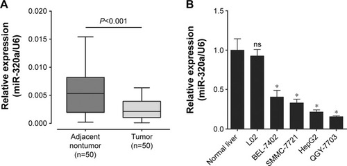
To determine the expression of miR-320a in HCC samples and cell lines, total RNA was extracted from HCC tissues, adjacent nontumor tissues, normal liver cell line, and HCC cell lines; quantitative reverse-transcription polymerase chain reaction (qRT-PCR) was used to analyze the expression levels of miR-320a, and normalized against an endogenous control (U6 RNA). MiR-320a was decreased significantly in HCC tissues versus adjacent nontumor tissues (). At the meantime, miR-320a was downregulated in four HCC cell lines compared with normal liver tissues and normal liver cell line (). Thus, these data demonstrated that the expression of miR-320a was downregulated in HCC samples and HCC cell lines.
Figure 1 miR-320a relative expression in HCC samples and cell lines.
Abbreviations: HCC, hepatocellular carcinoma; miR-320a, microRNA-320a; qRT-PCR, quantitative reverse-transcription polymerase chain reaction; ns, nonsignificant.
C-Myc mRNA contains two target sequences for miR-320a
Previous studies showed that miRNAs function by inhibiting target genes. To find the target gene of miR-320a correlated with HCC pathogenesis, the newly published CLASH data were further analyzed.Citation27 Almost 514 genes were found to be targeted by miR-320a in HEK293 cells. We focused on gene c-Myc among these 514 genes, and there are two potential targeting sequences for miR-320a within the coding DNA sequence of c-Myc mRNA (). These data showed that c-Myc may be a potential target of miR-320a in HCC cells.
Figure 2 miR-320a directly binds to the coding DNA sequence of c-Myc.
Abbreviations: CDS, coding DNA sequence; DNA, deoxyribonucleic acid; NC, negative control; miR-320a, microRNA-320a; UTR, untranslated regions.
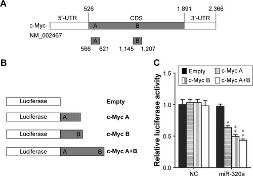
C-Myc is a potential target of miR-320a in HCC cells
There are data that showed that c-Myc may be a potential target of miR-320a; however, the correlation of c-Myc with miR-320a has not been clarified yet in HCC cells. To identify whether the c-Myc gene expression is regulated by miR-320a, luciferase reporter assay was performed in HEK-293 cells. First, we created luciferase reporter plasmid. Two sequences including two potential targeting sequences were individually or in combination cloned into luciferase reporter plasmid (), and miR-320a mimics or negative control was cotransfected into HEK293 cells for 48 h. Next, the luciferase activity was detected using Dual-Luciferase Reporter Assay. The results demonstrated that the reporter plasmid with targeting sequence of c-Myc mRNA lead to decrease of luciferase activity significantly in cells transfected with miR-320a compared with nonsequence of c-Myc reporter plasmid (). Hence, our findings proved that c-Myc is a direct target of miR-320a in HCC cells.
MiR-320a inhibited c-Myc expression in HCC cells
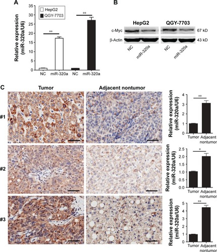
To determine the effect of miR-320a on c-Myc expression in HCC cell lines, HCC cell lines were transiently transfected with miR-320a mimics or negative control, and HCC cell lines showed low miR-320a expression. QRT-PCR was used to examine the expression of miR-320a. The results showed that miR-320a mimics transfection increased miR-320a expression in HepG2 and QGY-7703 cells compared with the negative control (). Then, the c-Myc protein level was verified by Western blotting. Our results demonstrated that the expression of c-Myc was consistently and substantially downregulated by miR-320a upregulation (). Consistently, Immunohistochemistry (IHC) assay demonstrated low miR-320a expression but high c-Myc expression in HCC tissues, whereas the normal liver tissues with high miR-320a expression showed lower c-Myc expression (). Taken together, these findings proved that the c-Myc expression is upregulated by low expression of miR-320a in HCC cells.
Figure 3 miR-320a downregulates endogenous c-Myc expression in HCC cells.
Abbreviations: HCC, hepatocellular carcinoma; IHC, immunohistochemistry; miR-320a, microRNA-320a; NC, negative control; qRT-PCR, quantitative reverse-transcription polymerase chain reaction.
MiR-320a inhibited HCC cells proliferation and invasion by repressing c-Myc expression
To examine the effects of miR-320a on HCC cells’ function through targeting c-Myc, we designed the small interfering RNA (siRNA) for c-Myc (c-Myc siRNA), which induced decrease of c-Myc expression at the protein levels in HepG2 and QGY-7703 cells, as shown in . At the same time, HepG2 and QGY-7703 cells were transfected with miR-320a mimics; we observed a similar decrease of c-Myc expression, as shown in . Next, the effect of miR-320a by targeting c-Myc on HCC cell viability was detected.
Figure 4 Upregulation of miR-320a inhibited c-Myc expression and hepatocellular carcinoma cells proliferation by targeting c-Myc in vitro.
Abbreviations: miR-320a, microRNA-320a; NC, negative control; OD, optical density; si or siRNA, small interfering RNA.
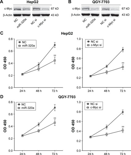
To identify the effects of miR-320a on HCC cell viability, 3-(4,5-dimethylthiazol-2-yl)-2,5-diphenyl tetrazolium bromide (MTT) assays were conducted, and the results showed that the proliferation ability of HepG2 () and QGY-7703 cells () transfected with miR-320a mimics were decreased obviously after 72 h compared with their respective controls. Similarly, we found that c-Myc siRNA reduced the proliferation ability of HepG2 () and QGY-7703 cells (), and the effect of c-Myc siRNA on cell proliferation was consistent with that of miR-320a in HepG2 and QGY-7703 cells.
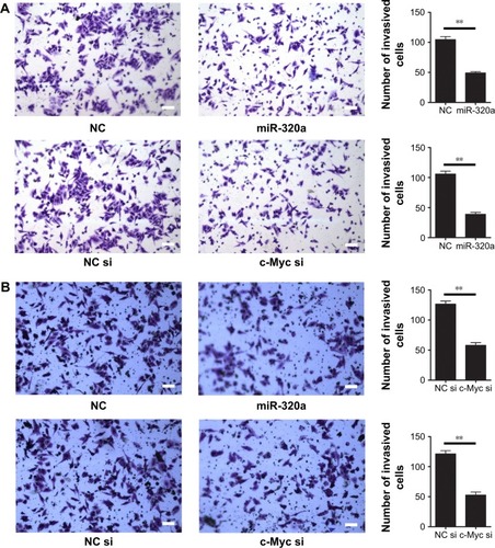
Next, the effect of miR-320a by targeting c-Myc on invasion capacity in HCC cells was detected. Invasion assay showed that invasion capacity of HepG2 () and QGY-7703 cells () transfected with miR-320a mimics was significantly decreased compared with their respective controls. Similarly, we found that c-Myc siRNA reduced the invasion capacity in HepG2 () and QGY-7703 cells (), and the effect of c-Myc siRNA on cell invasion was consistent with that of miR-320a mimics in HepG2 and QGY-7703 cells. These findings suggested that miR-320a inhibits the invasion of HCC cells through targeting c-Myc.
Figure 5 Upregulation of miR-320a inhibited hepatocellular carcinoma cells invasion by targeting c-Myc in vitro.
Abbreviations: miR-320a, microRNA-320a; NC, negative control; si or siRNA, small interfering RNA.
Discussion
miRNAs are proved to play key roles in gene regulation by multiple studies. Previous study found that the expression of miR-320a is upregulated significantly in the peripheral blood of coronary heart disease.Citation28 However, the expression of miRNA-320a was found to be downregulated in CRC,Citation29 primary BC,Citation30 and intrahepatic cholangiocarcinoma.Citation31 In our present study, we found that miR-320a was significantly repressed in HCC tissues and HCC cell lines.
Previous studies showed that miRNAs could conduct their tumor repression function by inhibiting correlated target genes. In this study, to find the target gene of miR-320a correlated with HCC pathogenesis, miRNA expression profiles analysis was used and showed that 514 genes were targeted by miR-320a in HEK293 cells. We focused on c-Myc gene, which plays an important role in the mitogen-activated protein kinase/extracellular signal–regulated kinase pathway. There are studies that proved that c-Myc is involved in cell growth, cell differentiation, apoptosis, invasion, and many other biological activities;Citation32–Citation34 thus, c-Myc is a very strong proto-oncogene. Pelengaris et al and Lin et al showed that the expression of C-Myc was found to be elevated in several types of cancers, such as HCC.Citation35,Citation36 Recent reports show that c-Myc targets miR-24,Citation37 miR-145,Citation38 let-7a,Citation39 and miR-185-3p.Citation40 A possible regulation of c-Myc by miR-320a was previously shown by Lu et al. They showed that progesterone (P4) promoted the expression of miR-320a in MCF-7 cell line by repressing c-Myc expression, while estrogen (E2) exerted the opposite effect.Citation23 However, correlation of c-Myc with miR-320a in HCC needs to be clarified. In this study, we found that c-Myc expression was upregulated by low expression of miR-320a in HCC tissues by IHC assay, and upregulation of miR-320a downregulated the c-Myc expression in HepG2 and QGY-7703 cell lines. Our results demonstrated that the c-Myc expression was inhibited by miR-320a in HCC tissues, HepG2, and QGY-7703 cell lines.
Lu et al found that the c-Myc/miR-320a axis provides a novel understanding of the mechanism of acquired tamoxifen resistance in BC.Citation23 In this study, the biological underlying mechanisms of miR-320a correlated with c-Myc were investigated in HepG2 and QGY-7703 cells. We found that miR-320a mimics or c-Myc siRNA all lead to the similar decrease of c-Myc expression level, and the cell viability and proliferation were all decreased at the same time. We hypothesize that correlation of c-Myc with miR-320a may stimulate the HCC tumor development. Invasion is also a crucial feature in tumor development and tumorigenesis. Evidence showed that miR-320a may inhibit cell invasion in some ways. Zhang Y, et al, 2012 demonstrated that miR-320a may inhibit angiogenesis and cancer cell invasion by targeting NRP-1 in CRC.Citation29 Nevertheless, the role of miR-320a by inhibiting c-Myc in HCC cells invasion remains unclear. Furthermore, our subsequent studies showed that the invasion capacity of HCC cells transfected with miR-320a mimics or c-Myc siRNA was significantly lower than that of the HCC cells treated with respective controls. Taken together, our findings demonstrated that the proliferation and invasion effects of miR-320a were consistent with those of c-Myc siRNA in HepG2 and QGY-7703 cell lines, implying that miR-320a represses the proliferation and invasion of HCC cell lines through targeting c-Myc.
Conclusion
In conclusion, miR-320a functions as a tumor suppressor through targeting c-Myc; our findings not only advance our understanding toward the molecular mechanism of carcinogenesis, but also provide evidence for further investigation that miR-320a acts as a novel biomarker or potential target for HCC treatment.
Acknowledgments
This work was financially supported by National Natural Science Foundation of China (No 81460435 and No 81372360), Key Project of Yunnan Applied Basic Research Plan (2014FA022), and Science Research Foundation of Education Department of Yunnan Province (2014Z004, 2014Y009).
Disclosure
The authors report no conflicts of interest in this work.
References
- HayesJPeruzziPPLawlerSMicroRNAs in cancer: biomarkers, functions and therapyTrends Mol Med20142046046925027972
- MartaGNGaricocheaBCarvalhoALRealJMKowalskiLPMicroRNAs, cancer and ionizing radiation: where are we?Revista da Associacao Medica Brasileira (1992)2015613275281 Portuguese26248252
- TutarLTutarEOzgurATutarYTherapeutic targeting of microRNAs in cancer: future perspectivesDrug Dev Res201576738238826435382
- BaoBWangZAliSMetformin inhibits cell proliferation, migration and invasion by attenuating CSC function mediated by deregulating miRNAs in pancreatic cancer cellsCancer Prev Res (Phila)20125335536422086681
- ChaiJWangSHanDDongWXieCGuoHMicroRNA-455 inhibits proliferation and invasion of colorectal cancer by targeting RAF proto-oncogene serine/threonine-protein kinaseTumour Biol2015361313132125355599
- ChuangKHWhitney-MillerCLChuCYMicroRNA-494 is a master epigenetic regulator of multiple invasion-suppressor microR-NAs by targeting ten eleven translocation 1 in invasive human hepatocellular carcinoma tumorsHepatology (Baltimore, Md)201562466480
- XieJTanZHTangXMiR-374b-5p suppresses RECK expression and promotes gastric cancer cell invasion and metastasisWorld J Gastroenterol20142046174391744725516656
- JemalABrayFCenterMMFerlayJWardEFormanDGlobal cancer statisticsCA Cancer J Clin2011612699021296855
- TsoulfasGRole of microRNA in the diagnosis and therapy of hepatocellular carcinoma: a new frontierMicroRNA (Shariqah, United Arab Emirates)201433137143
- SuHYangJRXuTMicroRNA-101, down-regulated in hepatocellular carcinoma, promotes apoptosis and suppresses tumorigenicityCancer Res20096931135114219155302
- FornariFGramantieriLFerracinMMiR-221 controls CDKN1C/p57 and CDKN1B/p27 expression in human hepatocellular carcinomaOncogene2008275651566118521080
- WangYLeeATCMaJZIProfiling microRNA expression in hepatocellular carcinoma reveals microRNA-224 up-regulation and apoptosis inhibitor-5 as a microRNA-224-specific targetJ Biol Chem200828319132051321518319255
- XuTZhuYXiongYGeYYunJZhuangSMicroRNA-195 suppresses tumorigenicity and regulates G1/S transition of human hepatocellular carcinoma cellsHepatology200950111312119441017
- GramantieriLFerracinMFornariFCyclin G1 is a target of miR-122a, a microRNA frequently down-regulated in human hepatocellular carcinomaCancer Res200767136092609917616664
- WongQWLungRWLawPTMicroRNA-223 is commonly repressed in repatocellular carcinoma and potentiates expression of stathmin1Gastroenterology2008135125726918555017
- MengFHensonRWehbe-JanekHGhoshalKJacobSTPatelTMicroRNA-21 regulates expression of the PTEN tumor suppressor gene in human hepatocellular cancerGastroenterology2007133264765817681183
- HouJLinLZhouWIdentification of miRNomes in human liver and hepatocellular carcinoma reveals miR-199a/b-3p as therapeutic target for hepatocellular carcinomaCancer Cell201119223224321316602
- AndersenHHDurouxMGazeraniPMicroRNAs as modulators and biomarkers of inflammatory and neuropathic pain conditionsNeurobiol Dis20147115916825119878
- AreebZStylliSSKoldejRMicroRNA as potential biomarkers in GlioblastomaJ Neurooncol2015125223724826391593
- KrauskopfJVerheijenMKleinjansJCde KokTMCaimentFDevelopment and regulatory application of microRNA biomarkersBiomark Med20159111137115126502281
- DongYZhaoJWuCWTumor suppressor functions of miR-133a in colorectal cancerMol Cancer Res20131191051106023723074
- VislovukhAKratassioukGPortoEProto-oncogenic isoform A2 of eukaryotic translation elongation factor eEF1 is a target of miR-663 and miR-744Br J Cancer2013108112304231123695020
- LuMDingKZhangGMicroRNA-320a sensitizes tamoxifen-resistant breast cancer cells to tamoxifen by targeting ARPP-19 and ERRgammaSci Rep20155873525736597
- ShangCZhangHGuoYHongYLiuYXueYMiR-320a down-regulation mediates bladder carcinoma invasion by targeting ITGB3Mol Biol Rep20144142521252724443232
- SongMyPanKfSuHjIdentification of serum microRNAs as novel non-invasive biomarkers for early detection of gastric cancerPLoS One201273e3360822432036
- WangHSuXYangMReciprocal control of miR-197 and IL-6/STAT3 pathway reveals miR-197 as potential therapeutic target for hepatocellular carcinomaOncoimmunology2015410e103144026451302
- HelwakAKudlaGDudnakovaTTollerveyDMapping the human miRNA interactome by CLASH reveals frequent noncanonical bindingCell2013153365466523622248
- ChenCWangYYangSMiR-320a contributes to atherogenesis by augmenting multiple risk factors and down-regulating SRFJ Cell Mol Med201519597098525728840
- ZhangYHeXLiuYMicroRNA-320a inhibits tumor invasion by targeting neuropilin 1 and is associated with liver metastasis in colorectal cancerOncol Rep201227368569422134529
- YanLXHuangXFShaoQMicroRNA miR-21 overexpression in human breast cancer is associated with advanced clinical stage, lymph node metastasis and patient poor prognosisRNA (New York, NY)2008141123482360
- SchaarDGMedinaDJMooreDFStrairRKTingYmiR-320 targets transferrin receptor 1 (CD71) and inhibits cell proliferationExp Hematol200937224525519135902
- SecombeJPierceSBEisenmanRNMyc: a weapon of mass destructionCell200411715315615084254
- AdhikarySEilersMTranscriptional regulation and transformation by Myc proteinsNat Rev Mol Cell Biol20056863564516064138
- GrandoriCCowleySMJamesLPEisenmanRNThe Myc/Max/Mad network and the transcriptional control of cell behaviorAnnu Rev Cell Dev Biol20001665369911031250
- PelengarisSKhanMEvanGc-MYC: more than just a matter of life and deathNat Rev Cancer200221076477612360279
- LinCPLiuCRLeeCNChanTSLiuHETargeting c-Myc as a novel approach for hepatocellular carcinomaWorld J Hepatol2010211621160952
- LalANavarroFMaherCAMiR-24 inhibits cell proliferation by targeting E2F2, MYC, and other cell-cycle genes via binding to “seedless” 3′UTR microRNA recognition elementsMol Cell200935561062519748357
- SachdevaMZhuSWuFp53 represses c-Myc through induction of the tumor suppressor miR-145Proc Natl Acad Sci U S A200910693207321219202062
- SampsonVBRongNHHanJMicroRNA let-7a down-regulates MYC and reverts MYC-induced growth in Burkitt lymphoma cellsCancer Res200767209762977017942906
- LiaoJMLuHAutoregulatory suppression of c-Myc by miR-185-3pJ Biol Chem201128639339013390921832077