
Abstract
Distant soft tissue tumor metastasis isolated in the digit, presenting as a primary esophageal squamous cell carcinoma is considered extremely rare. Herein, we present a rare case of a 44-year-old male patient with squamous cell carcinoma of the esophagus associated with the clinical symptoms of progressive dysphagia and hoarseness in the course of 2 days. During the second course of chemotherapy, the patient sustained a fall with scald to his right ring finger, while the swelling in the right ring finger was soft, cystic and 2.0×1.8 cm in size. Then, phalangectomy was performed in his right ring finger and pathologic diagnosis was considered metastasis from the esophagus. Unfortunately, the patient succumbed to this disease within 2 months of diagnosis of metastasis. In conclusion, detection of soft tissue metastasis may have prognostic implications, providing more accessible biopsy sites and helping avoid invasive procedures.
Introduction
Esophageal cancer (EC) is the sixth most common cause of cancer death worldwide and has become a major health concern, especially in Asia.Citation1 In China, esophageal squamous cell carcinoma (ESCC) is the predominant histologic subtype and accounts for nearly 90% of all types of ECs.
The liver, lung and the bones are the most common sites of metastases in EC, yet metastases to skin and subcutaneous soft tissue (ST) are very rare.Citation2,Citation3 For most common malignancies, metastasis to skin and subcutaneous ST often occurs in the breast, lung, kidney, colon, head and neck melanomas. Only 3 cases out of 4,020 cancer patients were confirmed with skin and subcutaneous metastases originating from squamous cell carcinoma of the esophagus in a large retrospective study from Pennsylvania State University College of Medicine.Citation4
Distant metastatic malignancies to the digit are extremely rare, accounting for ~0.1 % of all metastatic osseous involvement.Citation5 Previous work on this subject resulted in only a few reports, which play a guiding role in properly diagnosing metastases to the hand when the findings described are encountered by phalangectomy surgeons. Hsieh et al reported a patient with pathologically confirmed ESCC metastasis of the left thumb, which occurred 3 months after esophagectomy. Initially, the finger lesion on his left thumb was thought to be caused by accidental burns occurring before he had a whole body bone scan taken prior to an esophagectomy. As a result, physicians must be aware of the possibility of metastasis, even if there is no other concomitant metastatic area.Citation6 In Houston and Telepak’s study, a 56-year-old man had an 8-month history of dysphagia and was diagnosed with a large basaloid squamous cell carcinoma of the esophagus. The tumor work-up, including a three-phase bone scan, revealed no evidence of metastases. Seven months later, the patient returned with a fungating necrotic mass emanating from the tip of the left ring finger. The mass was removed and biopsy revealed metastasis of the esophageal carcinoma.Citation7 In addition, Dimri et al reported carcinoma of esophagus with unusual metastasis to the gingiva and phalanx as well.Citation8
In this report, we describe a patient diagnosed with ST metastasis from lower ESCC, who succumbed to the disease within 2 months.
Case presentation
A 44-year-old male with a history of high blood pressure was admitted with the clinical symptoms of progressive dysphagia and hoarseness in the course of 2 days.
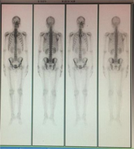
Esophago-gastro-duodenoscopy revealed irregular uplift lesions of 5 cm length, from 28 to 33 cm, and then histopathologic work-up of the specimens confirmed the diagnosis of squamous cell carcinoma of the esophagus ( and ). Computed tomography scan showed a circumferential middle-third esophageal growth, which was ~5 cm long, with significant regional lymphadenopathy but without extraesophageal spread. There were enlarged mediastinum and paracardiac lymph nodes in the computed tomography image, suggesting positivity for malignancy as well (). Unfortunately, the patient lost the opportunity to perform esophagectomy. Taking into account the poor prognostic factors such as transmural extension and lymph node positivity, the patient was given two cycles of Doxorubicin plus cisplatin. During the second course of chemotherapy, he discovered that his right ring finger had a protrusion for the first time, which was soft, cystic and 2.0×1.8 cm in size (). Initially, the patient thought that this protrusion was caused by swelling which occurred 1 day prior. Unfortunately, fine-needle aspiration biopsies from the site as well as the pathology revealed squamous cell carcinoma ( and ). Radiologic studies revealed no bony metastasis (). As a result, this lesion was considered metastatic, as it appeared simultaneously and was reported as squamous cell carcinoma on fine-needle aspiration biopsy. A subsequent metastatic work-up did not reveal any other site of metastases. Then, the patient underwent phalangectomy of his right ring finger. He was planned to undergo palliative chemotherapy and radiotherapy to the metastatic sites, but unfortunately his condition started deteriorating and he succumbed to the disease within 2 months of diagnosis of metastasis.
Figure 1 Esophago-gastro-duodenoscopy imaging.
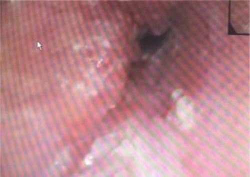
Figure 2 Histopathological imaging of lesions from the esophagus.
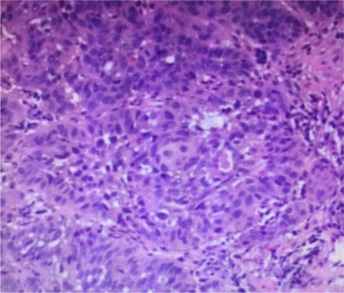
Figure 3 CT imaging of the esophageal and surrounding tissues.
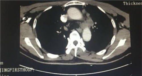
Figure 4 Imaging of the metastases in the right ring finger.
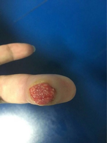
Figure 5 Fine-needle aspiration biopsies imaging from the métastases in the right ring finger.
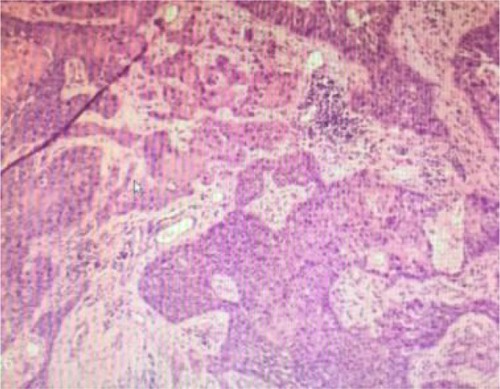
Figure 6 Pathology imaging from the metastases in the right ring finger.

Figure 7 Radiological bone imaging.
Discussion
Distant metastasis to the ST, defined as metastasis to skeletal muscle and subcutaneous tissues, has been rarely reported in previous studies. The most commonly reported primary carcinomas that result in clinically recognized ST metastasis are those of the lung, kidney and colon. A review of literature revealed the most frequently reported locations for ST metastasis as the back, chest wall and abdomen.Citation9 Metastatic tumors to the digit region are uncommon and mostly located in the hand, while only a few are in the ST of digit. Metastases to the digits of hand commonly arise from cancer of the lung, genitourinary tract and breast.Citation7 So far, only four cases of metastasis to the phalanx or digital pulp from an esophageal primary have been described.Citation6–Citation8,Citation10 Our report has provided a new case for the treatment of ESCC with digit metastases.
In addition, the prognosis in the presence of ST metastasis should be taken into account when weighing the merits of the findings. Distant ST metastasis was associated with poor prognosis, as demonstrated previously.Citation11 Our study showed that the patient died of ESCC within 2 months after ST metastasis was diagnosed, which was consistent with Damron and Heiner’s study which reported that the median survival ranged from <4 months to no more than 18 months after the diagnosis of ST metastasis.Citation12 The prognosis of such cases is poor, as the disease has generally disseminated by the time distant ST metastases are diagnosed. Therefore, primary esophageal carcinoma metastasizing simultaneously to distal finger of hand as reported here is an extremely rare presentation and is generally associated with a poor outcome.
Conclusion
Detection of ST metastasis may have prognostic implications, providing more accessible biopsy sites and helping avoid invasive procedures.
Acknowledgments
Written consent was taken from the patient for publishing his clinical details and histopathologic photomicrographs. This work was supported by the Natural Science Fundation of Jiangsu Province (BK20150092, J Shi).
Disclosure
The authors report no conflicts of interest in this work.
References
- FerlayJSoerjomataramIDikshitRCancer incidence and mortality worldwide: sources, methods and major patterns in GLOBOCAN 2012Int J Cancer20151365E359E38625220842
- LookingbillDPSpanglerNSextonFMSkin involvement as the presenting sign of internal carcinoma. A retrospective study of 7316 cancer patientsJ Am Acad Dermatol199022119262298962
- HedeshianMHWangXXuBFontaineJPPodbielskiFJSubcutaneous metastasis from oesophageal cancerAsian Cardiovasc Thorac Ann200614652052117130332
- LookingbillDPSpanglerNHelmKFCutaneous metastases in patients with metastatic carcinoma: a retrospective study of4020 patientsJ Am Acad Dermatol1993292 Pt 12282368335743
- KerinRMetastatic tumors of the handJ Bone Joint Surg Am1983659133113356654944
- HsiehCYBaiLYLoWCHuangHHChiuCFEsophageal squamous cell carcinoma with a solitary phalangeal metastasisSouth Med J2008101111159116019088530
- HoustonJDTelepakRJAn isolated digital metastasis of esophageal basaloid squamous cell carcinomaClin Nucl Med200025755755810885705
- DimriKRastogiNLalPCarcinoma of esophagus with unusual metastasis to gingiva and phalanxIndian J Cancer2003401373814716132
- SchwartzRACutaneous metastatic diseaseJ Am Acad Dermatol1995332 Pt 11611827622642
- SilfenRAmirATobarAHaubenDJThe digital pulp as a presenting site of metastatic esophageal carcinomaAnn Plast Surg200146218318411216618
- BalchCMBuzaidACSoongSJFinal version of the American Joint Committee on Cancer staging system for cutaneous melanomaJ Clin Oncol200119163635364811504745
- DamronTAHeinerJDistant soft tissue metastasis: a series of 30 new patients and 91 cases from the literatureAnn Surg Oncol20007752653410947022