
Abstract
Background
IRAK1 has been repoted to play an essential role in the development of multiple cancers. However, the clinical significance of IRAK1 in hepatocellular carcinoma (HCC) and the underlying molecular mechanism remain unclear. Therefore, we aimed to investigate the role of IRAK1 in the pathogenesis of HCC in this study.
Materials and methods
HCC tissues and para-carcinoma tissues were collected for immunohistochemistry (IHC) analysis to evaluate IRAK1 expression. Data of IRAK1 expression were downloaded from the cancer genome atlas (TCGA) for analyzing the clinical significance of IRAK1. Receiver operating characteristic (ROC) curve and survival analyses were carried out to assess the diagnostic and prognostic significance of IRAK1 in IHC and TCGA data. Additionally, we investigated the alteration of IRAK1 gene in HCC from cBioPortal to generate a network of the interaction between IRAK1 and the neighboring genes. The influence of IRAK1 gene alteration on the prognosis of HCC patients was evaluated by survival analysis.
Results
Analysis of both IHC and TCGA data revealed a significant upregulation of IRAK1 in HCC tissues. The IHC analysis revealed there was an increasing trend in IRAK1 expression among normal liver tissues, liver cirrhosis tissues, para-carcinoma tissues and HCC tissues. The ROC curves for IHC and TCGA data demonstrated that IRAK1 exhibited a significant diagnostic value for HCC. Moreover, IRAK1 expression was observed to be associated with tumor size, metastasis and T-stage. The survival analysis indicated that the upregulation of IRAK1 predicted a worse overall survival of HCC. Additionally, data from cBioPortal confirmed that 29% of HCC tissues possessed an alteration of the IRAK1 gene.
Conclusion
IRAK1 may act as an oncogene in the development of HCC with its overexpression in HCC. Moreover, IRAK1 might serve as a promising diagnostic and therapeutic target for HCC.
Introduction
Hepatocellular carcinoma (HCC) is one of the most common cancers and ranks second among the most fatal cancers.Citation1,Citation2 Approximately 250,000 new cases of HCC occur each year, with an estimated 600,000 HCC-related deaths.Citation3,Citation4 Surgery is currently the most common and effective therapy for HCC, although the prognosis of HCC patients remains unsatisfactory, with the 5-year survival rate ranging from 36% to 50%.Citation5,Citation6 Therefore, we aimed to determine an efficacious diagnostic and therapeutic target for HCC to improve the survival of HCC patients.
IRAK1, located on the X chromosome, belongs to the IRAK family.Citation7,Citation8 IRAK1 is a widely expressed serine/threonine kinase that plays a critical role in the TLR/IL1R signaling pathways.Citation9 The activation of the downstream receptors could cause the phosphorylation of IRAK1, which subsequently binds to the E3 ubiquitin ligase and TRAF6, leading to the activation of the NF-κB and MAPK pathways.Citation10–Citation13 There is mounting evidence that IRAK1 promotes the initiation and progression of various types of cancers, including acute myeloid leukemia (AML), melanoma, lung cancer and breast cancer, by aberrantly stimulating the downstream signaling pathways.Citation14–Citation19
To date, only one study on the role of IRAK1 in HCC has reported that IRAK1 could accelerate cell growth and suppress cell apoptosis via its overexpression in HCC.Citation8 However, that study did not provide information on the gene status of IRAK1 in HCC or analyze the significance of IRAK1 in the diagnosis and prognosis of HCC. Therefore, we carried out the present study to investigate the clinical significance of IRAK1, with a further investigation into the gene status of IRAK1 in the cBioPortal database of HCC, to shed light on the potential molecular mechanism of IRAK1 in the pathogenesis of HCC by analyzing the immunohistochemistry (IHC) and the cancer genome atlas (TCGA) data of IRAK1 expression in HCC.
Materials and methods
Patient samples
A total of 171 formalin-fixed and paraffin-embedded HCC tissues and 171 para-carcinoma tissues (153 males and 18 females) were obtained from the First Affiliated Hospital of Guangxi Medical University (Nanning, Guangxi, China) from March 2010 to December 2012. We also collected 37 cases of liver cirrhosis tissues and 33 normal tissues to compare the IRAK1 expression in different stages of HCC tissues. The research committee of Guangxi Medical University granted permission to conduct the study, and all participating patients signed the written informed consent for this study.
IHC
IHC analysis was performed on all formalin-fixed, paraffin-embedded tissue samples to identify the pattern of IRAK1 expression in HCC cells. The IRAK1 antibody (F-4, sc-5228, 1:50 dilution) provided by Santa Cruz Biotechnology (Heidelberg, Germany) was used for IHC detection. The detailed protocol of IHC was shown in our previous studies.Citation20,Citation21 The IRAK1 staining in HCC cells was analyzed through blind evaluation, which was performed by two pathologists who independently identified the intensity of staining and the positive staining ratio of IRAK1. The staining of IRAK1 was scored according to the following criteria: 1) scores of 0, 1, 2 and 3 indicated negative, weak, moderate and strong staining intensity of IRAK1, respectively, and 2) the proportion of cells positively stained for IRAK1 was graded as 0, 1, 2, 3 and 4 representing 0%, 1%–25%, 26%–50%, 51%–75% and 76%–100% positively stained cells, respectively. If the result of the two criteria was >2, the cells were considered to be positively immune reactive.
Extraction of TCGA data
TCGA data of IRAK1 expression in HCC and noncancerous tissues were downloaded to analyze IRAK1 expression in HCC and its correlations with the clinical parameters of HCC. Information about the alteration of the IRAK1 gene was available in cBioPortal OncoPrint (http://www.cBioPortal.org/index.do) and was analyzed to evaluate the status of IRAK1 gene in the development of HCC.Citation22,Citation23 Additionally, the genes associated with IRAK1 were accessed from cBioPortal OncoPrint to generate a network illustrating the interaction of the IRAK1-related genes. The Kaplan–Meier survival curves were drawn to examine the impact of the alteration of the IRAK1 gene on the prognosis of HCC patients.
Statistical analysis
All statistical analyses were performed using SPSS 22.0 (IBM Corp., Armonk, NY, USA). The difference in the level of IRAK1 expression by IHC was calculated using the chi-square test. The quantitative data on IRAK1 obtained from TCGA were analyzed by Student’s t-test. With regard to the diagnostic value of IRAK1 expression in the IHC and TCGA data, the receiver operating characteristic (ROC) curve was drawn to evaluate the diagnostic capability of IRAK1 for HCC. A graded area under the curve (AUC) value for the ROC curves of 0.5–0.7, 0.7–0.9 or 0.9–1.0 represented a poor, moderate or high diagnostic value of IRAK1, respectively. The influence of IRAK1 on the prognosis of HCC patients was assessed by Kaplan–Meier analysis for the TCGA and IHC data. Moreover, the Kaplan–Meier analysis was applied to analyze the impact of IRAK1 alterations on the prognosis of HCC patients. Two-tailed value of P<0.05 was considered as statistically significant.
Results
Validation of the clinical significance of IRAK1 expression in HCC by IHC
The IHC of normal liver, cirrhosis, normal liver para-HCC, cirrhosis para-HCC and HCC tissues is shown in –. To verify the aberrant expression of IRAK1 in HCC, we included a cohort of 171 samples of HCC. The result of the IHC showed the highest positive rate of IRAK1 in HCC tissues (48.5%) compared with para-HCC tissues (34.5%, P=0.008), liver cirrhosis tissues (24.3%, P=0.007) and normal liver tissues (15.2%, P<0.001; and ). Moreover, the IRAK1 expression increased noticeably in normal liver tissues, liver cirrhosis tissues, para-HCC tissues and HCC tissues (P<0.001; and ).
Figure 1 The IHC of normal liver tissues.
Abbreviation: IHC, immunohistochemistry.
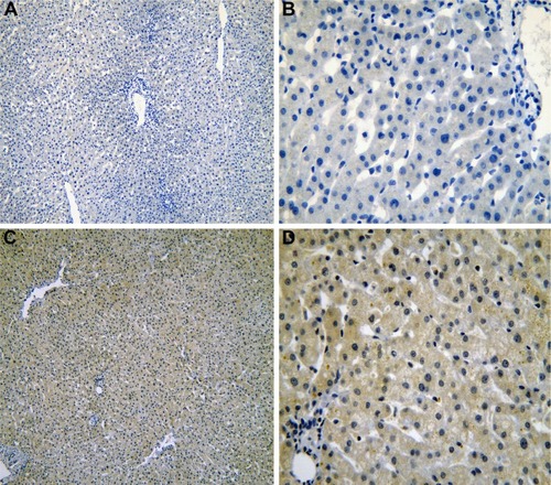
Figure 2 The IHC of liver tissues with cirrhosis.
Abbreviation: IHC, immunohistochemistry.
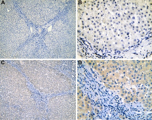
Figure 3 The IHC of para-HCC tissues with cirrhosis.
Abbreviations: IHC, immunohistochemistry; HCC, hepatocellular carcinoma.
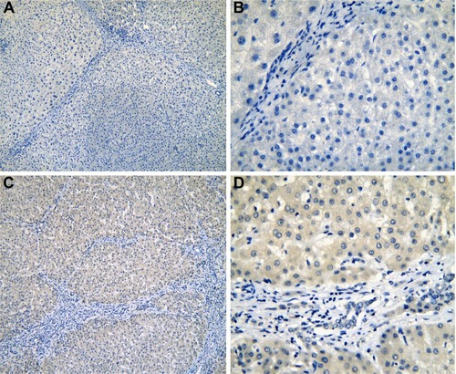
Figure 4 The IHC of normal liver tissues (para-HCC).
Abbreviations: IHC, immunohistochemistry; HCC, hepatocellular carcinoma.
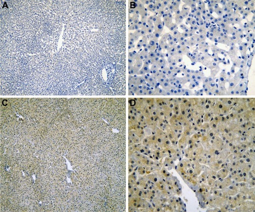
Figure 5 The IHC of HCC tissues.
Abbreviations: IHC, immunohistochemistry; HCC, hepatocellular carcinoma.
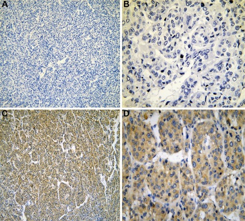
Figure 6 The expression of IRAK1 in IHC.
Abbreviations: IHC, immunohistochemistry; HCC, hepatocellular carcinoma.
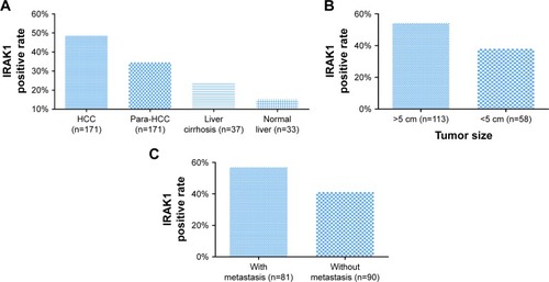
Table 1 Expression of IRAK1 in different types of liver tissues
Focusing on the relationships between IRAK1 and the clinicopathological features of HCC, we found a higher positive rate of IRAK1 expression in the groups of large tumor size (54.0%) and metastasis (56.8%) than that in the groups of small tumor size (37.9%, P=0.047; ) and no metastasis (41.1%, P=0.041; ). As shown in , the Spearman’s correlation test was utilized to evaluate the correlations between IRAK1 and the clinical features of HCC. The results indicated that IRAK1 expression was significantly correlated with tumor size (r=0.152, P=0.047) and metastasis (r=0.157, P=0.041). As for the diagnostic value of IRAK1 in HCC, the ROC curves indicated that IRAK1 showed a low diagnostic value for HCC (AUC =0.591, P=0.002; ).
Figure 7 The ROC curves of IRAK1 expression by IHC in HCC.
Abbreviations: ROC, receiver operating characteristic; IHC, immunohistochemistry; HCC, hepatocellular carcinoma; AUC, area under the curve.
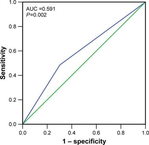
Table 2 Relationship between IRAK1 expression and clinicopathological features in HCC
IRAK1 expression detected by RNA-sequencing (RNA-seq) data in TCGA database
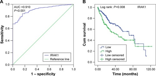
The clinical significance of IRAK1 expression in HCC based on TCGA data was investigated. The results revealed that an upregulation of IRAK1 was detected in 354 cases of HCC (10.3058±0.5212) compared with 50 adjacent normal liver tissues (11.5503±0.7946, P<0.001; ). Furthermore, a paired test of 49 pairs of HCC tissues and matched adjacent normal liver tissues showed a similar trend of IRAK1 expression between HCC and normal liver tissues (11.6174±0.8979 vs 10.2921±0.5174, respectively, P<0.001; ). We also used the ROC curves to estimate the diagnostic value of IRAK1 in HCC. An AUC value of 0.910 indicated a high diagnostic value (95% CI: 0.878–0.942, P<0.001; ).
Figure 8 The expression of IRAK1in HCC in the TCGA data.
Abbreviations: HCC, hepatocellular carcinoma; TCGA, the cancer genome atlas.
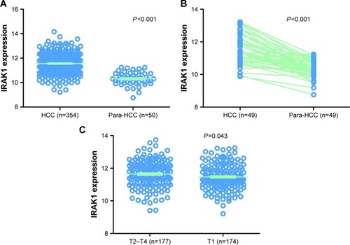
Figure 9 The ROC curve and Kaplan–Meier survival curve of IRAK1 expression for HCC from TCGA data.
Abbreviations: ROC, receiver operating characteristic; HCC, hepatocellular carcinoma; TCGA, the cancer genome atlas; AUC, area under the curve; Cum, cumulative.
With regard to the relationships between IRAK1 expression and clinicopathological characteristics, the expression of IRAK1 significantly increased only in the advanced tumor stage (T2–T4: 11.6363±0.8153 vs T1: 11.4647±0.7677, P=0.043; and ).
Table 3 Expression of IRAK1 detected by RNA sequencing and clinicopathological parameters in HCC in TCGA
As for the prognostic value of IRAK1 for HCC, the cohort of 354 HCC patients was separated into two groups according to the cutoff value of the mean level of IRAK1 expression. The median survival time was 45.7 months in the IRAK1 high-expression group and 71.0 months in the IRAK1 low-expression group (log rank: P=0.008; ).
IRAK1 alteration analyzed in cBioPortal database
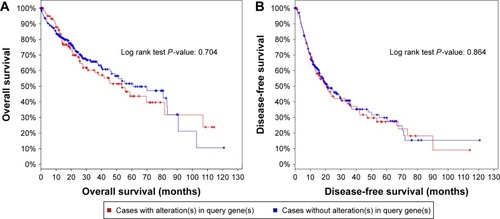
We investigated the genetic alteration of IRAK1 with a cohort of 440 cases of HCC available in the cBioPortal database. The result showed that 29% (126/440) cases of HCC in this cohort exhibited IRAK1 alteration, including 85 cases of mRNA upregulation, 35 cases of mRNA downregulation, eight cases of amplification, three cases of missense mutation, two cases of truncating mutation and one case of deep deletion. Among them, eight genes had the mixed type of alteration (). Moreover, a network of the interaction of IRAK1 with several frequently altered neighboring genes was drawn (). The network reflected clear interaction between IRAK1 and several genes, including MAP3K7, MAP2K4, NFKB1, AKT1 and ERC1. Moreover, Pearson correlation analysis was performed to investigate the correlations between IRAK1 expression and its potential targets in the network. The results revealed that the expressions of several target genes significantly correlated with IRAK1 expression (HMGB1: r=−0.13, MAP3K7: r=−0.15, ROCK1: r=−0.13, CSNK1A1: r=−0.15, CSNK1G3: r=−0.13, MAPKAPK5: r=0.18, ACVR2A: r=−0.19, MAPKAPK2: r=0.34, all P<0.05). Remarkably, MAPKAPK2 showed relatively a good correlation to IRAK1 (). The survival analysis (overall survival and disease-free survival) was performed to compare the prognosis of the patient group with and without IRAK1 alteration. However, the results did not show statistical significance ().
Figure 10 The alteration of IRAK1 and its interaction in altered neighboring genes in 440 HCC cases from the cBioPortal database.
Abbreviation: HCC, hepatocellular carcinoma.
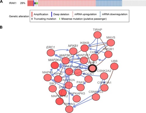
Figure 11 Correlation between IRAK1 and MAPKAPK2.
Abbreviation: RNA-seq, RNA sequencing.
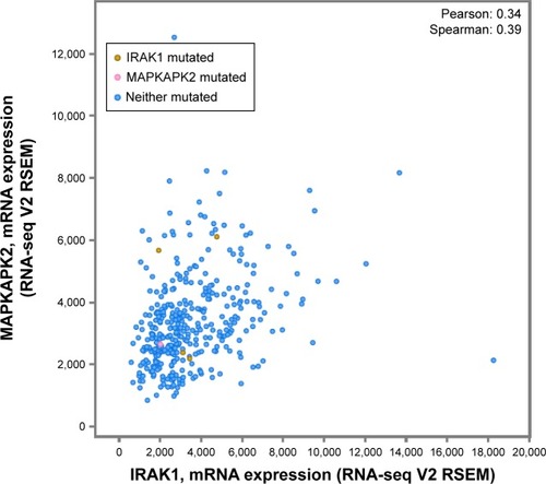
Figure 12 Analysis of the overall survival (A) and disease-free survival (B) of HCC patients with and without IRAK1 alteration.
Abbreviation: HCC, hepatocellular carcinoma.
Discussion
Using reverse transcription polymerase chain reaction (RT-PCR), Western blot and immunohistochemical staining, Li et alCitation8 confirmed that IRAK1 exhibited a higher expression in 33 HCC cases and noted that IRAK1 may augment the malignant potential of HCC by stimulating the proliferation of cells and suppressing the apoptosis of cells. However, no study has elaborated on the diagnostic and prognostic value of IRAK1 for HCC or the potential molecular mechanisms of IRAK1 in the pathogenesis of HCC. To date, this study is the first one to comprehensively investigate the clinical significance of IRAK1 in HCC with an analysis of IHC, TCGA and gene alteration data from cBioPortal.
In agreement with the study by Li et al,Citation8 the results of our study indicated that IRAK1 had a significantly higher expression in HCC tissues than in noncancer tissues of both the IHC and TCGA data. The large sample size of the current study may provide more powerful evidence to verify the upregulation of IRAK1 in HCC. We also observed an increasing trend of IRAK1 expression in normal liver tissues, liver cirrhosis tissues, para-HCC tissues and HCC tissues, which suggests that a higher expression of IRAK1 was closely associated with the malignant evolution of liver tissues. Thus, our results implied that IRAK1 may act as an oncogene in the tumorigenesis of HCC.
Consistent with an oncogenic effect of IRAK1 in HCC, the results obtained from the IHC and TCGA data in our study revealed that IRAK1 exhibited a significant diagnostic value for HCC, especially in TCGA data (AUC >0.9, P<0.001), which has not been reported in previous studies. Significantly, IRAK1 correlated well with the prognosis of HCC patients. Therefore, we anticipated that IRAK1 may serve as a potential diagnostic and prognostic tool for HCC. Further prospective study would be necessary to confirm the diagnostic and prognostic value of IRAK1 in HCC, especially in Asia.
To further explore the clinical significance of IRAK1 in HCC, we examined the relationships between IRAK1 expression and the clinicopathological variables of HCC. Analysis of our IHC and TCGA data revealed that IRAK1 expression was related to the malignant phenotype of HCC, including larger tumor sizes, tumor metastasis and advanced tumor stage, which was consistent with the previous results indicating that IRAK1 could strengthen the proliferative ability and inhibit the apoptosis of HCC cells.Citation8 It can be deduced from our result that IRAK1 may promote the progression of HCC by enhancing the growth, invasion and metastasis of cancer cells.
The oncogenic roles of IRAK1 in other cancers have been widely studied. In T-cell acute lymphoblastic leukemia (T-ALL), IRAK1/4 signaling promoted the progression of T-ALL by activating TRAF6, which subsequently strengthened MCL1, a protein with antiapoptotic effect.Citation24 In breast cancer, IRAK1 was reported to facilitate the growth of triple-negative breast cancer (TNBC) by inducing the expression of NF-κB-related cytokine.Citation19 Toll-like receptors (TLRs) are important molecules that regulate the innate immunity and participate in tissue repair, autoimmune disease and tumor microenvironment.Citation25,Citation26 The study by Fonte et al indicated that TLR1/2, TLR2/6 and TLR9 stimulated the proliferation of cancer cells in splenic marginal zone lymphoma (SMZL). Moreover, the inhibition of IRAK1/4, upstream kinases of the TLR signaling pathway, reduced the cell viability induced by TLRs.Citation27 Additionally, miR-146, which belongs to the miRNA family, has been shown to suppress the development of cancers, including gastric cancer, pancreatic cancer and breast cancer, which was implemented by targeting IRAK1.Citation28–Citation30 Thus, we assumed that IRAK1 may play an oncogenic role in HCC by regulating the expression of downstream molecules, such as MCL1 and NF-κB-related cytokine, and modulating the TLR signaling pathways. The aberrant expression of miR-146a may also lead to the carcinogenic function of IRAK1 in HCC. Further in vivo and in vitro studies on the molecular mechanisms of IRAK1 in HCC are needed to decipher the pathogenesis of HCC.
To facilitate the understanding of the function of IRAK1 in HCC, we further investigated the alteration of the IRAK1 gene in HCC. The results indicated that approximately one-third of the genes exhibited IRAK1 alterations, among which the mRNA upregulation was the predominant type of alteration, followed by mRNA downregulation, amplification and missense mutation. The upregulation of IRAK1 may cause an increased level of IRAK1 expression in HCC, as confirmed by our results. As for the missense mutation of IRAK1, a Phe196Ser change in IRAK1 was detected in primary effusion lymphoma (PEL),Citation31 which caused the constitutive phosphorylation of IRAK1 in PEL and promoted the progression of PEL. We postulated that the specific missense mutation of IRAK1 existed in HCC to facilitate the development of IRAK1 via the phosphorylation of IRAK1. Then, we attempted to trace the causes of the IRAK1 alteration by investigating the interaction between the IRAK1 gene and the neighboring frequently altered genes. From the network, we found that several genes, including MAP3K7, MAP2K4, NFKB1, AKT1 and ERC1, interact directly with IRAK1. Moreover, MAPKAPK2 showed relatively good correlation to IRAK1 and is worth be validated in future work. Thus, we speculated that these genes may influence the alteration of IRAK1 gene and exert a carcinogenic effect on IRAK1 to promote the occurrence and development of HCC. However, the bioinformatics analysis may produce a result with weak evidence. Thus, whether these genes were involved in the alteration of IRAK1 gene needs further in vitro and in vivo validation in future studies.
Conclusion
IRAK1 was significantly overexpressed in HCC and showed promise as a diagnostic and therapeutic target for HCC with its oncogenic function in HCC.
Acknowledgments
The study was supported by the fund of Youth Science Foundation of Guangxi Medical University (GXMUYSF201624). The authors thank the American Journal Experts (AJE) for editing the manuscript.
Disclosure
The authors report no conflicts of interest in this work.
References
- SiegelRLMillerKDJemalACancer statistics, 2016CA Cancer J Clin201666173026742998
- ChenWZhengRBaadePDCancer statistics in China, 2015CA Cancer J Clin201666211513226808342
- VenookAPPapandreouCFuruseJde GuevaraLLThe incidence and epidemiology of hepatocellular carcinoma: a global and regional perspectiveOncologist201015suppl 4513
- WhiteDLThriftAPKanwalFDavilaJEl-SeragHBIncidence of hepatocellular carcinoma in all 50 United States, from 2000 through 2012Gastroenterology Epub20161123
- BaloghJVictorD3rdAshamEHHepatocellular carcinoma: a reviewJ Hepatocell Carcinoma20163415327785449
- ColagrandeSInghilesiALAburasSTalianiGGNardiCMarraFChallenges of advanced hepatocellular carcinomaWorld J Gastroenterol201622347645765927678348
- JanssensSBeyaertRFunctional diversity and regulation of different interleukin-1 receptor-associated kinase (IRAK) family membersMol Cell200311229330212620219
- LiNJiangJFuJTargeting interleukin-1 receptor-associated kinase 1 for human hepatocellular carcinomaJ Exp Clin Cancer Res201635114027619757
- CaoZHenzelWJGaoXIRAK: a kinase associated with the interleukin-1 receptorScience19962715252112811318599092
- FlannerySBowieAGThe interleukin-1 receptor-associated kinases: critical regulators of innate immune signallingBiochem Pharmacol201080121981199120599782
- WescheHHenzelWJShillinglawWLiSCaoZPillars article: MyD88: an adapter that recruits IRAK to the IL-1 receptor complexImmunity199778378479430229 J Immunol2013190151523264669
- GuhaMMackmanNLPS induction of gene expression in human monocytesCell Signal2001132859411257452
- ZhangGGhoshSToll-like receptor-mediated NF-kappaB activation: a phylogenetically conserved paradigm in innate immunityJ Clin Invest20011071131911134172
- RhyasenGWBolanosLFangJTargeting IRAK1 as a therapeutic approach for myelodysplastic syndromeCancer Cell20132419010423845443
- SrivastavaRGengDLiuYAugmentation of therapeutic responses in melanoma by inhibition of IRAK-1,-4Cancer Res201272236209621623041547
- BehrensCFengLKadaraHExpression of interleukin-1 receptor-associated kinase-1 in non-small cell lung carcinoma and preneoplastic lesionsClin Cancer Res2010161344420028769
- ZhangXDangYLiPRongMChenGExpression of IRAK1 in lung cancer tissues and its clinicopathological significance: a microarray studyInt J Clin Exp Pathol20147118096810425550857
- ScheerenFAKuoAHvan WeeleLJA cell-intrinsic role for TLR2-MYD88 in intestinal and breast epithelia and oncogenesisNat Cell Biol201416121238124825362351
- WeeZNYatimSMKohlbauerVKIRAK1 is a therapeutic target that drives breast cancer metastasis and resistance to paclitaxelNat Commun20156874626503059
- LiJJLuoJLuJNRelationship between TRAF6 and deterioration of HCC: an immunohistochemical and in vitro studyCancer Cell Int2016167627708550
- LiJLiangLLiuYClinicopathological significance of STAT4 in hepatocellular carcinoma and its effect on cell growth and apoptosisOnco Targets Ther201691721173427051307
- GaoJAksoyBADogrusozUIntegrative analysis of complex cancer genomics and clinical profiles using the cBioPortalSci Signal20136269 pl1
- CeramiEGaoJDogrusozUThe cBio cancer genomics portal: an open platform for exploring multidimensional cancer genomics dataCancer Discov20122540140422588877
- LiZYoungerKGartenhausRInhibition of IRAK1/4 sensitizes T cell acute lymphoblastic leukemia to chemotherapiesJ Clin Invest201512531081109725642772
- O’NeillLAGolenbockDBowieAGThe history of toll-like receptors – redefining innate immunityNat Rev Immunol201313645346023681101
- Rakoff-NahoumSMedzhitovRToll-like receptors and cancerNat Rev Cancer200991576319052556
- FonteEAgathangelidisAReverberiDToll-like receptor stimulation in splenic marginal zone lymphoma can modulate cell signaling, activation and proliferationHaematologica2015100111460146826294727
- KogoRMimoriKTanakaFKomuneSMoriMClinical significance of miR-146a in gastric cancer casesClin Cancer Res201117134277428421632853
- LiYVandenboomTG2ndWangZmiR-146a suppresses invasion of pancreatic cancer cellsCancer Res20107041486149520124483
- BhaumikDScottGKSchokrpurSPatilCKCampisiJBenzCCExpression of microRNA-146 suppresses NF-kappaB activity with reduction of metastatic potential in breast cancer cellsOncogene200827425643564718504431
- YangDChenWXiongJSherrodCJHenryDHDittmerDPInterleukin 1 receptor-associated kinase 1 (IRAK1) mutation is a common, essential driver for Kaposi sarcoma herpesvirus lymphomaProc Natl Acad Sci U S A201411144E4762E476825341731