
Abstract
Resistance to chemotherapy is a primary problem for the effective treatment of ovarian cancer. Recently, increasing evidence has demonstrated that miRNAs modulate many important molecular pathways involved in chemotherapy. Previous studies demonstrated that miR-199a affected ovarian cancer cell resistance to cisplatin (DDP). However, the role of miR-199a and its target genes in determination of ovarian cancer sensitivity to DDP remains unclear. Quantitative reverse transcription polymerase chain reaction was used to detect the expression levels of miR-199a in ovarian cancer tissues and C13* and OV2008 cell lines. After transfection of miR-199a mimic or inhibitor, flow cytometry was used to detect cell apoptosis exposed to DDP. Enzyme-linked immunosorbent assay and Western blot assay were applied to detect tumor necrosis factor-α levels and protein expression levels of Bax, Fas, Fas-associated death domain, and caspase-8. The results indicated that the expression of miR-199a was downregulated and hypoxia-inducible factor 1α (Hif1α) upregulated in the ovarian tumors compared with those in the corresponding normal tissues. Besides, the expression levels of miR-199a were significantly higher in OV2008 cells compared with those in C13* cells. Moreover, suppression of Hif1α reversed the inhibiting function of miR-199a inhibitor on DDP-induced apoptosis in the OV2008 cells. However, overexpression of both miR-199a and Hif1α reduced DDP-induced apoptosis in C13* cells. In conclusion, miR-199a may change DDP resistance in ovarian cancer by regulating Hif1α.
Introduction
Ovarian cancer is one of the most common gynecologic malignancies and the primary cause of gynecologic cancer deaths for women.Citation1 Nowadays, ovarian cancer has become a serious threat to women’s health in China because of its high incidence and mortality.Citation2 One of the current standard treatments for women with ovarian cancer is platinum-based chemotherapy.Citation3 Cisplatin (DDP) is a platinum compound that is used as a first line of therapy in ovarian cancer.Citation4 Moreover, DDP directly covalently binds to nuclear DNA, leading to transcription inhibition processes.Citation5 More than 70% of patients initially respond to DDP, but the 5-year survival rate is only about 30%.Citation6 DDP resistance is the major cause of chemotherapy failure.Citation7 As the apoptotic response of cancer cells to drugs is used to evaluate the sensitivity to chemotherapy,Citation8 the decreased cellular uptake of chemotherapeutic drugs accompanied by increased antiapoptotic capacity may be important in DDP resistance in ovarian cancer.Citation9 Therefore, exploring the molecular mechanisms of DDP resistance in ovarian cancer cells has the potential to improve the therapy of the disease.
miRNAs are endogenous, noncoding RNAs that bind to complementary sequences on target mRNA transcripts to cause translational repression or target degradation.Citation10 miR-199a is located on human chromosome 19q13.2, and a low expression of miR-199a has been detected in human ovarian carcinoma.Citation11 Although recent reports demonstrate that miR-199a may affect ovarian cancer cell resistance to DDP,Citation12 the role of miR-199a and its target mRNAs in determination of ovarian cancer sensitivity to DDP still needs to be investigated.
Hypoxia-inducible factor 1 (Hif1) is overexpressed in human cancers under intratumoral hypoxia.Citation13 In addition, its subunit Hif1α has been widely studied as an endogenous hypoxia marker.Citation14 The Hif1α protein overexpression is more frequently observed in the ovarian cancer specimens than in noncancerous ovarian tissues.Citation15 Besides, a previous study demonstrated that Hif1α contained a potential binding site in its 3′ untranslated region for miR-199a in the ovarian cancer cells.Citation16
The purpose of this study was to investigate the regulatory mechanism of miR-199a in DDP drug resistance in the human OV2008 and C13* ovarian cancer cell lines. Also, miR-199a may be a new strategy for DDP resistance chemotherapy in patients with ovarian cancer.
Patients and methods
Patients
This study was approved by the Research Ethics Committee of The First Affiliated Hospital of Harbin Medical University. Written informed consent was obtained from all of the patients. The ovarian cancer tissue samples and corresponding normal tissues were collected from 23 patients diagnosed with primary ovarian cancer after operation at The First Affiliated Hospital of Harbin Medical University from January 2008 to December 2012. The normal tissues were >1.5 cm away from the edge of the ovarian cancer tissues in 23 patients. None of the patients received any chemotherapy or radiotherapy before operation. The samples were immediately snap-frozen in liquid nitrogen and kept at −80°C.
Cell lines and cell culture
The cisplatin-resistant ovarian cancer cell line (C13*) and its sensitive variant (OV2008) were gifts from Dr Rakesh Goel at Ottawa Regional Cancer Center (Ottawa, Canada). Cells were cultured in Roswell Park Memorial Institute (RPMI)-1640 media supplemented with 2 mM L-glutamine, 10% fetal bovine serum, 100 U/mL penicillin, and 100 mg/mL streptomycin at 37°C in a humidified atmosphere containing 5% CO2.
Transfection
The miR-199a mimics and inhibitors and the miR-mimic negative control (NC) and inhibitor NC were purchased from Thermo Fisher Scientific (Waltham, MA, USA). Lentiviral Hif1α shRNA and the nontargeting shRNA negative control (sh-NC) were obtained from The RNAi Consortium (Broad Institute, Cambridge, MA, USA). For gene overexpression, Hif1α full-length cDNAs were cloned into pcDNA3.1-vector (Thermo Fisher Scientific). For transfection, OV2008 and C13* cells were plated in six-well plates using Lipofectamine 2000™ (Thermo Fisher Scientific) according to the manufacturer’s instructions. After transfection for 48 h, cells were harvested and stored as cell pellets at −80°C until use.
Quantitative reverse transcription polymerase chain reaction (qRT-PCR)
To measure the relative level of transcript, qRT-PCR was performed. Briefly, total RNA was extracted by TRIzol reagent (Thermo Fisher Scientific). cDNA was obtained by reverse transcription using M-MLV reverse transcriptase (Promega). Also, qRT-PCR was performed using the Maxima SYBR Green qPCR master-mix (Fermentas) in a 7900 HT qPCR machine (Thermo Fisher Scientific). A specific forward primer of miR-199a (5′-CCCAGTGTTCAGACTACCTGTTC-3′) and a common universal reverse primer were used. Also, U6 or GAPDH was used for normalization using the comparative CT-method.Citation17 Each sample was performed in triplicate.
Measurement of tumor necrosis factor alpha (TNF-α) by enzyme-linked immunosorbent assay
To measure the amount of TNF-α in cell lysates, cells were lysed in PBS. After centrifugation, supernatant was taken for determination of intracellular TNF-α level by enzyme-linked immunosorbent assay (ELISA). The amount of TNF-α was measured using a Duoset ELISA (R&D Systems, Minneapolis, MN, USA) according to the manufacturer’s instructions.
Western blot analysis
Protein levels were measured by Western blot. Briefly, cells were lysed in ice-cold radioimmunoprecipitation assay (RIPA) buffer (Sigma-Aldrich, St Louis, MO, USA) and protein was quantified by Bradford assay. Then, an equal amount of protein was separated on sodium dodecyl sulfate polyacrylamide electrophoresis gel and transferred to a nitrocellulose membrane (Merck Millipore, Burlington, MA, USA). After being blocked with 10% skim milk, the membranes were incubated with primary antibodies overnight at 4°C. Then, the HRP-conjugated secondary antibodies were used. The bound antibodies were detected with enhanced chemiluminescence system (Bio-Rad Laboratories, Hercules, CA, USA). GAPDH was used as a loading control.
Apoptosis assay
After transfection as described above, the OV2008 and C13* cells were treated with DDP at a concentration of 40 μM for 48 h. After incubation, the cells were harvested and stained with 5 μL annexin V-fluorescein isothiocyanate (FITC) and 10 μL propidium iodide (PI; 20 μg/mL; Thermo Fisher Scientific) in the dark for 15 min at room temperature. Then, the apoptotic cells were analyzed by a FACScan (BD Biosciences, Franklin Lakes, NJ, USA). The signal of annexin V-FITC was detected using the FITC signal detector (FL1), and PI was measured with the phycoerythrin signal detector (FL2).
Statistical analysis
All experiments were repeated at least three times. Data are presented as the mean ± SD. Statistical significance was determined with Student’s t-test by using the SPSS 16.0 software (IBM Corp., Armonk, NY, USA). P<0.05 was considered as statistically significant.
Results
miR-199a was downregulated and Hif1α upregulated in ovarian cancer specimens
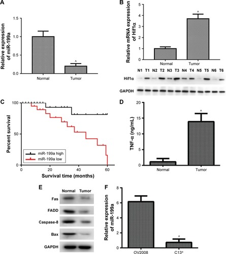
To detect the detailed relationship between miR-199a and ovarian cancer, the expression of miR-199a was determined in ovarian tumors and their corresponding normal tissues. We found that miR-199a levels were significantly lower in ovarian tumors compared with those in corresponding normal tissues (). Conversely, the relative mRNA and protein expression of Hif1α increased in ovarian tumors compared with the normal tissues ().
Figure 1 Expression levels of miR-199a, Hif1α and apoptosis-related proteins in ovarian tumor tissues and corresponding normal tissues detected by qPCR and Western blot.
Abbreviations: ELISA, enzyme-linked immunosorbent assay; FADD, Fas-associated death domain; Hif1α, hypoxia-inducible factor 1α; qPCR, quantitative polymerase chain reaction; TNF-α, tumor necrosis factor alpha.
Downregulation of miR-199a associated with poor prognosis of patients with ovarian tumors
Ovarian cancer patients with low miR-199a expression showed shorter survival time than those with high miR-199a expression ().
Apoptosis was involved in the development of ovarian cancer
Next, we further investigated the expression levels of apoptosis-related proteins in ovarian tumors and their corresponding normal tissues. illustrates that the levels of TNF-α were markedly increased in ovarian tumors compared to those in corresponding normal tissues. In addition, our data showed the protein expression levels of Fas, Fas-associated death domain (FADD), caspase-8, and Bax were significantly decreased in ovarian tumors compared to those in corresponding normal tissues (). These results revealed that the apoptosis-related proteins were involved in the development and progression of ovarian cancer.
Expression of miR-199a in DDP-resistant and -sensitive cell lines
Subsequently, we assessed the expression of miR-199a in cisplatin-sensitive and -resistant ovarian cancer cell lines OV2008 and C13*. The expression levels of miR-199a were significantly higher in OV2008 cells compared with to in C13* cells ().
Effect of miR-199a on DDP-induced apoptosis in the resistant and sensitive cell lines
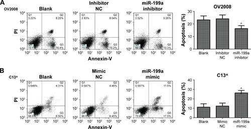
To investigate whether miR-199a could modulate the resistance of OV2008 and C13* cells to DDP, the OV2008 and C13* cells were transfected with miR-199a inhibitor and miR-199a mimic, respectively. Then, the cells were treated with 40 μM DDP for 48 h. reveals that the apoptosis assays show a significant decline of apoptosis ratios in the OV2008 cells transfected with miR-199a inhibitor compared to those in the blank and inhibitor NC groups. However, apoptosis assays using annexin V staining showed that miR-199a mimic in the C13* cells enhanced DDP-induced apoptosis compared with that in the blank and mimic NC groups (). These results revealed that miR-199a could change the resistance to DDP in the ovarian cancer cells through promoting DDP-induced apoptosis in vitro.
Figure 2 Effects of miR-199a on cisplatin-induced apoptosis in the OV2008 and C13* cells.
Abbreviations: NC, negative control; PI, propidium iodide.
miR-199a promoted DDP-induced apoptosis of ovarian cancer cells through targeting Hif1α
We further examined whether miR-199a was involved in the regulation of the expression of Hif1α. In Western blot, Hif1α expression exhibited a dramatic increase in the OV2008 cells transfected with miR-199a inhibitor compared to that in the cells transfected with inhibitor NC (). Next, we investigated whether the effect of miR-199a was involved in DDP-induced apoptosis through targeting Hif1α. As shown in , OV2008 cells transfected with the miR-199a inhibitor displayed lower levels of apoptosis in response to DDP. However, suppression of Hif1α reversed the inhibiting effects of miR-199a inhibitor on DDP-induced apoptosis in OV2008 cells. In contrast, the upregulation of miR-199a by the miR-199a mimic led to a significant downregulation of Hif1α protein expression in the C13* cells (). An inverse correlation between miR-199a and Hif1α protein expression levels existed in the ovarian cancer cells.
Figure 3 Effects of miR-199a on cisplatin-induced apoptosis were through the suppression of Hif1α expression.
Abbreviations: Hif1α, hypoxia-inducible factor 1α; NC, negative control; sh-NC, shRNA negative control.
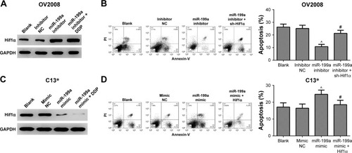
Moreover, overexpression of miR-199a promoted DDP-induced apoptosis in C13* cells. However, the effects were abolished by Hif1α (). Taken together, the results indicate that miR-199a may promote DDP-induced apoptosis through suppression of Hif1α in the ovarian cancer cells.
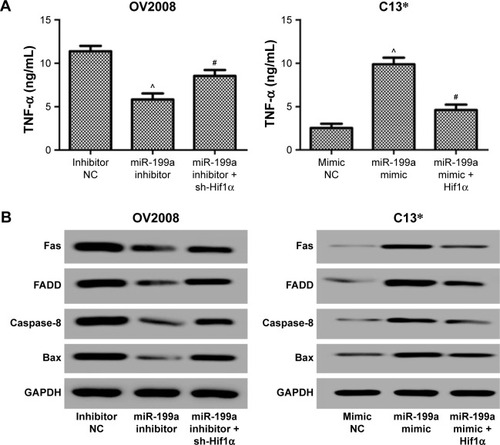
Finally, we investigated the expression levels of apoptosis-related proteins in the ovarian cancer cells treated with DDP. Inhibition of Hif1α abolished the suppression effect of miR-199a inhibitor on the levels of TNF-α and Fas, FADD, caspase-8, and Bax protein expression levels induced by DDP in the OV2008 cells (). However, overexpression of Hif1α blocked the acceleration effect of miR-199a mimic on the levels of TNF-α and Fas, FADD, caspase-8, and Bax protein expression levels induced by DDP in the C13* cells (). These results confirmed that miR-199a may increase the sensitivity of ovarian cancer cells to DDP through suppression of Hif1α.
Figure 4 Effects of miR-199a on apoptosis-related proteins induced by cisplatin were through the suppression of Hif1α expression.
Abbreviations: FADD, Fas-associated death domain; Hif1α, hypoxia-inducible factor 1α; NC, negative control; sh-NC, shRNA negative control; TNF-α, tumor necrosis factor alpha.
Discussion
Cisplatin results in the formation of DNA adducts to trigger apoptosis in cancer cells.Citation18 However, development of drug resistance is the main obstacle for DDP treatment.Citation19 A critical mechanism of drug resistance in the ovarian cancer cells is the defective apoptosis pathway.Citation20 Increasing evidence indicates that aberrant expression of miRNAs is related to the drug resistance of ovarian cancer cells through this mechanism. Kong et al found miR-125b conferred DDP resistance to ovarian cancer cells by blocking DDP-induced apoptosis.Citation21 Rao et al demonstrated that knockdown of miR-106a dramatically decreased DDP-induced apoptosis in DDP-sensitive cells A2780, while overexpression of miR-106a significantly increased DDP-induced apoptosis in DDP-resistant cells A2780/DDP.Citation22 However, the molecular mechanisms of miR-199a in DDP-resistant ovarian cancer remain unclear.
Previous studies indicated that miR-199a was deregulated in ovarian cancers through targeting IKKβ.Citation23 Besides, miR-199a expression is found to be significantly downregulated in patients with ovarian cancer in comparison with matched normal controls.Citation24 Moreover, miR-199a can reverse DDP resistance in human ovarian cancer cells through the inhibition of mTOR.Citation25 In this study, we found the miR-199a levels were significantly lower in ovarian tumors compared with those in corresponding normal tissues. In addition, miR-199a was downregulated in DDP-resistant C13* cells as compared with that in DDP-sensitive OV2008 cells. Furthermore, results from annexin V-FITC/PI double staining showed that inhibition of miR-199a in the OV2008 cells decreased the sensitivity and apoptosis induced by DDP, while the upregulation of miR-199a in C13* cells caused a notable increase of apoptotic index induced by DDP. These results suggested that miR-199a could change DDP resistance in ovarian cancer cells.
Apoptosis plays a key role in preventing tumorigenesis. Two main apoptotic pathways are reported, the intrinsic pathway and the extrinsic pathway.Citation26 The intrinsic apoptosis pathway contains the Bcl-2 family. Bcl-2 can limit the proapoptotic effects of Bax.Citation27 Furthermore, DDP activates Bax, which induces permeabilization of the outer mitochondrial membrane, leading to cytochrome c release and activation of caspases.Citation28 In addition, DDP upregulates the expression of TNF-α.Citation29 Also, activation of the extrinsic pathway triggers the activation of death receptors such as Fas and TNF-α–related apoptosis-inducing ligand receptors.Citation30 DDP also upregulates Fas ligand/receptor system. Besides, Fas can interact with FADD, which results in caspase-8 activation and cell death.Citation31 In this study, inhibition of miR-199a in the OV2008 cells significantly decreased the levels of TNF-α and the protein expression levels of Fas, FADD, caspase-8, and Bax induced by DDP. However, upregulation of miR-199a in the C13* cells dramatically increased the levels of TNF-α and the protein expression levels of Fas, FADD, caspase-8, and Bax induced by DDP. These results confirmed that miR-199a could change cell apoptosis and sensitivity to DDP in the ovarian cancer cells.
To investigate the possible mechanism by which miR-199a modulated DDP resistance in ovarian cancer, we predicted Hif1α might be a downstream target of miR-199a. It has been demonstrated that miR-199a directly targets and inhibits translation of Hif1α mRNA in cardiac myocytes.Citation32 Moreover, increased expression of miR-199a-5p was associated with decreased expression of Hif1α in the lungs from patients with COPD.Citation33 Furthermore, miR-199a inhibits the hypoxia-induced proliferation of non-small cell lung cancer cells by suppressing the expression of Hif1α.Citation34 Here, we proved that downregulation of miR-199a was associated with increased expression of Hif1α. In addition, we found that the inhibition or overexpression of both miR-199a and Hif1α changed the apoptosis ratios and the expression levels of apoptosis-related proteins induced by DDP. Thus, miR-199a may play a key role in modulating the DDP resistance of OV2008 and C13* cells through the regulation of Hif1α expression.
In conclusion, our data demonstrated that miR-199a might lead to change of DDP resistance by inhibiting the expression of Hif1α in the ovarian cancer cells. Additionally, the novel finding may provide a drug target for the drug resistance of ovarian cancer cells and be adopted to treat chemotherapy resistance in patients with ovarian cancer.
Acknowledgments
This work was supported by the Post Doctoral Foundation of Heilongjiang Province, People’s Republic of China (Grant No LBH-Z14141).
Disclosure
The authors report no conflicts of interest in this work.
References
- HanZFengJHongZSilencing of the STAT3 signaling pathway reverses the inherent and induced chemoresistance of human ovarian cancer cellsBiochem Biophys Res Commun2013435218819423665025
- ChenWZhengRZhangSReport of incidence and mortality in China cancer registries, 2009Chin J Cancer Res2012253171180
- YenMSJuangCMLaiCRChaoGCNgHTYuanCCIntraperitoneal DDP-based chemotherapy vs intravenous DDP-based chemotherapy for stage III optimally cytoreduced epithelial ovarian cancerInt J Gynaecol Obstet2001721556011146078
- BoulikasTVougioukaMCisplatin and platinum drugs at the molecular level. (Review)Oncol Rep20031061663168214534679
- ChuGCellular responses to DDP. The roles of DNA-binding proteins and DNA repairJ Biol Chem199426927877908288625
- ThigpenTduBoisAMcalpineJGynecologic Cancer Inter-GroupFirst-line therapy in ovarian cancer trialsInt J Gynecol Cancer201121475676221543937
- KigawaJNew Strategy for overcoming resistance to chemotherapy of ovarian cancerYonago Acta Med2013562435024031151
- AzmiASWangZBurikhanovRCritical role of prostate apoptosis response-4 in determining the sensitivity of pancreatic cancer cells to small-molecule inhibitor-induced apoptosisMol Cancer Ther2008792884289318790769
- RosenDGYangGLiuGOvarian cancer: pathology, biology, and disease modelsFront Biosci (Landmark Ed)2009142089210219273186
- BartelDPMicroRNAs: genomics, biogenesis, mechanism, and functionCell2004116228129714744438
- NamEJYoonHKimSWMicroRNA expression profiles in serous ovarian carcinomaClin Cancer Res20081492690269518451233
- WangZTingZLiYChenGLuYHaoXmicroRNA-199a is able to reverse DDP resistance in human ovarian cancer cells through the inhibition of mammalian target of rapamycinOncol Lett20136378979424137412
- SemenzaGLInvolvement of hypoxia-inducible factor 1 in human cancerIntern Med2002412798311868612
- SemenzaGLTargeting HIF-1 for cancer therapyNat Rev Cancer200331072173213130303
- ZhongHDe MarzoAMLaughnerEOverexpression of hypoxia-inducible factor 1α in common human cancers and their metastasesCancer Res199959225830583510582706
- JoshiHSubramanianIGhoshGZengYZhaoMRamakrishnanSAbstract 2070: Mir-199a is downregulated in hypoxia and targets HIF1α in ovarian cancer cellsCancer Res2010708 Suppl2070
- SchmittgenTDLivakKJAnalyzing real-time PCR data by the comparative CT methodNat Protoc2008361101110818546601
- MaternaVLiedertBThomaleJLageHProtection of platinum-DNA adduct formation and reversal of DDP resistance by anti-MRP2 hammerhead ribozymes in human cancer cellsInt J Cancer2005115339340215688364
- GalluzziLSenovillaLVitaleIMolecular mechanisms of DDP resistanceOncogene201231151869188321892204
- TanLKwokRPShuklaATrichostatin A restores Apaf-1 function in chemoresistant ovarian cancer cellsCancer2011117478479420925046
- KongFSunCWangZmiR-125b confers resistance of ovarian cancer cells to DDP by targeting pro-apoptotic Bcl-2 antagonist killer 1J Huazhong Univ Sci Technolog Med Sci201131454354921823019
- RaoYMShiHRJiMChenCHMiR-106a targets Mcl-1 to suppress DDP resistance of ovarian cancer A2780 cellsJ Huazhong Univ Sci Technol Med Sci201333456757223904379
- ChenRAlveroABSilasiDARegulation of IKKbeta by miR-199a affects NF-kappaB activity in ovarian cancer cellsOncogene200827344712472318408758
- ZuberiMKhanIGandhiGRayPCSaxenaAThe conglomeration of diagnostic, prognostic and therapeutic potential of serum miR-199a and its association with clinicopathological features in epithelial ovarian cancerTumor Biol20163781125911266
- WangZZhouTYaLIChenGYunpingLUHaoXmicroRNA-199a is able to reverse DDP resistance in human ovarian cancer cells through the inhibition of mammalian target of rapamycinOncol Lett20136378979424137412
- RameshGReevesWBTNF-alpha mediates chemokine and cytokine expression and renal injury in DDP nephrotoxicityJ Clin Invest2002110683584212235115
- AdamsJMCorySThe Bcl-2 apoptotic switch in cancer development and therapyOncogene20072691324133717322918
- CullenKJYangZSchumakerLGuoZMitochondria as a critical target of the chemotheraputic agent DDP in head and neck cancerJ Bioenerg Biomembr2007391435017318397
- Sánchez-GonzálezPDLópez-HernándezFJLópez-NovoaJMMoralesAIAn integrative view of the pathophysiological events leading to DDP nephrotoxicityCrit Rev Toxicol2011411080382121838551
- ThorburnADeath receptor-induced cell killingCell Signal200416213914414636884
- TsuruyaKNinomiyaTTokumotoMDirect involvement of the receptor-mediated apoptotic pathways in DDP-induced renal tubular cell deathKidney Int2003631728212472770
- RaneSHeMSayedDDownregulation of miR-199a derepresses hypoxia-inducible factor-1alpha and Sirtuin 1 and recapitulates hypoxia preconditioning in cardiac myocytesCirc Res2009104787988619265035
- MizunoSBogaardHJGomez ArroyoJMicroRNA-199a-5p is associated with hypoxia-inducible factor-1α expression in lungs from patients with COPDChest2012142366367222383663
- DingGHuangGLiuHDMiR-199a suppresses the hypoxia-induced proliferation of non-small cell lung cancer cells through targeting HIF1αMol Cell Biochem20133841–217318024022342