
Abstract
Objective
The purpose of this article is to explore the targeted treatment of malignant myofibroblastoma and evaluate the role of neoplasm metabolite markers in the evaluation of efficacy after targeted therapy.
Method
This report described a case of myofibroblastic sarcoma with rare mutation of ALK R401 in a 58-year-old man prescribed with crizotinib, to evaluate its curative effect by positron emission tomography coupled with computed tomography (PET-CT). After the progressive disease in the brain, bevacizumab combined with crizotinib was administered. The Response Evaluation Criteria in Solid Tumors (RECIST), metabolic tumor volume (MTV), and total lesion glycolysis (TLG) were used to assess the efficacy. The efficacy was assessed by comparing changes in MTV and TLG.
Result
After the treatment of crizotinib, the tumor volume was decreased. However, bevacizumab combined with crizotinib had not improved the prognosis. The change of MTV and TLG was consistent with the efficacy. The increase of MTV and TLG is an early indicator of the poor prognosis of patients.
Conclusion
The treatment of the crizotinib patient with the mutation of ALK R401 was effective. The values of MTV and TLG reflected the prognosis earlier than RECIST.
Introduction
Malignant myofibroblastoma is a rare tumor in adults and pediatric patients,Citation1–Citation4 the classification of which belongs to myofibroblastic tumors. The classification of the myofibroblastic tumors is myfibroblastic sarcoma (MS), inflammatory myofibroblastic, myofibromatosis, myofibroblastoma, myopericytoma, and angiomyofibrolastoma. This report is based on low-grade fibrous myofibroblasts, which are infrequently accompanied by metastasis, and surgical treatment is common. It has been known to arise mainly in the head and neck regions and the soft tissues of the extremities,Citation5,Citation6 although it could be rarely found at the bone and breast; only a small number of cases have been reported in the literature worldwide.Citation7 Inflammatory myofibroblastic tumor (IMT) and MS belong to soft tissue tumors, but the malignant level was different. The expression of anaplastic lymphoma kinase (ALK) protein occurs in 50%–70% of IMTs,Citation8,Citation9 while the ALK of MS are mostly immunonegative.Citation10,Citation11 ALK-R401 is a nonsense mutation, and its function is unknown.Citation12 In this case, the immunohistochemistry (IHC) of our patient did not reveal ALK gene mutation, but next-generation sequencing (NGS) supported ALK R401 mutation in exon five. Treatment with an ALK inhibitor was found to be effective in reducing tumor size and tumor metabolism.
Tumor-response assessments are still using Response Evaluation Criteria in Solid Tumors (RECIST), which evaluate the effectiveness using the maximum and rear diameters.Citation13–Citation15 Although tumor size changes often take time (up to several months), it is necessary in the clinical treatment process to be able to predict early clinical response and to adjust treatment plans accordingly. Furthermore, metabolic changes in the tumor may reflect changes in the condition earlier. Positron emission tomography coupled with computed tomography (PET-CT) has long been a reference imaging tool for the diagnosis and staging of tumors,Citation16 particularly in cases where the metastatic tumors of the primary lesion were unknown.Citation17 There are some studies confirming that metabolic tumor volume (MTV) and total lesion glycolysis (TLG) as a tumor metabolic index can be used to assess the prognosis of patients.Citation18 Herein, we reviewed the use of PET-CT to assess treatment response and found that the MTV can reflect the metabolic and volume changes, which are more conducive to predict the prognosis of patients.
Case report
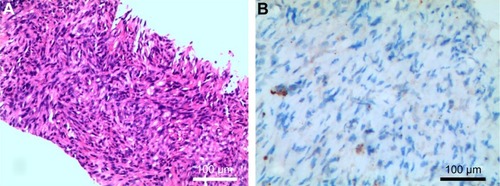
A 58-year-old male presented to the hospital with paroxysmal pain in the chest and discomfort in the head on August 10, 2015. The Karnofsky performance status (KPS) score was 60. PET-CT revealed a right upper chest mass was 51×41 mm, with a maximum standardized uptake value (SUV) of 5.6, accompanied with brain, bone, liver, and right subscapularis muscle metastases with high SUV. We recorded the tumor size of the primary tumor and metastases. The specimens showed malignant spindle cells, which displayed immunohistochemical positive staining for CK, SMA, CD34, CD99, and Bcl-2, and negative for Des, S-100, and B-Catenin. ALK was not supported (Ventana IHC) (). The patient was then diagnosed with malignant myofibroblastoma with brain, bone, and liver metastases.
Figure 1 Histopathological and immunohistological findings of the lymph nodes tissues samples. (A) Hematoxylin and eosin staining showed malignant spindle cells (200×), which displayed immunohistochemical positive staining for CK, SMA, CD34, CD99, and Bcl-2, and negative for Des, S-100, and B-Catenin. (B) IHC of our patient did not reveal supported ALK gene mutation (200×).
Considering the patient’s poor KPS score (60 at diagnosis) and metastases all over the body, chemotherapy, radio-chemotherapy, or surgery was excluded. Methylprednisolone was given at 800 mg/d, day (d)1-5 started from August 20, 2015. The patient then developed an aggravating headache. Fifteen days later, PET-CT imaging showed tumor volume enlargement. The MTV and TLG values are also increased. Subsequently, genetic tests revealed the fifth exon R401 mutation of the ALK gene. Considering the tumor histological and biological features, crizotinib was given at 250 mg daily from September 10, 2015. After 1 month of crizotinib at 250 mg daily, the headache was eased, and PET-CT (October 9, 2015) showed a dramatic reduction in tumor size and metabolism, resulting in stable disease (SD), based on criteria in RECIST 1.1 with KPS score of 70. Conversely, the MTV and TLG values were also significantly reduced.
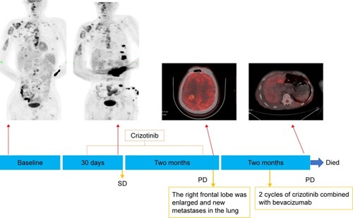
Two months later, the patient had headaches again, so we reviewed his PET-CT on December 7, 2015. The brain imaging showed that the primary tumor was enlarged with edema, and new metastases could be found in the lung (). The tumor size of the right upper chest wall, liver, and right subscapularis muscle metastases were stable, while the MTV and TLG values of them were almost doubled, representing progressive disease. The patient’s family rejected the second-generation ALK inhibitors. Thus, bevacizumab (500 mg/d, d1, every 21 days), combined with crizotinib at 250 mg daily, was administered. Twenty days later, the headache was eased, and the KPS score was 80. Two months after treatment with bevacizumab (500 mg/d, d1, every 21 days) combined with crizotinib 250 mg daily, the headache was aggravated, and the patient developed a poor diet, nausea, and vomiting. The KPS score was 50, PET-CT imaging showed right pleural effusion, all body metastases were enlarged, and the MTV and TLG values were significantly increased, indicating progressive disease (). The patient was administered nutritional supportive treatment. Unfortunately, the patient died 1 month later in March 2016 ().
Figure 2 The progression in the lung. (A1 and A2) were taken on October 9, 2015. (B1 and B2) were taken on December 10, 2015, and the green arrows refer to the new lesions.
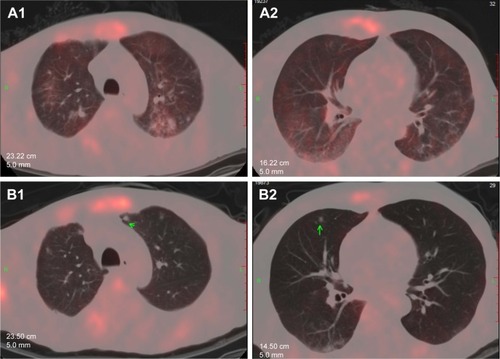
Figure 3 The dynamic evolution of the patient’s primary and metastases tumors during the treatment in the PET-CT images. (A) Images A1–A4 were taken at the first clinic visit on August 10, 2015. The right upper chest mass was 51×41 mm, with a the maximum of standardized uptake value (SUVmax) of 5.6, MTV value of 28.8, and TLG value of 105 (A1). The right subscapularis muscle mass was 27×12 mm, with a SUVmax of 4.9, MTV value of 2.1, and TLG value of 6.4 (A2). The liver mass was 12×11 mm, with a SUVmax of 6.2, MTV value of 2, and TLG value of 8.2 (A3). The larger mass in the occipital lobe of the brain mass was 26×17 mm (A4). (B) Images B1–B4 as baseline were taken after the methylprednisolone on September 10, 2015. The right upper chest mass was 53×52 mm, with a maximum SUV value of 7.2, MTV value of 39.9, and TLG value of 174 (B1). The right subscapularis muscle mass was 27×17 mm, with a SUVmax value of 4.8, MTV value of 5.2, and TLG value of 15.5 (B2). The liver mass was 12×11 mm, with a SUVmax value of 6.7, MTV value of 4, and TLG value of 15.4 (B3). The brain mass was 27×18 (B4). (C) Images C1–C4 were taken after 1 month treatment of oral crizotinib on October 9, 2015. The size of the tumor seemed to be stable. The right upper chest mass was 44×38 mm, with a maximum SUV value of 8, MTV value of 14.2, and TLG value of 24.9 (C1). The right subscapularis muscle mass was 27×17 mm, with a SUVmax value of 2.9, MTV value of 4.8, and TLG value of 11.3 (C2). The liver mass was 18×14 mm, with a SUVmax value of 4, MTV value of 2.2, and TLG value of 6.8 (C3). The brain mass was 27×18 mm (C4). (D) Images D1–D4 were taken on December 10, 2015, after 3 months of oral crizotinib, and there was progressive disease in the brain. The right upper chest mass was 46×43 mm, with a maximum SUV value of 3.6, MTV value of 28.5, and TLG value of 72.5 (D1). The right subscapularis muscle mass was 28×16 mm, with a SUVmax value of 2.9, MTV value of 9, and TLG value of 15.5 (D2). The liver mass was 13×8 mm, with a SUVmax value of 1.7, MTV value of 11, and TLG value of 14.1 (D3). The brain mass was 28×18 mm (D4). (E) Images E1–E4 were taken on February 2, 2016, after two cycles of bevacizumab with oral crizotinib, and all tumors were increased in bulk. The right upper chest mass was 60×56 mm, with a maximum SUV value of 3, MTV value of 88.4, and TLG value of 168.6 (E1). The right subscapularis muscle mass was 42×34 mm, with a SUVmax value of 2.4, MTV value of 49.8, and TLG value of 73.4 (E2). The liver mass was 17×13 mm, with a SUVmax value of 3.5, MTV value of 13.1, and TLG value of 25.2 (E3). The brain mass was 30×19 mm (E4). The change of the tumor of the right chest wall (A1–E1). The change of the right upper arm muscle space (A2–E2). The change of the liver (A3–E3). The change of the brain (A4–E4).
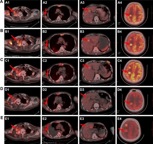
Figure 4 The clinical course of the diagnosis and treatment of the patient. On August 11, 2015, the PET-CT scan of the patient suggested right upper chest, brain, bone, liver, and right subscapularis muscle metastases. Methylprednisolone was given at 800 mg/d, d1-5, started on August 20, 2015. On September 10, 2015, PET-CT imaging showed tumor volume enlargement, and crizotinib was given at 250 mg daily. On October 9, 2015 (after one month of crizotinib treatment), PET-CT imaging showed a dramatic reduction in tumor size and metabolism, resulting in SD. (The concentrations in the left elbow and chest wall were radiocontamination.) On December 7, 2015, the brain imaging PET-CT showed that the primary tumor was enlarged, representing progressive disease in the brain, and bevacizumab (500 mg/d, d1, every 21 days), combined with crizotinib at 250 mg daily, was administered. On February 2, 2016 (after two cycles of bevacizumab with crizotinib), PET-CT imaging showed right pleural effusion, all body metastases were enlarged, and pleural cavity and peritoneal effusions could be found. Then, nutritional supportive treatment was given. The patient died in March 2016.
Written informed consent was obtained from the patient’s wife (the patient’s next of kin) for publication of this case report and accompanying images.
Discussion
Crizotinib is an ATP competitor that inhibits the multitarget protein kinase inhibitor of Met/ALK/ROS. Crizotinib has been used in ALK positive non-small cell lung cancer (NSCLC) to lengthen the period without progression. ALK inhibitors have been reported as effective treatments for ALK-rearranged IMT.Citation19,Citation20 It was reported that a patient who was diagnosed ALK-1-rearranged IMT with brain metastases was initially treated with crizotinib, and the disease progressed after 3 months.Citation21 After that, alectinib, the second-generation ALK inhibitor, was taken as the second-line therapy with 8-month progression-free survival. Our patient had malignant myofibroblastoma with the mutation of ALK-R401. There is no report about the function and treatment effect of the ALK-R401, but it was considered implicated in tumorigenesis. In this case, after the treatment of crizotinib, the progression-free survival was 3 months. It has been known that crizotinib has poor central nervous system (CNS) penetration, as evidenced by low concentrations detected in CNS samples during the treatment course,Citation22 which was likely the cause of treatment failure in our patient. Also, alectinib, which is a selective ALK inhibitor with high CNS penetration, is active against several secondary mutations that confer acquired resistance to crizotinib.Citation23 It may be effective to administer the second generation ALK inhibitor after drug resistance.
Bevacizumab, a recombinant, humanized monoclonal antibody against vascular endothelial growth factor (VEGF), is a vascular-targeted therapy that may inhibit neovascularization,Citation24 such as slower tumor development, reduced metastasis development, and improved drug delivery through vascular normalization.Citation25 Bevacizumab is more effective when combined with other conventional therapies, such as chemotherapy or radiotherapy, and may be added to treatment regimens to increase their efficacy or to reduce developing therapeutic resistance.Citation26,Citation27 Several studies have demonstrated that bevacizumab could reduce brain edema and intracranial pressure, reducing the symptoms of headache or dizziness. Currently, the clinical trials of the EGFR-TKI (Epidermal Growth Factor Receptor-Tyrosine Kinase Inhibitor), combined with bevacizumab, have shown good curative effects in the treatment of lung adenocarcinoma.Citation28,Citation29 So, in our treatment, when the disease progressed after oral crizotinib, bevacizumab combined with crizotinib was given. Then the headache was eased transient, but the treatment effect was unsatisfactory. Whether bevacizumab combined with an ALK inhibitor is efficacious is worth exploring.
PET-CT is not only a reference imaging tool for the diagnosis and staging of tumors, but can also reflect the metabolic status of the tumor, to be used for evaluation of efficacy and prognosis.Citation30 Currently, the prognostic information was deduced from the SUV, MTV, and TLG, which were significantly associated with an early metabolic response.Citation31 Additionally, once PET-CT was used to evaluate the effects of a patient with multiple metastases, clinicians can comprehensively assess patient’s condition changes. In targeted therapy, PET-CT can reflect the metabolic abilities of the cells to predict the curative effect early and adjust the treatment plan in time. At present, MTV is considered a prognostic indicator of tumor survival, being a volumetric and metabolic biomarker of the tumor.Citation32 Thus, unlike SUVmax, MTV can quantify the overall tumor burden. In some studies, higher MTV and TLG were significantly associated with shorter progression-free survival (PFS). In this case, the multiple growth of MTV may be related to the poor prognosis of the tumor.
Conclusion
Targeted therapy can be considered in the treatment of the poor KPS scores patient of advanced tumor with multiple metastases. Patients with mutated ALK-R401 are effective in the treatment of crizotinib. However, crizotinib has low penetration into the central nervous system, patient with head metastases is responded poorly to it. Bevacizumab, combined with crizotinib, had not improved the prognosis. Bevacizumab combined with crizotinib had not achieved excellent outcome in this advanced tumor patient with head metastases. PET-CT can be used to comprehensively assess the condition of patients with advanced tumor. MTV and TLG can reflect the metabolic changes of the tumor, and the sudden increase in its value may be related to the poor prognosis of the patient.
Acknowledgments
This study was supported, in part, by the National Natural Science Foundation of China (No 81272600), a project co-sponsored by the Henan Province and Ministry of Health of Medical Science and Technology Program (No 201601026), “51282 project”, leading talent of Henan Provincial Health Science and Technology Innovation Talents (No [2016]32), Natural Science Foundation of Henan Province (No 162300410300), Wu Jieping Medical Foundation for Clinical Research (No 320.6799.15018), Science and Technology Research Program of Henan Province (No 162102310327), Henan Province medical science and technology research project (No 201702249). The funders had no role in the study design, data collection and analysis, decision to publish, or preparation of the manuscript.
Disclosure
The authors report no conflicts of interest in this work.
References
- HadjigeorgiouGFSamarasVVarsosVLow-grade myofibroblastic sarcoma of the thoracic spine: report of an extreme rare caseBr J Neurosurg201731673173327535494
- SchurchWSeemayerTAGabbianiGThe myofibroblast: a quarter century after its discoveryAm J Surg Pathol19982221411479500214
- JoVYFletcherCDWHO classification of soft tissue tumours: an update based on the 2013 (4th) editionPathology20144629510424378391
- SinghSKRahejaAAJagdevanAKSinghPKPurkaitSSuriVAMalignant temporal muscle myofibroblastoma with an early recurrence in a childNeurol India201765240841028290416
- HumphriesWE3rdSatyanKBRelyeaKLow-grade myofibroblastic sarcoma of the sacrumJ Neurosurg Pediatr20106328629020809714
- MajnoGThe story of the myofibroblastsAm J Surg Pathol197936535542534390
- StarkMHoffmannAXiongZMammary myofibrosarcoma: case report and literature reviewBreast J201117330030421545434
- MontgomeryEAShusterDDBurkartALInflammatory myofibroblastic tumors of the urinary tract: a clinicopathologic study of 46 cases, including a malignant example inflammatory fibrosarcoma and a subset associated with high-grade urothelial carcinomaAm J Surg Pathol200630121502151217122505
- SukovWRChevilleJCCarlsonAWUtility of ALK-1 protein expression and ALK rearrangements in distinguishing inflammatory myofibroblastic tumor from malignant spindle cell lesions of the urinary bladderMod Pathol200720559260317396140
- LuZJZhouSHYanSXYaoHTAnaplastic lymphoma kinase expression and prognosis in inflammatory myofibroblastic tumours of the maxillary sinusJ Int Med Res20093762000200820146901
- QiuXMontgomeryESunBInflammatory myofibroblastic tumor and low-grade myofibroblastic sarcoma: a comparative study of clinicopathologic features and further observations on the immunohistochemical profile of myofibroblastsHum Pathol200839684685618400254
- YauNKFongAYLeungHFA pan-cancer review of ALK mutations: implications for carcinogenesis and therapyCurr Cancer Drug Targets201515432733625714698
- ChoiHRole of imaging in response assessment and individualised treatment for sarcomasClin Oncol (R Coll Radiol)201729848148828506521
- RiedlCCPinkerKUlanerGAComparison of FDG-PET/CT and contrast-enhanced CT for monitoring therapy response in patients with metastatic breast cancerEur J Nucl Med Mol Imaging20174491428143728462446
- TherassePArbuckSGEisenhauerEANew guidelines to evaluate the response to treatment in solid tumors. European Organization for Research and Treatment of Cancer, National Cancer Institute of the United States, National Cancer Institute of CanadaJ Natl Cancer Inst200092320521610655437
- BangJILimYPaengJCComparison of quantitative methods on FDG PET/CT for treatment response evaluation of metastatic colorectal cancerNucl Med Mol Imaging201751214715328559939
- SchaferJFGatidisSSchmidtHSimultaneous whole-body PET/MR imaging in comparison to PET/CT in pediatric oncology: initial resultsRadiology2014273122023124877983
- Ben BouallegueFTabaaYAKafrouniMCartronGVauchotFMariano-GoulartDAssociation between textural and morphological tumor indices on baseline PET-CT and early metabolic response on interim PET-CT in bulky malignant lymphomasMed Phys20174494608461928513853
- ButrynskiJED’AdamoDRHornickJLCrizotinib in ALK-rearranged inflammatory myofibroblastic tumorN Engl J Med2010363181727173320979472
- LiuQKanYZhaoYHeHKongLEpithelioid inflammatory myofibroblastic sarcoma treated with ALK inhibitor: a case report and review of literatureInt J Clin Exp Pathol2015811153281533226823889
- YuanCMaMJParkerJVMekhailTMMetastatic anaplastic lymphoma kinase-1 (ALK-1)-rearranged inflammatory myofibroblastic sarcoma to the brain with leptomeningeal involvement: favorable response to serial ALK inhibitors: a case reportAm J Case Rep20171879980428713152
- CostaDBKobayashiSPandyaSSCSF concentration of the anaplastic lymphoma kinase inhibitor crizotinibJ Clin Oncol20112915e443e44521422405
- HidaTNokiharaHKondoMAlectinib versus crizotinib in patients with ALK-positive non-small-cell lung cancer (J-ALEX): an open-label, randomised phase 3 trialLancet201739010089293928501140
- StefanouDStamatopoulouSSakellaropoulouABevacizumab, pemetrexed and carboplatin in first-line treatment of non-small cell lung cancer patients: focus on patients with brain metastasesOncol Lett20161264635464228101218
- AravantinosGPectasidesDBevacizumab in combination with chemotherapy for the treatment of advanced ovarian cancer: a systematic reviewJ Ovarian Res201475724864163
- HornLSandlerAChemotherapy and antiangiogenic agents in non-small-cell lung cancerClin Lung Cancer20078Suppl 2S68S7317382027
- TengLSJinKTHeKFWangHHCaoJYuDCAdvances in combination of antiangiogenic agents targeting VEGF-binding and conventional chemotherapy and radiation for cancer treatmentJ Chin Med Assoc201073628128820603084
- JiangTLiASuCAddition of bevacizumab for malignant pleural effusion as the manifestation of acquired EGFR-TKI resistance in NSCLC patientsOncotarget2017837626486265728977977
- OtsukaKHataATakeshitaJEGFR-TKI rechallenge with bevacizumab in EGFR-mutant non-small cell lung cancerCancer Chemother Pharmacol201576483584126349474
- KimKRShimHJHwangJEThe role of interim FDG PET-CT after induction chemotherapy as a predictor of concurrent chemoradiotherapy efficacy and prognosis for head and neck cancerEur J Nucl Med Mol Imaging201845217017828940101
- MatsumotoYBabaSEndoMMetabolic tumor volume by 18F-FDG PET/CT can predict the clinical outcome of primary malignant spine/spinal tumorsBiomed Res Int20172017813267628852650
- ChoiESHaSGKimHSHaJHPaengJCHanITotal lesion glycolysis by 18F-FDG PET/CT is a reliable predictor of prognosis in soft-tissue sarcomaEur J Nucl Med Mol Imaging201340121836184223880967