
Abstract
Background
Breast cancer is still one of the major public health burdens worldwide, although there is tremendous progress in early diagnosis and treatment of breast cancer. Tamoxifen was one of the most popular endocrine therapies for early-stage estrogen receptor (ER) + breast cancer patients. However, a high incidence of drug resistance develops along with poor prognosis in breast cancer. Currently, long noncoding RNAs (lncRNAs) are emerging and are well suited to play a role in the development of cancer and tamoxifen resistance. However, there is little reported about the relationship of breast cancer resistance to tamoxifen and lncRNA H19. Here, we validated that lncRNA H19 was highly expressed in breast cancer especially in patients with late stage (III and IV), compared to normal tissues and early stage cancers (I and II).
Methods
Quantitative polymerase chain reaction (qPCR) was utilized for comparison of lncRNA H19 expression level in breast cancers with different stages. qPCR and Western blotting were used to detect gene and protein, respectively.
Results
We found that lncRNA H19 expression level manipulated breast cancer cell proliferation both in parental breast cancer cell lines and tamoxifen-resistant cell lines. Knockdown of lncRNA H19 elevated tamoxifen sensitivity for promoting cell growth and inhibiting apoptosis in tamoxifen-resistant breast cancer cells. Moreover, knockdown of H19 inhibited Wnt pathway and epithelia–mesenchymal transition in tamoxifen-resistance breast cancer cells.
Conclusion
Taken together, the results of this study provided the evidence for H19 in regulating tamoxifen-resistant breast cancer and might provide novel options in the future treatment of tamoxifen-resistance breast cancer patients.
Background
Breast cancer is the most common invasive cancer in women which causes approximately 4 million deaths in worldwide each year.Citation1 For decades, classification of breast cancer stages was based on the tumor size and the metastasis of cancer to lymph nodes or other parts.Citation2 Recently, molecular classification, such as Her-2, estrogen receptor (ER), and progesterone receptor, was developed. It has great advances for personalized treatment of breast cancer with better outcomes than conventional classification.Citation3–Citation5 Tamoxifen is a nonsteroidal antiestrogen and as an adjuvant endocrine therapy has been extensively used for early ER positive and node negative breast cancer treatment. However, tamoxifen resistance can result in breast cancer recurrence and mortality.Citation6 Therefore, it is essential to explore molecular targets involved in tamoxifen resistance of breast cancer.
It was well known that long noncoding RNA (lncRNA) involved in diabetes, neurological diseases, and cancers.Citation7–Citation9 H19 was the first identified lncRNA in human breast cancer, and the expression of H19 is higher in ER positive cells.Citation10 Additionally, in ER-negative MDA-MB-231 cell line, highly proliferation ability of these cells was notably with ectopic overexpression of H19. The function of lncRNA in proliferation, invasion, and metastasis have become a hot spot of research.
Several studies have preliminarily revealed that lncRNA mediates the metastasis of tumor by regulating the epithelia– mesenchymal transition (EMT) process of tumor cells. Hu et al screened a series of high levels of lncRNAs in MCF10A (MCF10A/Twist) with high expression of twisted protein (TWIST) and proved that dysregulation lncRNAs was involved in the EMT process by regulating the activation of the target gene and the corresponding pathways of WNT and MAPK, inducing cell invasion and metastasis.Citation11 It was reported that that H19 can lead to de-repression of β-catenin, which eventually activated Wnt/β-catenin pathway and hence potentiated osteogenesis.Citation12 Moreover, H19 mediates methotrexate resistance in colorectal cancer through activating Wnt/β-catenin pathway.Citation13 lncRNA aquaporin can promote the migration and metastasis of EMT in intestinal cancer cells.Citation14 Zhou et al demonstrated H19 regulated EMT and mesenchymal–epithelial transition on breast cancer.Citation15 Therefore, expression level of lncRNAs plays an important role in Wnt pathway and EMT in tumor cells.
The aim of the present study was to identify new mechanisms underlying tamoxifen resistance in order to lay the foundation for therapies that could have greater efficacy. We focused on the mechanism of lncRNA H19 for drug sensitivity regulation in tamoxifen-resistance breast cancer. Taken together, this article might give a new hint for further clinical treatment of tamoxifen-resistant breast cancer patients.
Patients and methods
Patients’ recruitment and tissue sample collection
A total of 30 pairs of fresh breast cancer tissues and adjacent normal tissues from 30 treatment-naïve breast cancer patients were collected in two Chinese hospitals (The Fourth People’s Hospital of Jinan and Affiliated Hospital of Shandong Academy of Medical Sciences). The clinical information including tumor size, pathological stage, and lymphatic metastasis was collected and reviewed. For all the patients, sample were sampling intraoperatively and immediately frozen in liquid nitrogen with RNALater (Qiagen NV, Venlo, the Netherlands) and then stored at −80°C for further experimental using. This study was approved by the institutional Review Board for Human Research of The Fourth People’s Hospital of Jinan and the Affiliated Hospital of Shandong Academy of Medical Sciences. All recruited patients provided written informed consents.
Cell culture
Human breast cancer cell lines (MCF-7 and SK-BR-3) were obtained from Stem Cell Bank, Chinese Academy of Sciences (Shanghai, China). The tamoxifen-resistant MCF-7-R and SK-BR-3-R cell lines were developed by 70 days selection of MCF-7 and SK-BR-3 cells co-cultured with 800 nM 4-hydroxytamoxifen (4OH-TAM, Sigma-Aldrich Co., St Louis, MO, USA). The cells were cultured in RPMI-1640 (Sigma-Aldrich Co.) supplemented with 10% charcoal–dextran-stripped fetal bovine serum (Thermo Fisher Scientific, Waltham, MA, USA) and penicillin (100 U/mL, Sigma-Aldrich Co.) and streptomycin (100 U/mL, Sigma-Aldrich Co.) at 37°C with 5% CO2 in a humidified cell culture incubator (Sanyo Electric Co., Ltd., Osaka, Japan).
Cell transfection
siRNA specific for H19 was purchased from Invitrogen (Thermo Fisher Scientific). To minimize nonspecific effects of interfering RNAs, non-targeting control siRNA, also obtained from Invitrogen, was used as negative control (NC). The siRNA transfection reagent (Invitrogen) was used according to the manufacturer’s protocol. Briefly, 3 × 10⁴ cells in media without antibiotic were plated in 6-well plates and then transfected with 100 nM siRNA for H19 or NC when cells were 70% confluence the plate well at 24 hours using lipofectamine 2000 regent (Thermo Fisher Scientific). The cells were sampled at indicated time point for the different experiments with indicated purposes such as cell proliferation, invasion, and protein expression. For the validation of chemosensitivity experiment, cells with 24 hours siRNA transfection and then treated with tamoxifen in multiple concentrations and analyzed the cell proliferation and apoptosis rate.
Cell counting kit 8 assay
According to the manufacture instruction (Beyotime Institute of Biotechnology, Haimen, People’s Republic of China), the procedure briefly descripted as following, MCF-7, MCF-7-R, SK-BR-3, or SK-BR-3R cells at the logarithmic growth phase were plated in 96-well at the density of 5,000 cells/well and cultured for 24 hours. And then, siRNA or NC were added with transfection regents and cultured for another 72 hours. Cell counting kit solution (20 µL/well) was added to each well, and the cells were further incubated for another half hours. In the validation of chemosensitivity, following the transfection of siRNA or NC, cells were cultured with tamoxifen with dose escalation from 0 to 100 µM (0, 0.01, 0.1, 1, 10, and 100 µM). The living cell number was measured by the OD value at 450 nm wavelength using microplate reader.
Transwell cell migration and invasion assay
For in vitro cell migration and invasion assay, the published on-line available protocol was used. Briefly as following: 100 µL cells (3 × 10⁶ cells/well) transfected with H19 siRNA or NC were plated on the top of the membrane in a transwell inserted into the 24-well plate. When the cells settled down, 600 µL RPMI-1640 media with 20% FBS was added into the bottom of the lower chamber in a 24-well plate. Then, 48 hours later, cells that did not migrate from the top of the membrane were removed carefully and the migrated cells were fixed and stained with crystal violet. The cells in different fields of view were counted under microscope and using the average sum of cells to analyze the migration and invasion ability.
Flow cytometry analysis of apoptotic cells
Apoptosis was demonstrated by Annexin V-FITC and pro-pidium iodide (PI) (Thermo Fisher Scientific) according to the manufacturer’s protocol. Briefly, 1 × 10⁶ cells were plated in 100 mm dishes (Corning Incorporated, Corning, NY, USA) and then transfected with H19 or mock vector. Then, 72 hours later, the cells were lysed and collected following twice washing with cold PBS and re-suspended with 500 µL binding buffer. And then cells were stained with Annexin-FITC and PI with another further incubated for 5 minutes at room temperature before running the flow cytometry. For each group, the samples were triplicate.
Real-time PCR analysis
For indicated treatments, cells were collected, and total RNA was isolated from these cells or tumor tissues from patients using PicoPure™ RNA Isolation Kit (Arcturus, Sunnyvale, CA, USA) according to the manufacturer’s instructions. First, 2 µg of mRNA was transcribed into cDNA using SuperScript III RNase H Reverse Transcriptase (Thermo Fisher Scientific). Following the reverse transcription reactions, real-time PCR was performed with Taqman (Thermo Fisher Scientific) primers for lncRNA H19 and GAPDH and Vii™ 7 system (Bio-Rad Laboratories, Hercules, CA, USA) was used to run the qPCR procedure according the manufacturers instruments. Based on the real-time PCR results of Ct number, the expression of mRNA levels was calculated using the 2−ΔΔCt method and normalized to an internal reference gene GAPDH.
Western blot
Cells were collected at 72 hours with the treatment of H19 siRNA or NC and lysed in RIPA buffer (Sigma-Aldrich Co.). Protein concentration was quantified using BCA Protein Assay Kit (Beyotime). Then, 30 µg of total protein from each group was loaded and resolved with 10% Tris-SDS gel. After the electrophoresis, the gel was electro-transferred to polyvinylidene fluoride membranes (Thermo Fisher Scientific). For the following blocking and antibody incubation, iBind kit (Thermo Fisher Scientific) was used according the manufactures’ instrument. The primary antibodies: anti-B-catenin (Cell Signaling Technology, Inc., Danvers, MA, USA) (1:300), anti-E-Cadherin (Cell Signaling) (1:300) anti-vimentin (Santa Cruz Biotechnology Inc., Dallas, TX, USA) (1:1,000), anti-GAPDH (Santa Cruz Biotechnology Inc.) (1:1,000), and anti-Histone (Santa Cruz Biotechnology Inc.) (1:1,000). Horseradish peroxidase-conjugated anti-rabbit and anti-mouse, and anti-goat IgG was used as the secondary antibody (Santa Cruz Biotechnology Inc.) (1:3,000). The signals were detected with super sensitive regent (Thermo Fisher Scientific).
Statistical analysis
All statistical analysis was done with two-tailed Student’s t-test available in GraphPad Prism7 (GraphPad Software, Inc., La Jolla, CA, USA). In all assays, one-way ANOVA was used to determine the difference between groups and the P-value v0.01 was considered statistically significant difference. All experiments were performed in triplicate and all value was presented as the mean ± SD.
Results
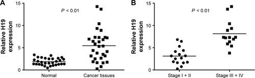
lncRNA H19 has been reported to express in multiple cancers and takes a key role in tumorigenesis and metastasis.Citation16 We first analyzed H19 mRNA level in breast cancer tissues and normal adjacent tissues from the 30 patients. RT-qPCR demonstrated that H19 mRNA was much higher in breast cancer tissues compared to normal tissues (, P < 0.01). Due to the fact that lncRNA H19 was associated with tumor metastasis, we investigated the correlation of lncRNA H19 level and tumor stage. As shown in , patients at late stage (stage III and IV) expressed relatively higher level of H19 compared to patients at early stage cancers (stage I and II) (P < 0.01). Taken together, these results demonstrated that highly expressed lncRNA H19 might be correlated with metastatic breast cancer.
Figure 1 Comparison of lncRNA H19 expression level in breast cancers with different TNM stage.
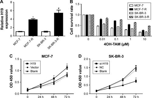
LncRNA H19 expression level manipulated breast cancer cell proliferation
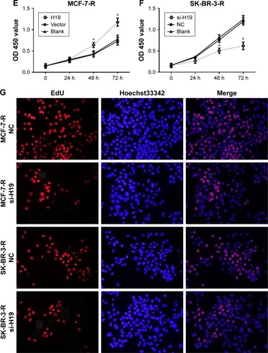
In order to investigate the relationship of lncRNA H19 and tamoxifen-resistance breast cancer cells, we detected the level of H19 in tamoxifen-resistant breast cancer cell lines. Results revealed that the level of lncRNA H19 in tamoxifen-resistance cells (MCF-7-R and SK-BR-3-R) was much higher than normal breast cancer cells (MCF-7 and SK-BR-3) (P < 0.01, ). Next, to focus on the association of H19 and breast cancer cell proliferation, we assessed the impact of H19 on cell proliferation using cell counting kit-8 (CCK8). As shown in , both MCF-7-R and SK-BR-3-R acquired tamoxifen resistance. In addition, compared with control group, MCF-7 and SK-BR-3 cells transfected with H19 displayed faster growth, especially 48 hours and 72 hours after transfection (, P < 0.01). Additionally, when we use siRNA of H19 to suppress H19 level of MCF-7-R and SK-BR-3-R cells, the cell proliferation was inhibited (). This observation was further confirmed by EdU staining, since siRNA H19-treated group contained less EdU-positive cells compared with NC group (), suggesting the strong correlation of lncRNA H19 level and cell proliferation.
Figure 2 lncRNA H19 affected breast cancer cell proliferation.
Abbreviation: NC, negative control.
Knockdown of lncRNA H19 improved tamoxifen sensitivity in terms of cell apoptosis in tamoxifen-resistant breast cancer cells
To investigate the contribution of lncRNA H19 to tamoxifen-resistance in breast cancer cells, we transfected H19 into tamoxifen-sensitive parental control cells (MCF-7 or SK-BR-3), as well as H19 siRNA into tamoxifen-resistant cells (MCF-7-R or SK-BR-3-R). All these cells were exposed to tamoxifen treatment. Under the same concentration of tamoxifen, sensitive cell lines displayed higher cell survival than resistant cell line (). We also observed that elevated level of H19 increased cell survival rate in sensitive cells, while knocking down of H19 decreased cell survival rate in resistant cells (P < 0.01). This indicated that in the presence of tamoxifen, suppression of lncRNA H19 could ameliorate the resistance to tamoxifen in tamoxifen-resistant cell lines.
Figure 3 lncRNA H19 expression manipulates the tamoxifen sensitivity of breast cancer cells.
Abbreviation: NC, negative control.
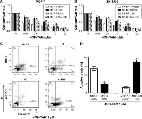
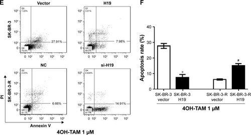
Next, we performed FACS assay to investigate the effect of H19 on cell apoptosis. Results showed that transfection of H19 can inhibit apoptosis in MCF-7 parental control cells, and knockdown of H19 by siRNA can ameliorate the low level of apoptosis status in tamoxifen-resistant cells (MCF-7-R) (). This observation was further confirmed in SK-BR-3 and SK-BR-3-R cells (). These findings indicated that knockdown of lncRNA H19 could improve tamoxifen sensitivity in tamoxifen-resistant breast cancer cells.
Knockdown of lncRNA H19 improved tamoxifen sensitivity in terms of cell invasion in tamoxifen-resistant breast cancer cells
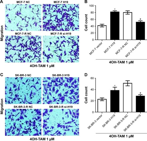
We performed Transwell experiments to interrogate the effect of lncRNA H19 on breast cancer cell invasion in the presence of tamoxifen (1 µM). Results showed that transfection of lncRNA H19 enhanced MCF-7 invasion to two folds compared to vector control. Tamoxifen-resistant MCF-7 cells with H19 knockdown displayed less invasion capability compared to control group under tamoxifen treatment (P < 0.01, ). This finding was further confirmed in SK-BR-3 and SK-BR-3-R cells (P < 0.01, ). This indicated that knockdown of lncRNA H19 improved tamoxifen sensitivity in terms of cell invasion in tamoxifen-resistant breast cancer cells.
Figure 4 Effect of lncRNA H19 expression manipulation on breast cancer cell migration.
Abbreviation: NC, negative control.
Knockdown of H19 inhibited Wnt pathway in tamoxifen-resistance breast cancer cells
As lncRNA H19 is thought to participate in the breast cancer cell metastasis and Wnt pathway has been shown to play a critical role in promoting metastasis.Citation15 Thus, we investigated the correlation of H19 and Wnt pathway. β-Catenin plays a central role mediating the canonical Wnt/β-catenin signaling pathway. We found that knockdown of H19 by siRNA transfection can promote the translocation of β-catenin from nuclear to cytoplasm in tamoxifen-resistant cells (). The re-localization of β-catenin prompted us to investigate Wnt-dependent transcription by using the Dual-luciferase reporter system and TOP/FOP reporter assays system. Upon knock-down of H19, the TOP/FOP ratio decreased to about 50% when compared to the control in both MCF-7-R and SK-BR-3-R cells (), indicating that loss of H19 inhibited Wnt-dependent transcription, in line with the loss of nuclear staining of beta-catenin observed in . We also measured the protein level of β-catenin, and results showed that knockdown of H19 reduced the expression of β-catenin in both MCF-7-R and SK-BR-3-R cells ().
Figure 5 The H19 knockdown deregulated Wnt pathway in tamoxifen-resistant breast cancer cells.
Abbreviation: NC, negative control.
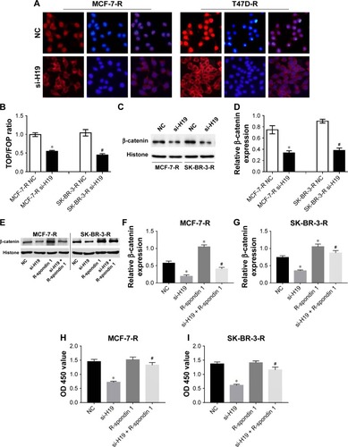
R-Spondin (RSPO) proteins are a family of secreted molecules that strongly potentiate Wnt/β-catenin signaling. We found that RSPO1 can reverse the β-catenin decrease induced by H19 knockdown in both two tamoxifen-resistant cell lines. (). In terms of cell proliferation, results showed that RSPO1 can block the inhibition of cell proliferation induced by H19 knockdown, both in MCF-7-R and SK-BR-3-R cells (). Taken together, these findings suggested that in tamoxifen-resistant cells, Wnt/beta-catenin signal pathway acted as a downstream of H19, and inhibition of H19 level can reduce the activity of Wnt/beta-catenin signal pathway and cell proliferation.
Knockdown of H19 inhibited EMT in tamoxifen-resistance breast cancer cells
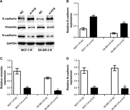
EMT is an important process in tumor development. EMT is essential for developmental processes and has also been shown to occur in wound healing, in organ fibrosis, and in the initiation of metastasis in cancer progression. Wnt/beta-catenin signal pathway is an inducer of EMT and plays an important role in EMT, which displayed an upregulation of N-cadherin and downregulation of E-cadherin and vimentin. In this study, we found that knockdown of H19 by siRNA transfection can significantly reduce the expression of N-cadherin, as well as increase E-cadherin and vimentin level, both in MCF-7-R and SK-BR-3-R cells (). This observation indicated that knockdown of H19 can inhibit EMT in tamoxifen-resistance breast cancer cells.
Figure 6 The H19 knockdown de-regulated EMT in tamoxifen-resistant breast cancer cells.
Abbreviation: NC, negative control.
Discussion
Breast cancer is the most common malignancy in women in worldwide and multiple advanced therapies have been developed. Tamoxifen is the widespread treatment for patients with ER positive breast cancer. Drug resistance is still a main obstacle preventing successful breast cancer treatment.Citation17,Citation18 Therefore, better understanding of the biological mechanisms for the acquired resistance to tamoxifen might suggest novel direction to overcome tamoxifen resistance.
lncRNA H19 is highly expressed in majority of human cancers and its overexpression is often correlated with tumor metastasis.Citation19,Citation20 Several studies about lncRNA H19 on drug resistance reported that H19 can reduce the sensitivity of breast cancer to paclitaxel via inactivation of BIK/NOXA, which were two important pro-apoptotic genes.Citation21 Moreover, H19 inactivated p53 via physically interacts with p53 in gastric cancer cells and finally regulated the cell cycle.Citation16 Stable expression of H19 significantly promotes EMT progression and accelerates in vivo and in vitro tumor growth in gastric cancer. H19 was an important factor contributing to drug resistance in breast cancer. lncRNA H19 overexpressed in various types of tumor cells such as bladder cancer, liver cancer, colon cancer, and breast cancer.Citation11,Citation22,Citation23 Adriaenssens found that the expression of H19 increased in 72.5% breast cancer tissues and overexpression of an ectopic H19 gene enhances the tumorigenic properties of breast cancer cells.Citation24 Berteaux et al also found that over expression of H19 in breast cancer is the reason for promoting cell proliferation.Citation25 Similar to these studies, the expression of lncRNA H19 in breast cancer cells was investigated, and the underlying possible molecular mechanism of H19 overexpression regulated tamoxifen resistant was interrogated. It was found that the expression level of H19 in tamoxifen-resistant breast cancer cells was significantly higher than that of tamoxifen-sensitive breast cancer cells.
More and more studies have shown that lncRNA H19 is involved in the development of tumor invasion and metastasis. In bladder cancer cells, the high expression of lncRNA H19 promotes the expression of the transcription factor EZH2, down the expression of E-cadherin, and then enhances the invasion ability of the cells in the hepatoma cells.Citation26 lncRNA H19 is expressed by upregulation of Slug to induce the tumor metastasis.Citation27 The above research results have certain reference significance for this study. The migration and invasion ability of tumor cells is a manifestation of their malignant biological behavior and is also one of the main factors that affect tumor progression and prognosis. The metastatic risk of breast cancer is high because of the failure of endocrine therapy, and the expression of H19 in tamoxifen-resistant breast cancer is significantly improved, and our results also suggests that H19 is associated with TAM resistance in breast cancer. Therefore, we further study the effect of H19 on the metastasis of TAM breast cancer and the underlying involved pathways. We found that knockdown of lncRNA H19 can ameliorate tamoxifen resistance and promote cell apoptosis and inhibit cell proliferation in tamoxifen-resistant breast cancer cells. Moreover, knockdown of H19 can inhibited Wnt pathway and EMT in tamoxifen-resistance breast cancer cells. Therefore, in this study, we provide evidence for a novel mechanism that is employed by the lncRNA H19 to ameliorate tamoxifen resistance in breast cancer cells. Due to the crucial role of H19 in the amelioration of drug resistance, it may be used as a useful biomarkers and potential therapeutic target for breast cancer patients.
Over the last decade, researchers put efforts to illuminate the complex network of ER transcriptional effects such as ER/ERK/MAPK pathway and stress-activated protein kinase/c-junNH2 terminal kinase pathway and suggested that alterations of the elements in these signaling pathway play an important role in resistance to tamoxifen in breast cancer treatment.Citation28–Citation30 Moreover, with the development of molecular research, several critical importance targets of breast cancer cells have been identified, such as HER2, other receptor tyrosine kinases, and components of the PI3K/AKT/mTOR and Raf/MEK/ERK pathways.Citation31,Citation32 In the current study, H19 overexpression may be related to the poor prognosis of tamoxifen-resistance breast cancer. Therefore, we present a rationale for dual inhibition of these pathways in the treatment of breast cancer patients with acquired resistance.
Taken together, the reduction of H19 expression level inhibits the expression of transcription factors related to the Wnt pathway and EMT, thus inhibiting the invasion ability of tamoxifen-resistance breast cancer, suggesting that H19 may be related to the poor prognosis of tamoxifen-resistance breast cancer, and is expected to be a drug target for the treatment of metastatic breast cancer after tamoxifen-resistance. This provided a theoretical basis for further studies about targeted drugs investigation.
Disclosure
The authors report no conflicts of interest in this work.
References
- BartschRBergenEASCO 2017: highlights in breast cancerMemo201710422823229250202
- AgarwalaVChoudharyNGuptaSA risk-benefit assessment approach to selection of adjuvant chemotherapy in elderly patients with early breast cancer: a mini reviewIndian J Med Paediatr Oncol201738452653429333024
- BaşaranGATwelvesCDiérasVCortésJAwadaAOngoing unmet needs in treating estrogen receptor-positive/HER2-negative metastatic breast cancerCancer Treat Rev20186314415529329006
- CalhounBCCollinsLCPredictive markers in breast cancer: an update on ER and HER2 testing and reportingSemin Diagn Pathol201532536236925770732
- LeeADjamgozMBATriple negative breast cancer: emerging therapeutic modalities and novel combination therapiesCancer Treat Rev20186211012229202431
- WuYZhangZCenciariniMETamoxifen resistance in breast cancer is regulated by the EZH2-ERα-GREB1 transcriptional axisCancer Res201878367168429212856
- HuangXLuoYLMaoYSJiJLJlJThe link between long noncoding RNAs and depressionProg Neuropsychopharmacol Biol Psychiatry201773737827318257
- MirzaAHKaurSPociotFLong non-coding RNAs as novel players in β cell function and type 1 diabetesHum Genomics20171111728738846
- LuoQChenYLong noncoding RNAs and Alzheimer’s diseaseClin Interv Aging20161186787227418812
- ColletteJLe BourhisXAdriaenssensERegulation of human breast cancer by the long non-coding RNA H19Int J Mol Sci201718112319
- HuPYangJHouYLncRNA expression signatures of twist-induced epithelial-to-mesenchymal transition in MCF10A cellsCell Signal2014261839324113349
- LiangWCFuWMWangYBH19 activates Wnt signaling and promotes osteoblast differentiation by functioning as a competing endogenous RNASci Rep201662012126853553
- WuKFLiangWCFengLH19 mediates methotrexate resistance in colorectal cancer through activating Wnt/β-catenin pathwayExp Cell Res2017350231231727919747
- ChenJWangTZhouYCAquaporin 3 promotes epithelial-mesenchymal transition in gastric cancerJ Exp Clin Cancer Res2014333824887009
- ZhouWYeXLXuJThe lncRNA H19 mediates breast cancer cell plasticity during EMT and MET plasticity by differentially sponging miR-200b/c and let-7bSci Signal201710483eaak955728611183
- LiTMoXFuLXiaoBGuoJMolecular mechanisms of long non-coding RNAs on gastric cancerOncotarget2016788601861226788991
- FedelePCiccareseMSuricoGCinieriSAn update on first line therapies for metastatic breast cancerExpert Opin Pharmacother201819324325229336185
- Gonzalez-AnguloAMMorales-VasquezFHortobagyiGNOverview of resistance to systemic therapy in patients with breast cancerAdv Exp Med Biol200760812217993229
- LiCXLiHGHuangLTH19 lncRNA regulates keratinocyte differentiation by targeting miR-130b-3pCell Death Dis2017811e317429192645
- WeiYLiuZFangJH19 functions as a competing endogenous RNA to regulate human epidermal growth factor receptor expression by sequestering let-7c in gastric cancerMol Med Rep20181722600260629207111
- SiXZangRZhangELncRNA H19 confers chemoresistance in ERα-positive breast cancer through epigenetic silencing of the pro-apoptotic gene BIKOncotarget2016749814528146227845892
- YuMBardiaAWittnerBSCirculating breast tumor cells exhibit dynamic changes in epithelial and mesenchymal compositionScience2013339611958058423372014
- GunasingheNPWellsAThompsonEWHugoHJMesenchymal-epithelial transition (MET) as a mechanism for metastatic colonisation in breast cancerCancer Metastasis Rev2012313–446947822729277
- LottinSAdriaenssensEDupressoirTOverexpression of an ectopic H19 gene enhances the tumorigenic properties of breast cancer cellsCarcinogenesis200223111885189512419837
- OhanaPKopfEBibiOThe expression of the H19 gene and its function in human bladder carcinoma cell linesFEBS Lett19994541–2818410413100
- LuoMLiZWangWZengYLiuZQiuJLong non-coding RNA H19 increases bladder cancer metastasis by associating with EZH2 and inhibiting E-cadherin expressionCancer Lett2013333221322123354591
- MatoukIJRavehEAbu-LailROncofetal H19 RNA promotes tumor metastasisBiochim Biophys Acta2014184371414142624703882
- RingADowsettMMechanisms of tamoxifen resistanceEndocr Relat Cancer200411464365815613444
- YamaguchiNNakayamaYYamaguchiNDown-regulation of Forkhead box protein A1 (FOXA1) leads to cancer stem cell-like properties in tamoxifen-resistant breast cancer cells through induction of interleukin-6J Biol Chem2017292208136814828270510
- ChenSYaoFXiaoQEZH2 inhibition sensitizes tamoxifen- resistant breast cancer cells through cell cycle regulationMol Med Rep20181722642265029207119
- SainiKSLoiSde AzambujaETargeting the PI3K/AKT/mTOR and Raf/MEK/ERK pathways in the treatment of breast cancerCancer Treat Rev201339893594623643661
- Herter-SprieGSGreulichHWongKKActivating mutations in ERBB2 and their impact on diagnostics and treatmentFront Oncol201338623630663