
Abstract
Background
CK1 is involved in regulating Wnt/β-catenin signaling and represents a promising target for the treatment of breast cancer. A purine derivative longdaysin has recently been identified as a novel modulator of cellular circadian rhythms through targeting the protein kinases CK1δ, CK1α, and ERK2. However, the antitumor activity of longdaysin and its underlying mechanisms remain unclear.
Methods
The inhibitory effect of longdaysin on Wnt/β-catenin signaling was investigated using the SuperTOPFlash reporter system. The levels of phosphorylated LRP6, total LRP6, DVL2, active β-catenin, and total β-catenin were examined by Western blot. The expression of Wnt target genes was determined using real-time PCR. The ability of colony formation of breast cancer cells was measured by colony formation assay. The effects of longdaysin on cancer cell migration and invasion were assessed using transwell assays. The effect of longdaysin on cancer stem cells was tested by sphere formation assay. The in vivo antitumor effect of longdaysin was evaluated using MDA-MB-231 breast cancer xenografts.
Results
Longdaysin suppressed Wnt/β-catenin signaling through inhibition of CK1δ and CK1ε in HEK293T cells. In breast cancer Hs578T and MDA-MB-231 cells, micromolar concentrations of longdaysin attenuated the phosphorylation of LRP6 and DVL2 and reduced the expression of active β-catenin and total β-catenin, leading to the downregulation of Wnt target genes Axin2, DKK1, LEF1, and Survivin. Furthermore, longdaysin inhibited the colony formation, migration, invasion, and sphere formation of breast cancer cells. In MDA-MB-231 breast cancer xenografts, treatment with longdaysin suppressed tumor growth in association with inhibition of Wnt/β-catenin signaling.
Conclusion
Longdaysin is a novel inhibitor of the Wnt/β-catenin signaling pathway. It exerts antitumor effect through blocking CK1δ/ε-dependent Wnt signaling.
Background
CK1 is a family of serine/threonine-specific protein kinases that is conserved in eukaryotes from yeast to humans.Citation1,Citation2 It regulates diverse cellular processes including Wnt signaling, circadian rhythms, and DNA repair.Citation3,Citation4 The CK1 family consists of six human isoforms (α, δ, ε, γ1, γ2, γ3). The amino-terminal kinase domain is highly conserved in all CK1 family members.Citation5–Citation7
The Wnt/β-catenin signaling pathway plays a pivotal role in embryonic development, tumorigenesis, and stem cell self-renewal.Citation8,Citation9 This pathway is activated by the binding of secreted Wnt proteins to a receptor complex containing a member of the FZD family and LRP5/6.Citation8,Citation10 CK1 family members phosphorylate several important components in the Wnt/β-catenin signaling pathway and act as either negative or positive regulators of the pathway.Citation11,Citation12 In the destruction complex, CK1α phosphorylates β-catenin and subsequently leads to degradation of β-catenin.Citation13 Moreover, CK1δ or CK1ε can phosphorylate the APC protein to enhance the binding of APC to β-catenin for β-catenin degradation.Citation14 However, in the presence of Wnt, CK1δ and CK1ε can phosphorylate LRP6 and DVL to disrupt the formation of the β-catenin degradation complex, resulting in β-catenin accumulation and nuclear translocation, as well as the expression of Wnt target genes, including DKK1, LEF1, Axin2, and Survivin.Citation10,Citation15–Citation17
CK1δ and CK1ε play a critical role in circadian pacemaking because they phosphorylate the core clock protein PER, leading to proteasome-mediated degradation and nuclear translocation.Citation18 The human familial advanced sleep-phase syndrome is associated with a T44A missense mutation in CK1δ.Citation19,Citation20 In Syrian hamsters and mice, the Tau mutation of CK1ε (CK1εTau) accelerates the circadian pacemaker by selectively destabilizing PER proteins.Citation21,Citation22 Hirota et al identified a purine derivative longdaysin as a novel modulator of cellular circadian rhythms using high-throughput chemical screening.Citation23 This compound exerts a strong period-lengthening effect both in cultured mammalian cells and in living zebrafish through targeting the protein kinases CK1δ, CK1α, and ERK2.Citation23 However, the antitumor activity of longdaysin and its underlying mechanisms are still unclear. In this study, we demonstrated that longdaysin suppressed Wnt/β-catenin signaling via targeting CK1δ and CK1ε. Administration of longdaysin slowed tumor growth with a concomitant inhibition of the Wnt/β-catenin signaling pathway in MDA-MB-231 breast cancer xenografts. Our results indicate that longdaysin may be a promising agent for breast cancer treatment.
Methods
Reagents and plasmids
Longdaysin was purchased from Sigma-Aldrich. The construction of SuperTOPFlash reporter and the expression plasmids encoding Wnt1, LRP6, DVL2, CK1δ, CK1ε, and β-gal have been described previously.Citation24
Cell culture
All cell lines were obtained from the American Type Culture Collection (ATCC). HEK293T and Hs578T cells were maintained in DMEM supplemented with 10% FBS and 1% penicillin–streptomycin in a humidified incubator at 37°C with 5% CO2. MDA-MB-231 cells were grown in Leibovitz’s L-15 medium supplemented with 10% FBS and 1% penicillin–streptomycin at 37°C in a humidified incubator without CO2.
Transfection and luciferase reporter analysis
HEK293T cells were transfected with SuperTOPFlash reporter along with control plasmid pCMXβgal, and the indicated expression plasmids, using the X-tremeGENE HP DNA Transfection Reagent (Roche) according to the manufacturer’s instructions. Cells were then incubated with DMSO or the indicated concentrations of longdaysin. Luciferase activity was evaluated using a luciferase assay kit (Promega), and the luciferase values were normalized to β-gal activities.
Western blot analyses
Lysates of cells or tumor tissues were prepared with lysis buffer containing 20 mM Tris⋅HCl, pH 7.4, 150 mM NaCl, 1 mM EDTA, 1 mM ethylene glycol tetraacetic acid, 1% Triton X-100, 2.5 mM sodium pyrophosphate, 1 mM β-glycerol phosphate, 1 mM sodium orthovanadate, 2 µg/mL leupeptin, and 1 mM phenylmethylsulfonyl fluoride. Equal amounts of proteins were separated by SDS-PAGE and transferred to polyvinylidene fluoride membranes. Immunoblotting was performed with anti-phosphorylated LRP6 (Ser1490) (2568; Cell Signaling), anti-LRP6 (2560; Cell Signaling), DVL2 (3216; Cell Signaling), anti-active β-catenin (8814; Cell Signaling), anti-β-catenin (sc-7963; Santa Cruz), anti-CK1α (108296; Abcam), anti-CK1δ (sc-55554; Santa Cruz), anti-CK1ε (12448; Cell Signaling), anti-CD44 (3570; Cell Signaling), anti-Slug (9585; Cell Signaling), anti-Snail (3879; Cell Signaling), anti-GAPDH (HC-301; TransGen Biotech), and anti-β-actin (HC-201; TransGen Biotech).
Real-time PCR analyses
Total RNA was extracted using RNAiso Plus (TaKaRa) and was then reverse-transcribed into cDNA using the Prime-Script RT Reagent Kit (TaKaRa) according to the manufacturer’s instructions. The prepared cDNA was then subjected to quantitative PCR analysis using GoTaq® qPCR Master Mix (Promega). The primer pairs used for quantitative PCR amplification were as follows: Axin2: sense, 5′-TACACTCCTTATTGGGCGATCA-3′ and antisense, 5′-TTG GCTACTCGTAAAGTTTTGGT-3′; DKK1: sense, 5′-AT AGCACCTTGGATGGGTATTCC-3′ and antisense, 5′-CT GATGACCGGAGACAAACAG-3′; LEF1: sense, 5′-AGGAACATCCCCACACTGAC-3′ and antisense, 5′-AGGTCTTTTTGGCTCCTGCT-3′; Survivin: sense, 5′-AGGACCACCGCATCTCTACAT-3′ and antisense, 5′-AAGTCTGGCTCGTTCTCAGTG-3′; CD44: sense, 5′-CTGCCGCTTTGCAGGTGTA-3′ and antisense, 5′-CATTGTGGGCAAGGTGCTATT-3′; Slug: sense, 5′-CGAACTGGACACACATACAGTG-3′ and antisense, 5′-CTGAGGATCTCTGGTTGTGGT-3′; Snail: sense, 5′-TCGGAAGCCTAACTACAGCGA-3′ and antisense, 5′-AGATGAGCATTGGCAGCGAG-3′; GAPDH: sense, 5′-CCAGAACATCATCCCTGCCTCTACT-3′ and antisense, 5′-GGTTTTTCTAGACGGCAGGTCAGGT-3′.
Lentiviral shRNA
The sequences of CK1 shRNAs were as follows: shCK1δ: sense, GATCCGTGATCAGTCGCATCGAATATTCAAGAGATATTCGATGCGACTGATCATTTTTTGGAAA and antisense, AGCTTTTCCAAAAAATGATCAGTCGCATCGAATATCTCTTGAATATTCGATGCGACTGATCACG; and shCK1ε: sense, GATCCGTATATCCACTCCAAGAACTTCAAGAGAGTTCTTGGAGTGGATATACTTTTTTGGAAA and antisense, AGCTTTTCCAAAAAAGTATATCCACTCCAAGAACTCTCTTGAAGTTCTTGGAGTGGATATACG. For infection with lentivirus, the cells were cultured with lentiviral solution for 24 hours in the presence of 5 µg/mL Polybrene (Sigma).
Colony formation assays
Cells were seeded onto a six-well plate at a density of 1×10³ cells per well and then incubated with the indicated concentrations of longdaysin. After incubation for 7 days, cells were stained with crystal violet and photographed.
In vitro migration and invasion analyses
As previously described,Citation24 2×10⁵ cells were suspended in 100 µL serum-free medium containing the indicated concentrations of longdaysin, and then seeded in 24-transwell chambers with 8 µm pore membrane. The lower chamber contained medium with 20% FBS. After incubation at 37°C for 6 hours, the unmigrated cells on the upper side of membrane were removed by a cotton swab, and the migrated cells were stained with crystal violet and stained cells were photomicrographed. For invasion assays, the transwell chambers with 8 µm pore membranes were coated with Matrigel.
Sphere formation assays
Cells were seeded in DMEM/F12 medium containing 2% B-27, 10 ng/mL EGF, 10 ng/mL fibroblast growth factor, and 10 µg/mL insulin with the indicated concentrations of longdaysin in a 24-well plate with an Ultra-Low Attachment surface (250 cells per well). After incubation for 10 days, spheres were photographed.
Animal model study
All animal experiments were performed according to the protocols approved by the Administrative Committee on Animal Research of Shenzhen University, and the Committee also approved all the animal experiments performed in this study. Female BALB/c nude mice were obtained from Beijing Vital River Laboratory Animal Technology Co, Ltd. MDA-MB-231 cells were injected s.c. into the right flank of nude mice (1×10⁷ cells per mouse), and tumor growth was closely observed and measured every 3 days. When the tumors reached approximately 50 mm3, the mice were randomly divided into two groups (eight mice per group) and i.p. injected with the vehicle (0.8% DMSO/12% Cremophor/8% ethanol in normal saline) or 5 mg/kg longdaysin in vehicle every 3 days. This longdaysin dosage was selected based on results from preliminary experiments, and was well tolerated in the mouse model. Subsequently, tumor volumes were measured with a caliper and calculated as follows: 0.523×(length)×(width)Citation2. After treatment for 3 weeks, the mice were sacrificed and the tumor tissues were collected and weighed before being fixed in buffered formalin.
Histologic analyses
As previously described,Citation24 formalin-fixed tumors were embedded with paraffin and sectioned, after which immunocytochemistry and H&E staining were performed. Immunocytochemistry was performed using the following primary antibodies: anti-Ki-67 (350501; BioLegend), anti-active β-catenin (8814; Cell Signaling), and anti-β-catenin (sc-7963; Santa Cruz).
Statistical analyses
Statistical analyses were performed with the GraphPad Prism software (version 5.0). Results are presented as mean ± SD. A P-value <0.05 was considered statistically significant.
Results
CK1δ/ε-dependent suppression of Wnt/β-catenin signaling by longdaysin
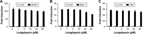
CK1 family members play both positive and negative roles in the Wnt/β-catenin signaling pathway.Citation25 A novel compound longdaysin () exhibited a drastic effect on the circadian period through inhibition of CK1δ, CK1α, and ERK2.Citation23 To determine the effect of longdaysin on Wnt/β-catenin signaling, HEK293T cells were transfected with a SuperTOPFlash reporter vector together with expression plasmids for Wnt1, LRP6, DVL2, CK1δ, CK1ε, CK1δ/LRP6, CK1ε/LRP6, CK1δ/DVL2, CK1ε/DVL2, and β-catenin. Longdaysin dramatically blocked Wnt signaling activated by Wnt1, LRP6, DVL2, CK1δ, CK1ε, CK1δ/LRP6, CK1ε/LRP6, CK1δ/DVL2, and CK1ε/DVL2 in a dose-dependent manner (). Moreover, increased Wnt reporter activity induced by Wnt3A-conditioned medium was also inhibited by longdaysin (). However, this compound had little effect on β-catenin-induced reporter activity (). These results indicate that longdaysin may target the upstream components of Wnt signaling. In control experiments, longdaysin at Wnt-inhibitory concentrations had little effect on the luciferase activity of NFAT reporter (NFAT-Luc), AP-1 reporter (AP-1-Luc), or Hippo reporter (8× GTIIC-Luc) ().
Figure 1 Longdaysin inhibits the activity of the Wnt/β-catenin pathway.
Notes: (A) Structure of longdaysin. HEK293T cells were transfected with the SuperTOPFlash reporter plasmid along with empty vector or (B) Wnt1, (C) LRP6, (D) DVL2, (E) CK1δ, (F) CK1ε, (G) CK1δ/LRP6 and CK1ε/LRP6, (H) CK1δ/DVL2 and CK1ε/DVL2, and (I) β-catenin expression plasmids. The transfected cells were then treated with DMSO or 1–50 µM longdaysin for 24 hours. (J) The SuperTOPFlash reporter plasmid was transfected into HEK293T cells. After transfection for 24 hours, the cells were incubated with control or Wnt3A-CM containing DMSO or the indicated amounts of longdaysin for another 24 hours. The luciferase values were normalized to β-gal activities. The results were the average from three independent experiments done by duplicates. Statistical significance between groups was calculated by Student’s t-test. *P<0.05 vs the corresponding vehicle control.
Abbreviation: Wnt3A-CM, Wnt3A-conditioned medium.
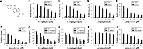
Figure 2 Longdaysin exhibits little inhibitory effect on the luciferase activity of NFAT, AP-1, or 8*GTIIC reporter gene.
Notes: HEK293T cells were transfected with (A) NFAT-Luc reporter together with control vector or NFATc expression plasmid, (B) AP-1-Luc reporter along with control vector or a constitutively active Rasv12 expression plasmid, or (C) 8*GTIIC-Luc reporter along with control vector or YAP expression plasmid. The luciferase values were normalized to β-gal activities. The results were the average from three independent experiments done by duplicates.
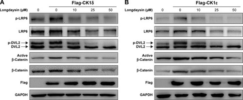
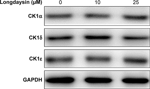
Since longdaysin has been demonstrated to inhibit CK1δ activity,Citation23 we speculate this compound may suppress the Wnt/β-catenin signaling cascade through targeting CK1δ and CK1ε. To investigate whether the Wnt-inhibitory effect of longdaysin is dependent on CK1δ/ε, HEK293T cells were transfected with Flag-tagged CK1δ or CK1ε expression plasmid and then treated with the indicated concentrations of longdaysin. As expected, CK1δ () or CK1ε () expression increased the level of phosphorylated LRP6 and DVL2 and activated β-catenin. Treatment with longdaysin significantly reduced the levels of phosphorylated LRP6, total LRP6, phosphorylated DVL2, activated β-catenin, and total β-catenin in a dose-dependent manner (), indicating that longdaysin may inhibit the Wnt/β-catenin pathway by suppressing CK1δ/ε. In addition, Western blot analysis was performed to examine the basal expression of CK1 isoforms in HEK293T cells. As shown in Figure S1, the basal expression of CK1α, CK1δ, and CK1ε was detected in HEK293T cells. Longdaysin treatment had no effect on their basal expression (Figure S1).
Figure 3 Longdaysin suppresses the Wnt/β-catenin pathway induced by CK1δ and CK1ε in HEK293T cells.
Notes: HEK293T cells were transfected with (A) Flag-tagged CK1δ expression plasmid or (B) Flag-tagged CK1ε expression plasmid. After transfection for 24 hours, the cells were treated with DMSO or the indicated concentrations of longdaysin for another 24 hours. Then, phosphorylated LRP6, total LRP6, DVL2, active β-catenin, total β-catenin, and Flag-tagged CK1δ or CK1ε were detected by immunoblotting. The phosphorylated DVL2 showed slower mobility following SDS/PAGE.
CK1δ/ε-dependent inhibition of the Wnt/β-catenin signaling cascade in breast cancer cells by longdaysin
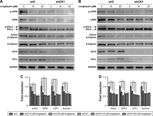
To determine the effect of CK1δ and CK1ε on Wnt/β-catenin signaling in breast cancer cells, lentivirus-mediated shRNAs were used to suppress the expression of CK1δ and CK1ε in Hs578T and MDA-MB-231 cells. Similar to longdaysin, shRNA-mediated silencing of CK1δ and CK1ε resulted in decreased protein levels of phosphorylated LRP6, total LRP6, phosphorylated DVL2, active β-catenin, and total β-catenin (). Importantly, depletion of CK1δ and CK1ε abolished the effects of longdaysin on LRP6, DVL2, and β-catenin in both cell lines (). Furthermore, knockdown of CK1δ and CK1ε downregulated the mRNA expression of Wnt target genes Axin2, DKK1, LEF1, and Survivin, while longdaysin treatment had no further effects on the expression of these Wnt target genes in shRNA-silenced cells ().
Figure 4 Longdaysin represses the Wnt/β-catenin pathway via targeting CK1δ and CK1ε in breast cancer cells.
Notes: (A and C) Hs578T or (B and D) MDA-MB-231 breast cancer cells were infected with lentivirus containing control shRNA (shC) or shRNAs targeting CK1δ and CK1ε (shCK1), treated with DMSO or the indicated concentrations of longdaysin for 24 hours, and then analyzed by (A and B) immunoblotting to detect the indicated proteins. (C and D) Real-time PCR was used to determine mRNA levels of Wnt target genes Axin2, DKK1, LEF1, and Survivin. Data are expressed as mean ± SD of three replicates. *P<0.05 vs the corresponding vehicle control (Student’s t-test).
Repression of colony formation, migration, and invasion in breast cancer cells by longdaysin via targeting CK1δ/ε
To examine the effect of longdaysin on the colony formation ability of breast cancer cells, a colony formation assay was performed. Longdaysin treatment inhibited colony formation of Hs578T and MDA-MB-231 cells in a dose-dependent manner, while knockdown of CK1δ/ε abrogated the effect of longdaysin ().
Figure 5 Longdaysin inhibits breast cancer cell colony formation, migration, and invasion through suppressing CK1δ and CK1ε. Hs578T or MDA-MB-231 breast cancer cells were infected by lentivirus containing control shRNA (shC) or shRNAs targeting CK1δ and CK1ε (shCK1).
Notes: (A) Hs578T and MDA-MB-231 cells were seeded and treated with the indicated concentrations of longdaysin for 7 days. Cells were then stained with crystal violet, and stained cells were photographed. The right panel is a graphical illustration of quantitative data of the relative colony numbers per well. (B) Hs578T and MDA-MB-231 cells were transferred into transwells with the indicated concentrations of longdaysin for 6 hours. The migrated cells were stained and photomicrographed. The right panel is a graphical illustration of quantitative cells migrated per field. (C) Hs578T and MDA-MB-231 cells were transferred into transwells coated with Matrigel and incubated with the indicated concentrations of longdaysin for 24 hours. The invaded cells were stained and photomicrographed. The right panel shows a graphical illustration of quantitative cells invaded per field. Data were collected from three independent experiments. *P<0.05.
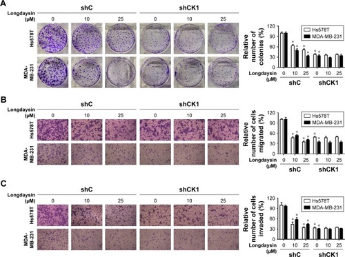
Considering the important role of Wnt/β-catenin signaling in migration and invasion of malignant cells, we investigated the effect of longdaysin on migration and invasion in breast cancer cells using transwell assay. As shown in , longdaysin treatment dose-dependently suppressed the migration and invasion of Hs578T and MDA-MB-231 cells (). However, the effects of longdaysin on the migration and invasion of breast cancer cells were abolished in CK1δ/ε-knockdown cells (). These results suggest that longdaysin may repress breast cancer cell colony formation, migration, and invasion through targeting CK1δ/ε.
Blockage of breast cancer cell stemness by longdaysin through inhibition of CK1δ/ε
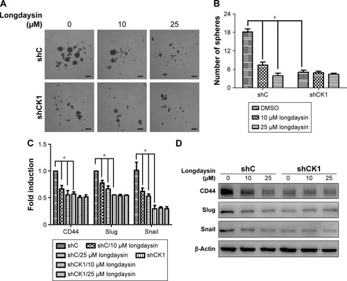
Since Wnt/β-catenin signaling plays a crucial role in the survival and maintenance of CSCs, we examined the effect of longdaysin on the stemness of breast cancer cells using a sphere formation assay. The breast cancer Hs578T cells were incubated with longdaysin at different concentrations for 1 week. The images of tumor spheres of Hs578T cells showed markedly decreased number and size of tumor spheres in cells treated with longdaysin (). Furthermore, longdaysin treatment downregulated the expression of stemness marker genes, CD44, Slug, and Snail, in Hs578T cells (). As expected, CK1δ/ε knockdown attenuated tumor sphere formation and inhibited expressions of stemness genes CD44, Slug, and Snail in mRNA and their protein levels in Hs578T cells (). Longdaysin had little effects on the sphere formation, and expression of stemness marker genes in CK1δ/ε-silenced cells (). It has been well established that CD44, Slug, and Snail are the target genes of Wnt/β-catenin signaling.Citation26,Citation27 Thus, it is fairly reasonable to assume longdaysin-induced inhibition of stemness may be associated with its antagonistic effects on Wnt/β-catenin signaling through targeting CK1δ/ε.
Figure 6 Longdaysin suppresses the sphere-forming ability and the expression of stemness marker genes through inhibition of CK1δ/ε in breast cancer cells.
Notes: (A) Hs578T breast cancer cells were infected with control shRNA (shC) or shRNAs targeting CK1δ and CK1ε (shCK1). Cells were then cultured in Ultra-Low Attachment dishes to examine the ability of sphere formation in the absence or presence of the indicated longdaysin. The number of spheres (diameter >50 µm) was counted under a microscope. (B) Graphical illustration of quantitative data of the relative number of spheres. (C) Hs578T cells were infected with control shRNA (shC) or shRNAs targeting CK1δ and CK1ε (shCK1). Cells were then treated with the indicated concentrations of longdaysin for 24 hours. Real-time PCR was used to determine mRNA levels of stemness marker genes CD44, Slug, and Snail. (D) Hs578T cells were infected by lentivirus containing control shRNA (shC) or shRNAs targeting CK1δ and CK1ε (shCK1). Cells were then treated with the indicated concentrations of longdaysin for 24 hours. CD44, Slug, and Snail were detected by immunoblotting. Scale bar, 100 µm. The results are shown as mean ± SD from three independent experiments. *P<0.05.
Suppression of tumor growth in vivo and inhibition of Wnt/β-catenin signaling in MDA-MB-231 xenografts by longdaysin
To determine the inhibitory effect of longdaysin on the Wnt/β-catenin pathway in breast cancer cells in vivo, MDA-MB-231 cells were injected s.c. into nude mice. The tumors were measured every 3 days. Once the volumes of the tumors reached approximately 50 mm3, mice were randomly divided into two groups, and i.p. injected with vehicle or long-daysin at 5 mg/kg every 3 days, respectively. After 3-week treatment, the mice were sacrificed and tumors were removed and weighed. The treatment regimen with longdaysin obviously inhibited tumor growth (), and decreased tumor cell density () and proliferation as indicated by Ki-67 staining (). However, longdaysin did not influence body weight, suggesting minimal toxicity.
Figure 7 Longdaysin represses tumor growth in MDA-MB-231 xenograft mouse model.
Notes: Established MDA-MB-231 xenografts were treated with 5 mg/kg longdaysin every 3 days for 3 weeks. Eight mice per group were used. (A) Tumor images. (B) Mean tumor volumes. (C) Mean tumor weights. (D) H&E staining; original magnification ×40. Scale bar, 100 µm. (E) Ki-67 staining; original magnification ×40. Scale bar, 100 µm. (F) Active β-catenin antibody staining; original magnification ×40. Scale bar, 100 µm. (G) Total β-catenin antibody staining; original magnification ×40. Scale bar, 100 µm. (H) The expression levels of phosphorylated LRP6, total LRP6, DVL2, active β-catenin, total β-catenin, CD44, Slug, and Snail in tumor samples were detected by immunoblotting. (I) The mRNA expression levels of Axin2, DKK1, LEF1, Survivin, CD44, Slug, and Snail were quantitated by real-time PCR. The results are shown as mean ± SD from three independent experiments. *P<0.05.
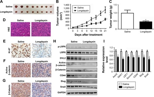
To investigate the effect of longdaysin on Wnt/β-catenin signaling in MDA-MB-231 xenografts, immunohistochemical staining, immunoblot analyses, and real-time PCR were performed. As indicated by immunohistochemical staining, administration of longdaysin markedly reduced the expression of active β-catenin and total β-catenin (). Moreover, Western blot demonstrated that longdaysin treatment decreased the expression of phosphorylated and total LRP6, phosphorylated DVL2, and active and total β-catenin (). The Wnt-inhibitory effect of longdaysin was further confirmed by real-time PCR, which showed reduced mRNA expression of the Wnt target genes including Axin2, DKK1, LEF1, and Survivin in longdaysin-treated group compared with control group ().
We further explored the effect of longdaysin on the expression of stemness-related Wnt target genes CD44, Slug, and Snail. Longdaysin treatment inhibited both mRNA and protein levels of CD44, Slug, and Snail ().
Discussion
Longdaysin is a novel chemical regulator of circadian clock. This compound markedly slows the circadian clock both in a variety of mammalian cells and in zebrafish in vivo through inhibition of the protein kinases CK1δ, CK1α, and ERK2. Longdaysin may form the basis for therapeutic strategies directed toward circadian disorders, such as shift-work fatigue and jet lag.Citation23 However, little is known about the effect of longdaysin on the Wnt/β-catenin pathway. In this study, longdaysin was identified as a potent inhibitor of Wnt/β-catenin signaling through targeting CK1δ and CK1ε. In MDA-MB-231 breast cancer xenografts, longdaysin suppressed in vivo tumor growth, concomitant with reduced expression of phosphorylated and total LRP6, phosphorylated DVL2, active and total β-catenin, and Wnt target genes. Our results indicate that longdaysin may have therapeutic potential for the treatment of breast cancer.
Aberrant regulation of CK1δ and CK1ε is involved in the pathogenesis of several human cancers including breast cancer.Citation2 CK1ε has been found to be an essential regulator of β-catenin activity and proliferation in breast cancer cells. The expression of CK1ε could promote oncogenic transformation of mammary epithelial cells in a β-catenin-dependent manner.Citation28 Hirner et al reported that inactivating mutations in CK1δ could attenuate SV40-mediated cellular transformation in vitro and SV40-induced mouse mammary carcinogenesis in vivo.Citation29 Rosenberg et al demonstrated that CK1δ is frequently amplified and over-expressed in a subset of human breast cancers. Knockdown or inhibition of CK1δ induced breast tumor regression in patient-derived and cell line orthotopic xenograft models of TNBC and HER2+ breast cancer.Citation30 Recently, Janovska et al identified CK1δ/ε as a novel therapeutic target in chronic lymphocytic leukemia.Citation31 These studies indicate that CK1δ and CK1ε may be therapeutic targets for cancer drug development. Our results demonstrated that long-daysin could inhibit the Wnt/β-catenin signaling through inhibition of CK1δ/ε (). LRP6 and DVL2 are well known as specific substrates for CK1. Longdaysin markedly decreased phosphorylation of LRP6 and DVL2, and reduced the levels of active β-catenin and total β-catenin protein, finally leading to the transcriptional downregulation of Wnt target genes. Consistently, knockdown of CK1δ/ε also decreased protein levels of phosphorylated and total LRP6, phosphorylated DVL2, active β-catenin, and total β-catenin in breast cancer cells, resulting in downregulation of the mRNA expression of Wnt target genes. Moreover, the effects of longdaysin on Wnt/β-catenin occurred at concentrations comparable to those required for inhibiting colony formation, migration, and invasion in breast cancer cells. These results suggest that the antitumor activity of longdaysin is associated with its inhibitory effects on the Wnt/β-catenin signaling pathway.
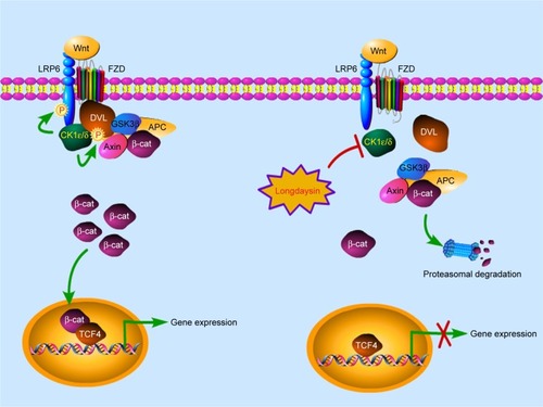
Figure 8 A novel mechanism by which longdaysin inhibits Wnt/β-catenin signaling.
Notes: The Wnt/β-catenin signaling cascade is initiated when a Wnt molecule binds to the Wnt coreceptor LRP6 and a member of the FZD receptor family. CK1δ/ε activity is rapidly upregulated and DVL and LRP6 are phosphorylated, resulting in accumulation of cytoplasmic β-catenin. Longdaysin could decrease phosphorylation of LRP6 and DVL2 through inhibition of CK1δ/ε, and reduce the levels of cytoplasmic β-catenin, finally leading to the transcriptional downregulation of Wnt target genes.
Our results showed that longdaysin treatment dose-dependently inhibited colony formation, migration, invasion, and formation of tumor spheres in breast cancer cells, while knockdown of CK1δ/ε abrogated its effects. Additionally, we did not observe any synergistic or additive effect after combined CK1δ/ε knockdown and longdaysin treatment. These results strongly suggest that longdaysin exhibits its antitumor activity against breast cancer via targeting CK1δ/ε.
Increasing evidence has demonstrated that the Wnt/β-catenin pathway regulates self-renewal and stemness properties of CSCs.Citation32 Several Wnt target genes have been proposed as CSC markers including CD44, Slug, and Snail in breast cancer.Citation33,Citation34 Thus, targeting the Wnt/β-catenin pathway can potentially eliminate CSC populations in breast cancer. Our results showed that longdaysin significantly inhibited sphere formation of breast cancer cells and decreased the expression of stemness marker genes CD44, Slug, and Snail. In the MDA-MB-231 xenografts, longdaysin suppressed tumor growth in vivo and reduced both mRNA and protein levels of CD44, Slug, and Snail. These results suggest that longdaysin may be an efficient inhibitor of breast CSCs. Further investigation is needed to characterize the inhibitory action of longdaysin on breast CSCs.
Conclusion
Our results showed that longdaysin is able to inhibit the Wnt/β-catenin pathway by targeting CK1δ/ε. This compound markedly decreased phosphorylation of LRP6 and DVL2, and reduced the levels of active β-catenin and total β-catenin protein, finally leading to the transcriptional downregulation of Wnt target genes. We further demonstrated that long-daysin could repress breast cancer cell colony formation, migration, and invasion in a CK1δ/ε-dependent manner. In breast cancer xenografts, longdaysin suppressed in vivo tumor growth with concurrent inhibition of Wnt/β-catenin signaling. To our knowledge, this is the first study providing evidence that longdaysin is a potent antitumor agent. It exhibited antitumor activity against breast cancer via inhibition of CK1δ/ε-dependent Wnt signaling.
Data sharing statement
All data underlying the findings described in this manuscript are fully available without restrictions.
Abbreviations
| CSC | = | scancer stem cells |
| DMSO | = | dimethyl sulfoxide |
| i.p. | = | intraperitoneally |
| s.c. | = | subcutaneously |
Acknowledgments
The authors would like to thank the Cancer Research Center, Department of Pharmacology, Shenzhen University Health Science Center, for providing the facilities to carry out this study. This work was supported by the National Natural Science Foundation of China (Grants 31870754, 81802662 and 81372342), the Natural Science Foundation of Guangdong Province (Grant 2017A030310329), the Shenzhen Peacock Innovation Team Project (Grant KQTD20140630100658078), the Key Laboratory Project of Shenzhen (Grant ZDSY20130329101130496), the Shenzhen Peacock Plan (Grants 827000183 and 827000186), and the Shenzhen Basic Research Program (Grants JCYJ20150525092941006, JCYJ20170302143447936 and JCYJ20170817094611664).
Supplementary material
Figure S1 The basal expression of CK1α, CK1δ, and CK1ε in HEK293T cells detected by immunoblotting.
Disclosure
The authors report no conflicts of interest in this work.
References
- KnippschildUGochtAWolffSHuberNLöhlerJStöterMThe casein kinase 1 family: participation in multiple cellular processes in eukaryotesCell Signal200517667568915722192
- KnippschildUWolffSGiamasGThe role of the casein kinase 1 (CK1) family in different signaling pathways linked to cancer developmentOnkologie2005281050851416186692
- CheongJKVirshupDMCasein kinase 1: complexity in the familyInt J Biochem Cell Biol201143446546921145983
- PriceMACKI, there’s more than one: casein kinase I family members in Wnt and Hedgehog signalingGenes Dev200620439941016481469
- SchittekBSinnbergTBiological functions of casein kinase 1 isoforms and putative roles in tumorigenesisMol Cancer201413123125306547
- KnippschildUKrugerMRichterJThe CK1 family: contribution to cellular stress response and its role in carcinogenesisFront Oncol2014419624904820
- RichterJUllahKXuPEffects of altered expression and activity levels of CK1delta and varepsilon on tumor growth and survival of colorectal cancer patientsInt J Cancer2015136122799281025404202
- NusseRCleversHWnt/beta-catenin signaling, disease, and emerging therapeutic modalitiesCell2017169698599928575679
- CleversHNusseRWnt/beta-catenin signaling and diseaseCell201214961192120522682243
- MacdonaldBTTamaiKHeXWnt/β-catenin signaling: components, mechanisms, and diseasesDev Cell200917192619619488
- CruciatCMCasein kinase 1 and Wnt/β-catenin signalingCurr Opin Cell Biol201431465525200911
- Del Valle-PérezBArquésOVinyolesMde HerrerosAGDuñachMCoordinated action of CK1 isoforms in canonical Wnt signalingMol Cell Biol201131142877288821606194
- LiuCLiYSemenovMControl of beta-catenin phosphorylation/degradation by a dual-kinase mechanismCell2002108683784711955436
- KlimowskiLKGarciaBAShabanowitzJHuntDFVirshupDMSite-specific casein kinase 1epsilon-dependent phosphorylation of dishevelled modulates beta-catenin signalingFEBS J2006273204594460216965538
- SwiatekWTsaiICKlimowskiLRegulation of casein kinase I epsilon activity by Wnt signalingJ Biol Chem200427913130111301714722104
- CongFSchweizerLVarmusHCasein kinase Iepsilon modulates the signaling specificities of dishevelledMol Cell Biol20042452000201114966280
- NiehrsCShenJRegulation of LRP6 phosphorylationCell Mol Life Sci201067152551256220229235
- EtchegarayJPMachidaKKNotonECasein kinase 1 delta regulates the pace of the mammalian circadian clockMol Cell Biol200929143853386619414593
- BrennanKCBatesEAShapiroRECasein kinase iδ mutations in familial migraine and advanced sleep phaseSci Transl Med20135183183ra56111
- MengQJMaywoodESBechtoldDAEntrainment of disrupted circadian behavior through inhibition of casein kinase 1 (CK1) enzymesProc Natl Acad Sci U S A201010734152401524520696890
- MengQJLogunovaLMaywoodESSetting clock speed in mammals: the CK1ε tau mutation in mice accelerates circadian pacemakers by selectively destabilizing period proteinsNeuron2008581788818400165
- MaywoodESCheshamJESmyllieNJHastingsMHThe Tau mutation of casein kinase 1epsilon sets the period of the mammalian pacemaker via regulation of Period1 or Period2 clock proteinsJ Biol Rhythms201429211011824682205
- HirotaTLeeJWLewisWGHigh-throughput chemical screen identifies a novel potent modulator of cellular circadian rhythms and reveals CKIα as a clock regulatory kinasePLoS Biol2010812e100055921179498
- WangZLiBZhouLProdigiosin inhibits Wnt/β-catenin signaling and exerts anticancer activity in breast cancer cellsProc Natl Acad Sci U S A201611346131501315527799526
- JiangJCK1 in developmental signaling: hedgehog and WntCurr Top Dev Biol201712330332928236970
- KatohMCanonical and non-canonical WNT signaling in cancer stem cells and their niches: cellular heterogeneity, omics reprogramming, targeted therapy and tumor plasticity (Review)Int J Oncol20175151357136929048660
- HeubergerJBirchmeierWInterplay of cadherin-mediated cell adhesion and canonical Wnt signalingCold Spring Harb Perspect Biol201022a00291520182623
- KimSYDunnIFFiresteinRCK1ε is required for breast cancers dependent on β-catenin activityPLoS One201052e897920126544
- HirnerHGünesCBischofJImpaired CK1 delta activity attenuates SV40-induced cellular transformation in vitro and mouse mammary carcinogenesis in vivoPLoS One201271e2970922235331
- RosenbergLHLafitteMQueredaVTherapeutic targeting of casein kinase 1δ in breast cancerSci Transl Med20157318318318ra202
- JanovskaPVernerJKohoutekJCasein kinase 1 is a therapeutic target in chronic lymphocytic leukemiaBlood2018131111206121829317454
- De FrancescoEMSotgiaFLisantiMPCancer stem cells (CSCs): metabolic strategies for their identification and eradicationBiochem J201847591611163429743249
- LeeSYJeongEKJuMKInduction of metastasis, cancer stem cell phenotype, and oncogenic metabolism in cancer cells by ionizing radiationMol Cancer20171611028137309
- WuYSarkissyanMVadgamaJVEpithelial-mesenchymal transition and breast cancerJ Clin Med201652E1326821054