
 ?Mathematical formulae have been encoded as MathML and are displayed in this HTML version using MathJax in order to improve their display. Uncheck the box to turn MathJax off. This feature requires Javascript. Click on a formula to zoom.
Abstract
Purpose
Chemotherapy after surgery can prolong the survival of patients with gliomas. Dimethylaminomicheliolide (DMAMCL), a novel chemotherapeutic agent, exhibited antitumor properties in acute myeloid leukemia stem cells and showed an increased drug concentration in the brain. This study aims to investigate the specific anticancer activities and mechanisms of DMAMCL in glioma cells.
Materials and methods
In this study, the effects of DMAMCL were evaluated and characterized in U87-MG and U251 glioma cells. Cell viability was assessed by Cell Counting Kit-8. Apoptosis, mitochondrial membrane potential, and intracellular reactive oxygen species (ROS) generation were assessed by fluorescence microscopy. Autophagosome formation was observed with transmission electron microscopy, and the autophagy flux was measured by transfecting cells with mRFP-GFP-LC3 adenoviral vectors. Immunofluorescence and Western blot analyses were used to determine the expression of proteins.
Results
In the present study, treatment with DMAMCL decreased cell viability and induced apoptosis in U87-MG and U251 glioma cells. Additionally, DMAMCL activated autophagy-mediated cell death as evidenced by the formation of autophagosomes, accumulation of LC3B-II, inhibition of autophagy flux, and increase in cell viability after cotreatment with an autophagy inhibitor. Subsequent experiments showed that the DMAMCL-induced apoptosis and autophagy were possibly mediated by ROS generation and Akt/mTOR signaling pathway inhibition. On the other hand, the ROS scavenger N-acetyl-L-cysteine and the Akt activator insulin-like growth factor-1 attenuated the DMAMCL-induced autophagy and cell death.
Conclusion
Our findings revealed that DMAMCL induced apoptosis and autophagic cell death by regulating the ROS/mitogen-activated protein kinase signaling pathway and suppressing the Akt/mTOR signaling pathway in human glioma cells. DMAMCL may be a novel effective anticancer agent, which can target gliomas.
Introduction
Gliomas are one of the most malignant and lethal primary brain cancers, which can occur in any part of the central nervous system. The overall incidence rate is 6.03 per 100,000.Citation1 Due to the high invasiveness of gliomas, it is difficult to completely resect them. People with these neoplasms generally have a poor prognosis and poor quality of life as the disease progresses.Citation2,Citation3 Even after considerable research, there is little progress in limiting the development of gliomas and prolonging the median overall survival of patients. The high recurrence rate and the development of drug resistance contribute to this situation.Citation4 The current clinical treatments for malignant gliomas are surgery, adjuvant postoperative radiotherapy, and chemotherapy. Although these therapies can prolong overall survival, the poor curative effects due to the inherent apoptosis-resistant phenotype of the malignancy and the extremely negative systemic side effects result in unsatisfactory outcomes.Citation5,Citation6 Hence, more effective treatments are still urgently needed.
Recent studies have revealed that sesquiterpene lactone compounds have many antitumor and anti-inflammatory effects.Citation7–Citation9 Parthenolide (PTL), derived from Tanacetum parthenium, is one of these compounds. Many reports have revealed that PTL and its soluble analog dimethylamino parthenolide have cytotoxic effects on breast, prostate, pancreatic, glioma, lung, and bladder cancer cells, in addition to hematologic malignancies. As these compounds are unstable in both acidic and basic conditions, their therapeutic applications have been restricted.Citation10–Citation16 Micheliolide, which is much more stable than PTL, was isolated from the Michelia compressa and Michelia champaca plants and showed remarkable therapeutic efficacy in nonobese diabetic/severe combined immunodeficiency AML models.Citation17 Dimethylaminomicheliolide (DMAMCL), as a novel chemotherapeutic agent, has been reported to suppress inflammation in cases of intestinal disease and sepsis.Citation18 In addition, it was proven to prolong the lifespan of a mouse model of human acute myelogenous leukemia.Citation19 The distribution analysis in the DMAMCL-treated rats showed that the drug concentration in the brain was higher than in the plasma, and it was innocuous to the main organs.Citation20
Apoptosis, also called type I programmed cell death, plays an important role in the progression of chemotherapy. Apoptosis is caspase-dependent and is characterized by some conspicuous changes in the cell death process; for example, cell membrane blebbing, cell shrinkage, nuclear fragmentation, chromatin condensation, DNA fragmentation, and apoptotic body formation.Citation21 However, in many cases, chemotherapy can induce autophagic cell death by activating the lysosome-dependent proteolytic pathway.Citation22 Autophagy, a conservative process, enables cells to isolate the damaged or surplus organelles into autophagosomes and deliver them to lysosomes to degrade. However, autophagy has conflicting roles in various cell types under different cellular states.Citation23
Reactive oxygen species (ROS) play an important role in the development of cancers. However, superfluous ROS have cytotoxicity against diverse targets, such as proteins, DNA, and lipids. In many exogenous stress conditions, ROS are important signaling molecules that induce apoptosis and autophagy and activate cellular signaling kinases.Citation24 Some chemical drugs targeting ROS-related signaling pathways were proven to be effective in the treatment of human cancers, including ROS/mitogen-activated protein kinase (MAPK) signaling pathways, which was a momentous discovery. However, the effects of DMAMCL-induced ROS damage and the regulation of related signaling pathways in human glioma cancer cells remain unclear.
In the present study, we aimed to determine the anticancer activities and potential mechanisms of DMAMCL in two different human glioma cell lines. We found that DMAMCL could induce not only apoptosis through ROS generation, mitochondrial dysfunction, and caspase activation but also autophagy through the inhibition of the Akt/mTOR signaling pathways in the U87-MG and U251 cell lines. These novel findings provide a new perspective for DMAMCL in glioma chemotherapeutic interventions.
Materials and methods
Cell lines and cell culture and DMAMCL preparation
The human glioma cell lines U87-MG and U251 were obtained from the Chinese Academy of Sciences Cell Bank. These cell lines were both cultured in Dulbecco’s Modified Eagle’s Medium/HIGH glucose culture medium supplemented with 10% FBS, 100 U/mL penicillin, and 100 mg/mL streptomycin. Cells were kept in the exponential growth phase and cultured at 37°C in a humidified atmosphere containing 5% CO2 and 95% air. DMAMCL was a gift provided by Accendatech Co., Ltd. (Tianjin, China). The formula is C17H27NO3·C4H4O4. For the following experiments, DMAMCL was dissolved in water at a concentration of 10 mM as a stock solution and diluted to the indicated concentration with medium before use.
Cell viability assay
Cell viability was determined by Cell Counting Kit-8 (CCK-8) assays. Briefly, cells in the exponential phase of growth were harvested and seeded into 96-well plates at a density of 5,000 cells per well. After 24 hours of incubation, the cells were treated with different concentrations of DMAMCL or medium for another 24, 48, and 72 hours. Then, 10 μL CCK-8 solution was added into each well. One hour later, the absorbance was determined using a microplate reader (EL340; BioTek Instruments, Waltham, MA, USA) at 450 nm.
Cell apoptosis analysis by flow cytometry
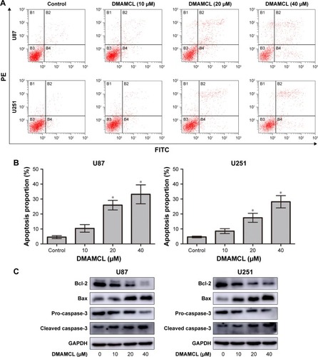
The Annexin V-fluorescein isothiocyanate (FITC)/propidium iodide (PI) kit was used to determine the effect of DMAMCL on apoptosis. First, the cells were exposed to different concentrations of DMAMCL for 48 hours, and then the cells were collected. Next, the cells were resuspended in binding buffer and incubated with 5 μL Annexin V-FITC and 5 μL PI for 15 minutes in the dark. The results were analyzed using a FACS Calibur or an EPICS XL flow cytometer (BD Biosciences).
Live/dead assay
U87-MG and U251 cells were treated with various concentrations of DMAMCL for 48 hours and then the LIVE/DEAD™ Cell Imaging Kit was used according to the instructions. After another 15 minutes of culture in the dark, the results were observed using fluorescence microscopy (Olympus).
Transmission electron microscopy
U87-MG and U251 cells were treated with DMAMCL (40 μM) for 48 hours. Afterward, the cells were fixed with 3% glutaraldehyde and 2% paraformaldehyde in a 0.1 M phosphate-buffered saline buffer for 30 minutes and postfixed with 1% osmium tetroxide for 1.5 hours. Then, the cells were washed and stained in 3% aqueous uranyl acetate for 1 hour. Subsequently, the cells were dehydrated in a graded series of ethanol and acetone and then embedded in Epon-Araldite resin. Ultrathin sections were cut by a Reichert ultramicrotome, double-stained with 0.3% lead citrate, and examined using a JEOL 1200EX electron microscope (Japan).
Confocal microscopy
U87-MG and U251 cells were seeded into 96-well plates, and when they reached 70% confluence, they were transfected with mRFP-GFP-LC3 adenoviral vectors (purchased from HanBio Technology, Shanghai, China) following the manufacturer’s instructions. Then, the cells were treated with DMAMCL for 48 hours. Finally, the LC3 puncta were examined with a confocal microscope (USA).
Measurement of intracellular ROS generation
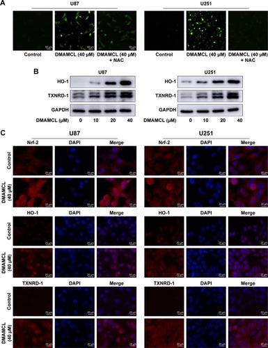
Intracellular ROS were detected by the Total ROS Assay Kit. Briefly, 5,000 cells per well were cultured for 24 hours in a 96-well plate, and then the cells were incubated with 2′,7′-dichlorofluorescein-diacetate (DCFH-DA) for 1 hour. Following the treatment, the cells were washed with serum-free medium three times and exposed to the indicated treatments. The level of ROS was analyzed and imaged using a fluorescence microscope (Olympus).
Measurement of mitochondrial membrane potential
The mitochondrial membrane potential (MMP) was measured with the JC-1 Assay Kit. A total of 5 × 10⁵ cells per well were cultured for 24 hours in a six-well plate. Then, the cells were treated with different concentrations of DMAMCL, ranging from 10 to 40 μM, for another 48 hours. The cells were washed three times, collected, and then incubated for 20 minutes with the JC-1 probe at 37°C. The MMPs were measured using a flow cytometer (BD Biosciences).
Western blot analysis
Cells were lysed in cold RIPA lysis buffer containing 1% phosphatase inhibitor and 1% protease inhibitor for 30 minutes. Protein samples were collected and centrifuged at 12,000 rpm for another 30 minutes at 4°C. The supernatants were collected for continued analysis. The Bradford protein method and the bicinchoninic acid protein assay kit were used to determine the protein concentration according to the manufacturer’s directions. A total of 40 μg protein was separated, electrophoresed using a bis-Tris polyacrylamide gel and transferred onto a polyvinylidenedifluoride membrane. Membranes were incubated with primary anti-bodies overnight at 4°C after being blocked in 5% nonfat dry milk. Then, the antigen–antibody binding reaction was performed using horseradish peroxidase-conjugated secondary antibodies for 1 hour at room temperature. Immunoblots were visualized using enhanced chemiluminescence (LAS-4000).
Immunofluorescence
Cell slides were prepared and exposed to various treatments. Cells were then fixed with 4% paraformaldehyde for 20 minutes and permeabilized with 0.3% Triton for 15 minutes at room temperature. Next, the smears were blocked with goat serum solution for 1 hour, followed by primary antibody incubation at 4°C overnight in a wet box. Cells were thoroughly rinsed and stained with a fluorescent secondary antibody for 1 hour. Finally, nuclei were stained with DAPI for 10 minutes and imaged using a fluorescence microscope (Olympus).
Statistical analysis
The experiments were performed independently at least three times. The data were statistically analyzed using t-test, chi-squared test, or Fisher’s exact tests using SPSS version 19.0 software (IBM SPSS, NY, USA). P-values <0.05 were considered statistically significant.
Results
DMAMCL inhibited the viability of glioma cells and induced cell death
To investigate the inhibitory effects of DMAMCL on human glioma cells, U87-MG and U251 cells were treated with different concentrations of DMAMCL. Cell viability was determined by the CCK-8 assay (). The results showed that the viability of these cells was reduced in a dose-dependent manner at 24, 48, and 72 hours. The ratios between the live and dead cells were further determined with the LIVE/DEAD Cell Imaging Kit. As shown in , the number of live cells (green) was significantly reduced, and the density of dead cells (red) was increased compared with the control group. Next, we further examined the effects of different treatments on cell viabilities. As shown in , co-treated 3-MA or IGF-1 with DMAMCL and pretreated with NAC could increase the cell viability compared with DMAMCL alone.
Figure 1 DMAMCL inhibits the viability of glioma cells and induces cell death.
Notes: (A) Dose- and time-dependent effects of DMAMCL on cell viability. U87-MG and U251 glioma cells were administered different concentrations of DMAMCL (10, 20, 30, 40, 50, and 60 μM) for 24, 48, and 72 hours, and cell viability was assessed via CCK-8 assays. *Indicates statistically significant difference (P<0.05) vs untreated control cells. (B, C) Effects of DMAMCL, ROS scavenger NAC, and Akt activator IGF-1 on U87-MG and U251 cells. The percentage of dead cells (red) increased compared with the control. *Indicates statistically significant difference (P<0.05) vs untreated control cells. #Indicates significantly different (P<0.05) compared with DMAMCL treatment. Scale bars =50 μm. (D) The CCK-8 proliferation assay using U87-MG and U251 cells was performed with different treatments (40 μM DMAMCL for 48 hours; 3-MA [100 nM] for 48 hours; 40 μM DMAMCL and 3-MA [100 nM] for 48 hours; pretreatment with NAC [5 mM] for 2 hours and then culture media for 48 hours; pretreatment with NAC [5 mM] for 2 hours, followed by 40 μM DMAMCL for 48 hours; IGF-1 [100 ng/mL] for 48 hours; 40 μM DMAMCL and IGF-1 [100 ng/mL] for 48 hours). *Indicates statistically significant (P<0.05) vs untreated control cells. #Indicates significantly different (P<0.05) compared with DMAMCL treatment. Data are expressed as the mean ± SD of three independent experiments.
Abbreviations: 3-MA, 3-methyladenine; CCK-8, Cell Counting Kit-8; DMAMCL, dimethylaminomicheliolide; IGF-1, insulin-like growth factor-1; NAC, N-acetyl-L-cysteine; ROS, reactive oxygen species.
![Figure 1 DMAMCL inhibits the viability of glioma cells and induces cell death.Notes: (A) Dose- and time-dependent effects of DMAMCL on cell viability. U87-MG and U251 glioma cells were administered different concentrations of DMAMCL (10, 20, 30, 40, 50, and 60 μM) for 24, 48, and 72 hours, and cell viability was assessed via CCK-8 assays. *Indicates statistically significant difference (P<0.05) vs untreated control cells. (B, C) Effects of DMAMCL, ROS scavenger NAC, and Akt activator IGF-1 on U87-MG and U251 cells. The percentage of dead cells (red) increased compared with the control. *Indicates statistically significant difference (P<0.05) vs untreated control cells. #Indicates significantly different (P<0.05) compared with DMAMCL treatment. Scale bars =50 μm. (D) The CCK-8 proliferation assay using U87-MG and U251 cells was performed with different treatments (40 μM DMAMCL for 48 hours; 3-MA [100 nM] for 48 hours; 40 μM DMAMCL and 3-MA [100 nM] for 48 hours; pretreatment with NAC [5 mM] for 2 hours and then culture media for 48 hours; pretreatment with NAC [5 mM] for 2 hours, followed by 40 μM DMAMCL for 48 hours; IGF-1 [100 ng/mL] for 48 hours; 40 μM DMAMCL and IGF-1 [100 ng/mL] for 48 hours). *Indicates statistically significant (P<0.05) vs untreated control cells. #Indicates significantly different (P<0.05) compared with DMAMCL treatment. Data are expressed as the mean ± SD of three independent experiments.Abbreviations: 3-MA, 3-methyladenine; CCK-8, Cell Counting Kit-8; DMAMCL, dimethylaminomicheliolide; IGF-1, insulin-like growth factor-1; NAC, N-acetyl-L-cysteine; ROS, reactive oxygen species.](/cms/asset/569f51fa-dee8-4e4c-9ebe-7855128e20f8/dott_a_195329_f0001_c.jpg)
![Figure 1 DMAMCL inhibits the viability of glioma cells and induces cell death.Notes: (A) Dose- and time-dependent effects of DMAMCL on cell viability. U87-MG and U251 glioma cells were administered different concentrations of DMAMCL (10, 20, 30, 40, 50, and 60 μM) for 24, 48, and 72 hours, and cell viability was assessed via CCK-8 assays. *Indicates statistically significant difference (P<0.05) vs untreated control cells. (B, C) Effects of DMAMCL, ROS scavenger NAC, and Akt activator IGF-1 on U87-MG and U251 cells. The percentage of dead cells (red) increased compared with the control. *Indicates statistically significant difference (P<0.05) vs untreated control cells. #Indicates significantly different (P<0.05) compared with DMAMCL treatment. Scale bars =50 μm. (D) The CCK-8 proliferation assay using U87-MG and U251 cells was performed with different treatments (40 μM DMAMCL for 48 hours; 3-MA [100 nM] for 48 hours; 40 μM DMAMCL and 3-MA [100 nM] for 48 hours; pretreatment with NAC [5 mM] for 2 hours and then culture media for 48 hours; pretreatment with NAC [5 mM] for 2 hours, followed by 40 μM DMAMCL for 48 hours; IGF-1 [100 ng/mL] for 48 hours; 40 μM DMAMCL and IGF-1 [100 ng/mL] for 48 hours). *Indicates statistically significant (P<0.05) vs untreated control cells. #Indicates significantly different (P<0.05) compared with DMAMCL treatment. Data are expressed as the mean ± SD of three independent experiments.Abbreviations: 3-MA, 3-methyladenine; CCK-8, Cell Counting Kit-8; DMAMCL, dimethylaminomicheliolide; IGF-1, insulin-like growth factor-1; NAC, N-acetyl-L-cysteine; ROS, reactive oxygen species.](/cms/asset/220c3b44-4be7-4dd1-b4fe-465498171c62/dott_a_195329_f0001a_b.jpg)
DMAMCL induced apoptosis in glioma cells
To determine whether DMAMCL induced cell death via apoptosis, we measured the MMPs of the U87-MG and U251 cells with different concentrations of DMAMCL through a flow cytometric analysis using JC-1 as a mitochondrial fluorescent probe. As shown in , DMAMCL reduced the MMP in the U87-MG and U251 cells in a dose-dependent manner, as indicated by a decrease in the red–green fluorescence intensity ratio. To quantify the level of apoptosis, we used Annexin V-FITC/PI double staining. The results demonstrated that DMAMCL increased the numbers of both early and late apoptotic cells in a dose-dependent manner (). We also determined the expression of the main mitochondrion-dependent apoptotic proteins, including caspase-3, Bcl-2, and Bax, by Western blotting analysis. As shown in , the levels of procaspase-3 and Bcl-2 were decreased, while the levels of cleaved caspase-3 and Bax were significantly increased in the DMAMCL-treated U87-MG and U251 cells. Overall, these results indeed suggested that DMAMCL contributed to apoptosis in human glioma cells.
Figure 2 DMAMCL induces a decrease in MMPs in glioma cells.
Notes: (A, B) The MMP of U87-MG and U251 cells with ascending concentrations of DMAMCL as determined by flow cytometric analysis using JC-1 as a mitochondrial fluorescent probe. *Indicates statistically significant (P<0.05) vs untreated control cells.
Abbreviations: DMAMCL, dimethylaminomicheliolide; MMPs, mitochondrial membrane potentials.
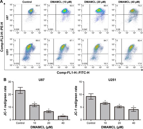
Figure 3 DMAMCL induces apoptosis in glioma cells.
Notes: (A, B) After treatment with the indicated concentrations of DMAMCL for 48 hours, apoptosis was detected using Annexin V-FITC/PI staining and the percentage of apoptotic cells compared with the untreated control groups was quantitatively analyzed. (C) The apoptosis biomarkers Bcl-2, Bax, and caspase-3 were measured after cells were treated with different concentrations of DMAMCL (10, 20, 40 μM) for 48 hours by Western blot analysis. GAPDH is shown as a loading control. The results shown are one representative of three independent experiments. *P<0.05 vs the control group.
Abbreviations: DMAMCL, dimethylaminomicheliolide; FITC, fluorescein isothiocyanate; GAPDH, glyceraldehyde 3-phosphate dehydrogenase; PI, propidium iodide.
DMAMCL induced autophagic activity in glioma cells, which subsequently promoted cell death
Autophagy is also an important type of programmed cell death; therefore, we next examined whether DMAMCL-induced cell death was due to autophagy. Transmission electron microscopy (TEM) was used to directly detect the formation of the autophagosome after DMAMCL treatment (). We found that the level of double membrane autophagosome formation was increased in the DMAMCL-treated group compared with the untreated group. Next, the expression of autophagy-related proteins was investigated by Western blotting after treatment with different concentrations of DMAMCL. As shown in , DMAMCL increased the levels of LC3B-II and beclin-1 in the U87-MG and U251 cells compared with the control cells, while p62 expression was upregulated. To determine whether the change in LC3B resulted from an increase in autophagosome formation or the inhibition of autophagosome–lysosome fusion, we used mRFP-GFP-LC3 adenovirus transfection to determine the autophagic flux with or without DMAMCL treatment. The results showed that the levels of autophagosomes (yellow point in merged images) were significantly increased, while autophagosome–lysosomes (red point in merged images) were not significantly changed after DMAMCL treatment in the U87-MG and U251 cells (). This suggested that the increased expression of LC3B resulted from the formation and accumulation of autophagosomes. Additionally, DMAMCL treatment might inhibit late-stage autophagy.
Figure 4 DMAMCL induces autophagy activity but inhibits autophagy flux in glioma cells.
Notes: (A) The representative ultrastructure of autophagic vesicles (red arrows) was observed after U87-MG and U251 cells were treated with DMAMCL (40 μM) for 48 hours with transmission electron micrographs. Scale bars =4 μm. (B) Effects of DMAMCL on LC3B-II, Beclin-1, and P62 levels in U87-MG and U251 cells. (C–F) mRFP-GFP-LC3 adenovirus infection showed that being culturing with DMAMCL (40 μM) for 48 hours enhanced autophagosome accumulation and inhibited the fusion of autophagosomes with lysosomes. The green puncta represented autophagosomes, and the red puncta represented autolysosomes. Scale bars =10 μm. (G) Effects of 3-MA on DMAMCL-mediated expression levels of LC3B-II, Bcl-2 and BAX. All experiments were conducted in triplicate, and the results are displayed as the mean±SD. *P<0.05 versus the control group.
Abbreviations: 3-MA, 3-methyladenine; DMAMCL, dimethylaminomicheliolide.
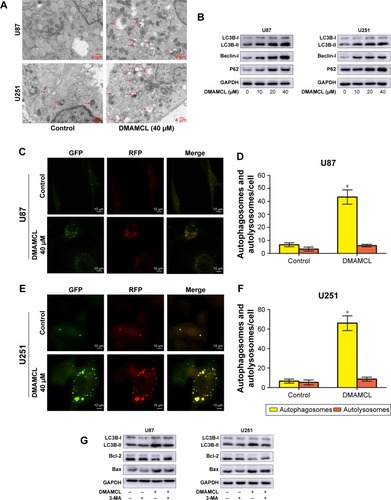
Autophagy is a double-edged sword in the process of cell death;Citation25 thus, the autophagy inhibitor 3-methyladenine (3-MA) was also used in the CCK-8 assays to elucidate the exact role of DMAMCL in the autophagic activity of glioma cells. The cell viabilities, after combined treatment with DMAMCL and 3-MA increased 18.21%±3.34% and 16.08%±1.75% than DMAMCL treatment in U87-MG and U251 cells, respectively; cotreatment with 3-MA indeed alleviated the effects on the cell viability induced by DMAMCL treatment. Also, treatment with 3-MA reduced the apoptotic effects of DMAMCL (). All the above results indicated that DMAMCL could induce autophagy-related cell death.
DMAMCL induced ROS generation
The balance of intracellular ROS is of vital importance in determining the fate of cancer cells, and this is done by regulating different important signaling pathways. Since our previous report clarified that MCL, the active ingredient of DMAMCL, exerted cytotoxic effects by generating ROS, we determined the levels of ROS in U87-MG and U251 cells after treatment with DCFH-DA probes. The results showed that DMAMCL dramatically increased ROS accumulation compared with the control group (). Subsequent analysis indicated that the levels of Nrf-2, heme oxygenase 1 (HO-1), and TXNRD expressions were significantly elevated, further suggesting the activation of oxidative stress response-related signaling pathways (). Additionally, when the cells were pretreated with N-acetyl-L-cysteine (NAC) for 2 hours, the DMAMCL-induced inhibition of cell viability was significantly alleviated. The same results were also obtained with the live/dead assay (). Overall, these experiments confirmed the results that the DMAMCL-induced cell death was ROS-dependent in U87-MG and U251 cells.
Figure 5 DMAMCL induces ROS generation.
Notes: (A) After treatment with DMAMCL (40 μM) for 6 hours, the ROS level was increased compared with the control group, and pretreatment with the antioxidant NAC can reduce the ROS level. Scale bars =50 μm. (B) The representative antioxidant response elements increased after treatment with increasing concentrations of DMAMCL (10, 20, 40 μM) for 48 hours as determined by Western blot analysis. (C) DMAMCL promotes the Nrf-ARE antioxidant pathway. Immunofluorescence analyses were performed to assess Nrf-2, HO-1, and TXNRD-1 expression levels after DMAMCL administration. The results showed that Nrf-2 showed clear nuclear translocation, and the levels of the downstream HO-1 and TXNRD-1 were also evaluated. Scale bars =20 μm. Data are expressed as the mean ± SD of three independent experiments.
Abbreviations: ARE, antioxidant response elements; DMAMCL, dimethylaminomicheliolide; HO-1, heme oxygenase 1; NAC, N-acetyl-L-cysteine; Nrf2, nuclear factor erythroid 2-related factor 2; ROS, reactive oxygen species.
DMAMCL regulated the ROS/MAPK signaling pathways and blocked the Akt/mTOR signaling pathway
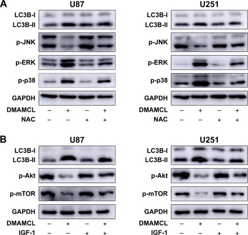
As shown in , Western blotting results showed that DMAMCL dose-dependently downregulated the phosphorylation levels of Akt and mTOR. Additionally, the MAPK pathway was also affected, that is, the phosphorylation of extracellular signal-regulated kinase (ERK) and p38 was increased, but the phosphorylation of c-Jun N-terminal kinase (JNK) was suppressed. To investigate whether the ROS/MAPK and Akt/mTOR signaling pathways were involved in DMAMCL-induced cell death, NAC (an ROS scavenger) and insulin-like growth factor-1 (IGF-1; an Akt activator) were introduced for further exploration. As shown in , after pretreatment with NAC for 2 hours and cotreatment with IGF-1 for 48 hours, the cell viability was significantly elevated. The same results could also be obtained with the live/dead assay (). Simultaneously, the level of LC3B was downregulated compared with the DMAMCL-only treatment group (). All the results shown above proved that DMAMCL-induced autophagic cell death might be associated with the ROS/MAPK and Akt/mTOR signaling pathways.
Figure 6 DMAMCL regulates the ROS/MAPK signaling pathway and inhibits the Akt/mTOR signaling pathway.
Notes: DMAMCL suppressed the Akt/mTOR pathway and regulated the ROS/MAPK pathway in human glioma cells in a dose-dependent manner. The cells were treated with various concentrations of DMAMCL (10, 20, 40 μM) for 6 and 48 hours to measure the effects on MAPK signaling pathway and Akt/mTOR signaling pathway, respectively. GAPDH served as the loading control. The blots shown are one representative of three independent experiments (P<0.05).
Abbreviations: DMAMCL, dimethylaminomicheliolide; GAPDH, glyceraldehyde 3-phosphate dehydrogenase; MAPK, mitogen-activated protein kinase; ROS, reactive oxygen species.
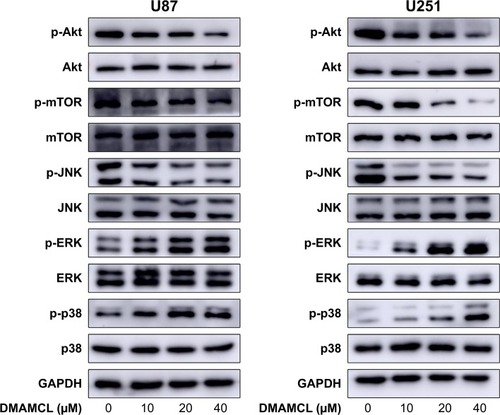
Figure 7 The relationship between the autophagy induced by DMAMCL and the ROS/MAPK and Akt/mTOR signaling pathways.
Notes: (A) Effects of NAC on DMAMCL-induced autophagy and MAPKs in U87-MG and U251 cells. The cells were treated with DMAMCL (40 μM) in the absence or presence of NAC for 6 hours; (B) Effects of IGF-1 on DMAMCL-induced autophagy and the Akt/mTOR signaling pathway in U87-MG and U251 cells. The cells were treated with DMAMCL (40 μM) in the absence or presence of IGF-1 for 48 hours. GAPDH is shown as a loading control. The results shown are one representative of three independent experiments (P<0.05).
Abbreviations: DMAMCL, dimethylaminomicheliolide; GAPDH, glyceraldehyde 3-phosphate dehydrogenase; IGF-1, insulin-like growth factor-1; MAPK, mitogen-activated protein kinase; NAC, N-acetyl-L-cysteine; ROS, reactive oxygen species.
Discussion
In the present study, we examined whether DMAMCL, as a novel anticancer chemotherapeutic reagent, could exhibit cytotoxic activities in U87-MG and U251 cells and investigated the possible mechanisms. Briefly, we studied cell growth, apoptosis, autophagy, and the possible pathways impacted by different doses of DMAMCL in two glioma cell lines. Our results clearly indicated that DMAMCL induced cytotoxicity by interrupting autophagy flux, mediating autophagosome accumulation, inducing mitochondrial dysfunction, and stimulating excessive ROS generation. Collectively, we confirmed that DMAMCL inhibited the growth of U87-MG and U251 cells by promoting apoptosis and autophagosome accumulation through the ROS/MAPK and Akt/mTOR pathways.
Many anticancer compounds can induce not only apoptosis but also autophagic cell death. Both are well-known mechanisms of cell death. In many previous studies, PTL, the natural compound of DMAMCL, induced cancer cell death through apoptosis, autophagy, or both.Citation26–Citation28 However, the possible anticancer mechanisms of DMAMCL in glioma cells have rarely been reported. Apoptosis is the main process of antitumor, drug-induced cell death. Recent studies have shown that apoptosis can be induced by multiple signaling pathways, but the mitochondria-mediated pathways are also of vital importance.Citation29 In our study, we found that the MMP was strikingly decreased following DMAMCL treatment for 48 hours, illustrating that mitochondrial membrane depolarization occurred. It is well known that mitochondrial membrane depolarization can induce cytochrome c release and activate the cytosolic caspases.Citation30 Our subsequent experiments confirmed these results by showing that the ratio of apoptotic cells was increased after DMAMCL administration, while the pro-apoptotic proteins Bax and cleaved caspase-3 were upregulated and the anti-apoptotic proteins Bcl-2 and pro-caspase-3 were downregulated.
Autophagy, on the other hand, is an evolutionarily conserved pathway involved in cellular homoeostasis that is activated during stress and includes two main processes: autophagosome formation to sequester misfolded proteins and damaged organelles and autophagosome-to-lysosome fusion and degradation.Citation31 Therefore, we next investigated, using TEM, whether autophagy was involved in DMAMCL-induced U87-MG and U251 cell death. In our study, we first detected an increased level of double membrane autophagosome formation after treatment with 40 μM DMAMCL for 48 hours. Subsequently, we found by Western blotting analyses that the expression levels of the quantitative autophagic biomarkers LC3B-II and Beclin-1 proteins were also upregulated in a dose-dependent manner. The p62/SQSTM1 protein, as an autophagic cargo protein, was degraded when the autophagosomes fused with the lysosomes to form autolysosomes. Surprisingly, p62/SQSTM1 was upregulated after DMAMCL administration, indicating that the final step of autophagy was inhibited and autophagosome degradation was reduced. According to Bernadette’s research, p62 not only acted as a cargo carrier but also stimulated the progress of autophagosome formation.Citation32 For this reason, levels of autophagosomes could be further increased. This hypothesis was further confirmed by mRFP-GFP-LC3 adenovirus transfection. Consistent with the previous results,Citation33–Citation35 late-stage autophagy flux inhibition and autophagosome accumulation led to glioma cell death. To further illustrate this point, 3-MA, the most commonly used autophagy inhibition reagent, was used. 3-MA can interfere with the PI3KC3 signaling pathway during the formation of autophagosomes. Notably, our research data showed that the combined treatment of DMAMCL and 3-MA could alleviate U87-MG and U251 cell death. Consequently, we concluded that DMAMCL indeed played a key role in inducing both apoptosis and autophagy-related cell death in U87-MG and U251 cells.
Many anticancer drugs can induce oxidative stress, which is an important mechanism during autophagy and apoptosis.Citation36 A surplus of ROS, including hydroxyl radical (•OH), superoxide , and hydrogen peroxide (H2O2), can be generated to induce the oxidative damage of various macromolecules, including proteins, lipids, and DNA, which in turn promotes cell death.Citation37 Increased ROS generation in cancer cells could therefore be a strategy for cancer therapy. However, the role of ROS in the induction of DMAMCL-treated cell death remains to be illustrated. Thorpe et alCitation38 demonstrated that ROS can upregulate autophagy genes. Nuclear factor erythroid 2-related factor 2 (Nrf2), a redox-sensitive transcription factor, has an important function under oxidative stress by translocating into the nucleus and interacting with antioxidant response elements (ARE) to induce the subsequent expression of downstream genes, such as HO-1, thioredoxin reductase (TXNRD-1), NADPH dehydrogenase, and thioredoxin. P62, a selective autophagy adaptor, could activate the Nrf-ARE antioxidant pathway by degrading Kelch-like-ECH-associated protein (keap1).Citation39 Consistent with the previous results, we found that DMAMCL administration in the U87-MG and U251 cells could induce ROS generation, as detected by the DCFH-DA probe. Furthermore, the nuclear translocation of Nrf2 and the upregulation of the downstream genes HO-1 and TXNRD-1 enhanced ROS generation in response to DMAMCL treatment. To determine whether ROS generation was related to autophagy and apoptosis, NAC, an ROS scavenger, was used before DMAMCL treatment. As the results show, NAC alleviated the DMAMCL-induced autophagy and cell death, suggesting that ROS mediated DMAMCL-induced autophagic cell death and apoptosis.
Among the many signaling pathways, the Akt-mTOR pathway is usually suppressed and autophagy is stimulated by anticancer agents.Citation40,Citation41 Targeting the Akt-mTOR signaling pathway has therefore been a novel therapeutic approach to curing gliomas.Citation42 In our study, after DMAMCL treatment for 48 hours, phosphorylated Akt and phosphorylated mTOR were markedly suppressed along with the upregulation of autophagy markers and apoptosis. To confirm the biological effects of the Akt-mTOR pathway within these processes, IGF-1, an Akt activator, was coadministered with DMAMCL in U87-MG and U251 cells. We found that IGF-1 could not only reverse the levels of phosphorylated Akt and mTOR but also inhibit the induction of autophagy by DMAMCL. These findings indicated that the suppression of the Akt-mTOR pathway was associated with DMAMCL-mediated autophagy in inducing cell death.
The MAPKs include three main groups of proteins: ERK1/2, the JNK/stress-activated protein kinases (SAPK), and the p38 kinases. MAPKs are generally involved in response to stress stimuli.Citation43 Chemopreventive compounds may inhibit cell proliferation by modulating these pathways. Different MAPK pathways are diversely regulated in different cells and compounds. For example, in gliomas, CIL-102 inhibited human astrocytoma cell growth by ERK1/2 activity.Citation44 Evodiamine inhibited cell growth by activating the JNK and p38 pathways.Citation45 On the other hand, nobiletin suppressed glioma cell proliferation by inhibiting p38 and JNK phosphorylation.Citation46 We examined the activity of the MAPK pathway after DMAMCL treatment for 6 hours and found that the phosphorylation levels of p38 and ERK1/2 were increased, but the phosphorylation of JNK was significantly decreased. When the cells were pretreated with NAC, the changes in MAPKs could be reversed. We, therefore, concluded that the ROS/MAPK pathway played a role in DMAMCL-induced growth inhibition. Further studies are required to understand the detailed mechanism.
Conclusion
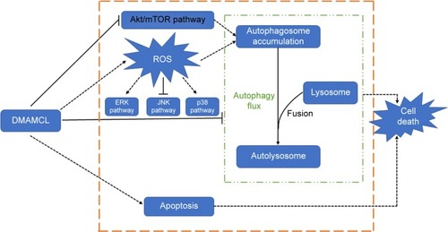
Our findings indicate that DMAMCL can induce autophagy, autophagosome accumulation, and apoptosis by regulating the Akt/mTOR and ROS/MAPK pathways, leading to inhibited proliferation of the U87-MG and U251 glioma cells (as shown in ). These novel findings provide a new perspective for DMAMCL in glioma chemotherapeutic interventions.
Figure 8 Proposed mechanism of DMAMCL-mediated cell death in human glioma cells.
Abbreviations: DMAMCL, dimethylaminomicheliolide; ROS, reactive oxygen species.
Acknowledgments
This work was supported by Natural Science Foundation of China (Grant No 81771270), Shandong Provincial Natural Science Foundation (Grant No ZR2015HM015), and Jinan Science and Technology Bureau (Grant No 201602170).
Disclosure
The authors report no conflicts of interest in this work.
References
- WangGLiuMWangHCentrosomal protein of 55 regulates glucose metabolism, proliferation and apoptosis of glioma cells via the Akt/mTOR signaling pathwayJ Cancer20167111431144027471559
- HuseJTHollandECTargeting brain cancer: advances in the molecular pathology of malignant glioma and medulloblastomaNat Rev Cancer201010531933120414201
- AlexanderBMCloughesyTFAdult glioblastomaJ Clin Oncol201735212402240928640706
- AlifierisCTrafalisDTGlioblastoma multiforme: pathogenesis and treatmentPharmacol Ther2015152638225944528
- JiangYJiaoYWangZSinomenine hydrochloride inhibits human glioblastoma cell growth through reactive oxygen species generation and autophagy-lysosome pathway activation: an in vitro and in vivo studyInt J Mol Sci2017189 pii:E1945
- WangYWangKZhaoJMultifunctional mesoporous silica-coated graphene nanosheet used for chemophotothermal synergistic targeted therapy of gliomaJ Am Chem Soc2013135124799480423495667
- ZhaoYChenSJWangJCSesquiterpene lactones inhibit advanced oxidation protein product-induced MCP-1 expression in podocytes via an IKK/NF-κB-dependent mechanismOxid Med Cell Longev20152015113
- RenYYuJKinghornADDevelopment of anticancer agents from plant-derived sesquiterpene lactonesCurr Med Chem201623232397242027160533
- ViennoisEXiaoBAyyaduraiSMicheliolide, a new sesquiterpene lactone that inhibits intestinal inflammation and colitis-associated cancerLab Invest201494995096525068660
- MendoncaMSTurchanWTAlpucheMEDMAPT inhibits NF-κB activity and increases sensitivity of prostate cancer cells to x-rays in vitro and in tumor xenografts in vivoFree Radic Biol Med201711231832628782644
- FuchsOTranscription factor NF-κB inhibitors as single therapeutic agents or in combination with classical chemotherapeutic agents for the treatment of hematologic malignanciesCurr Mol Pharmacol2010339812220594187
- ShanmugamRKusumanchiPAppaiahHA water soluble parthenolide analog suppresses in vivo tumor growth of two tobacco-associated cancers, lung and bladder cancer, by targeting NF-κB and generating reactive oxygen speciesInt J Cancer2011128102481249420669221
- CarlisiDButtittaGdi FioreRParthenolide and DMAPT exert cytotoxic effects on breast cancer stem-like cells by inducing oxidative stress, mitochondrial dysfunction and necrosisCell Death Dis20167e219427077810
- HexumJKBeckerCMKempemaAMOhlfestJRLargaespadaDAHarkiDAParthenolide prodrug LC-1 slows growth of intracranial gliomaBioorg Med Chem Lett201525122493249525978958
- ShiCWangYGuoYChenYLiuNCooperative down-regulation of ribosomal protein L10 and NF-κB signaling pathway is responsible for the anti-proliferative effects by DMAPT in pancreatic cancer cellsOncotarget2017821350093501828388532
- JinPMadiehSAugsburgerLLThe solution and solid state stability and excipient compatibility of parthenolide in feverfewAAPS PharmSciTech200784E10518181526
- ZhangQLuYDingYGuaianolide sesquiterpene lactones, a source to discover agents that selectively inhibit acute myelogenous leukemia stem and progenitor cellsJ Med Chem201255208757876922985027
- QinXJiangXJiangXMicheliolide inhibits LPS-induced inflammatory response and protects mice from LPS challengeSci Rep2016612324026984741
- JiQDingYHSunYAntineoplastic effects and mechanisms of micheliolide in acute myelogenous leukemia stem cellsOncotarget2016740650126502327542251
- AnYGuoWLiLMicheliolide derivative DMAMCL inhibits glioma cell growth in vitro and in vivoPLoS One2015102e011620225658946
- BurgessDJApoptosis: refined and lethalNat Rev Cancer20131327923344537
- WangGZhangTSunWArsenic sulfide induces apoptosis and autophagy through the activation of ROS/JNK and suppression of Akt/mTOR signaling pathways in osteosarcomaFree Radic Biol Med2017106243728188923
- DebnathJBaehreckeEHKroemerGDoes autophagy contribute to cell death?Autophagy200512667416874022
- ZouJZhangYSunJDeoxyelephantopin induces reactive oxygen species-mediated apoptosis and autophagy in human osteosarcoma cellsCell Physiol Biochem20174251812182128750364
- LinLBaehreckeEHAutophagy, cell death, and cancerMol Cell Oncol201523e98591327308466
- CarlisiDLauricellaMD’AnneoAThe synergistic effect of SAHA and parthenolide in MDA-MB231 breast cancer cellsJ Cell Physiol201523061276128925370819
- D’AnneoACarlisiDLauricellaMParthenolide generates reactive oxygen species and autophagy in MDA-MB231 cells. A soluble parthenolide analogue inhibits tumour growth and metastasis in a xenograft model of breast cancerCell Death Dis20134e89124176849
- LiuWWangXSunJYangYLiWSongJParthenolide suppresses pancreatic cell growth by autophagy-mediated apoptosisOnco Targets Ther20171045346128176967
- BoothLATavallaiSHamedHACruickshanksNDentPThe role of cell signalling in the crosstalk between autophagy and apoptosisCell Signal201426354955524308968
- FesikSWPromoting apoptosis as a strategy for cancer drug discoveryNat Rev Cancer200551187688516239906
- DikicIElazarZMechanism and medical implications of mammalian autophagyNat Rev Mol Cell Biol201819634936429618831
- CarrollBOttenEGManniDOxidation of SQSTM1/p62 mediates the link between redox state and protein homeostasisNat Commun20189125629343728
- DolmaSSelvaduraiHJLanXInhibition of dopamine receptor D4 impedes autophagic flux, proliferation, and survival of glioblastoma stem cellsCancer Cell201629685987327300435
- LiCLiuYLiuHImpact of autophagy inhibition at different stages on cytotoxic effect of autophagy inducer in glioblastoma cellsCell Physiol Biochem20153541303131625721868
- WangXQiuYYuQEnhanced glioma therapy by synergistic inhibition of autophagy and tyrosine kinase activityInt J Pharm2018536111028887220
- ChenYMcmillan-WardEKongJIsraelsSJGibsonSBOxidative stress induces autophagic cell death independent of apoptosis in transformed and cancer cellsCell Death Differ200815117118217917680
- HelfingerVSchröderKRedox control in cancer development and progressionMol Aspects Med201863889829501614
- ThorpeGWFongCSAlicNHigginsVJDawesIWCells have distinct mechanisms to maintain protection against different reactive oxygen species: oxidative-stress-response genesProc Natl Acad Sci U S A2004101176564656915087496
- LobodaADamulewiczMPyzaEJozkowiczADulakJRole of Nrf2/HO-1 system in development, oxidative stress response and diseases: an evolutionarily conserved mechanismCell Mol Life Sci201673173221324727100828
- WangJLiuXHongYIbrutinib, a Bruton’s tyrosine kinase inhibitor, exhibits antitumoral activity and induces autophagy in glioblastomaJ Exp Clin Cancer Res20173619628716053
- PoornimaPWengCFPadmaVVNeferine from Nelumbo nucifera induces autophagy through the inhibition of PI3K/Akt/mTOR pathway and ROS hyper generation in A549 cellsFood Chem201314143598360523993526
- SamiAKarsyMTargeting the PI3K/Akt/mTOR signaling pathway in glioblastoma: novel therapeutic agents and advances in understandingTumour Biol20133441991200223625692
- GenestraMOxyl radicals, redox-sensitive signalling cascades and antioxidantsCell Signal20071991807181917570640
- TengC-CKuoH-CChengH-CWangT-CSzeC-IThe inhibitory effect of CIL-102 on the growth of human astrocytoma cells is mediated by the generation of reactive oxygen species and induction of ERK1/2 MAPKToxicol Appl Pharmacol20122631738022683510
- WangXZouSLanY-LXingJ-SLanX-QZhangBSolasonine inhibits glioma growth through anti-inflammatory pathwaysAm J Transl Res2017993977398928979674
- LienL-MWangM-JChenR-JNobiletin, a polymethoxylated flavone, inhibits glioma cell growth and migration via arresting cell cycle and suppressing MAPK and Akt pathwaysPhytother Res201630221422126560814