
Abstract
Objective
The objective of the present investigation was to explore the expression and significance of Gal-3 and MUC1 in colorectal cancer tissue and tissue adjacent to carcinoma.
Methods
In this study we collected colorectal cancer tissues and the tissues adjacent to carcinoma from 45 cases from the Colorectal Cancer Surgery Department of Zhengzhou People’s Hospital from December of 2009 to June of 2010. At the same time, this study also collected nontumor tissues adjacent to carcinoma from 20 cases as the control group. The expression of Gal-3 and MUC1 of these tissues was detected by using immunohistochemistry streptavidin-peroxidase method, and the correlation between colorectal cancer and expression of Gal-3 and MUC1 was analyzed.
Results
The positive expression rates of Gal-3 in the tissues adjacent to carcinoma and colorectal cancer were 15.0% and 73.3%, respectively. The positive expression rate of Gal-3 in colorectal cancer was significantly higher than that in the tissue adjacent to carcinoma. The positive expression rate of Gal-3 of the patients without lymph node metastasis was 61.5% (16/26). The positive expression rate of Gal-3 in the patients with lymph node metastasis was 89.5% (17/19), and the difference was statistically significant (P=0.0363). The positive expression rates of MUC1 in the tissues adjacent to carcinoma and in colorectal cancer tissues were 0.0% and 54.5%, respectively. The positive expression rate of MUC1 in colorectal cancer tissues was significantly higher than that in the normal tissues adjacent to carcinoma (P<0.05); the positive expression rate of MUC1 in the patients without lymph node metastasis was 34.6% (9/26). The positive expression rate of MUC1 in the patients with lymph node metastasis was 84.2% (16/19), and the expression difference was statistically significant (P=0.0009).
Conclusion
The expression of Gal-3 and MUC1 was significantly higher than that in the nontumor tissue adjacent to carcinoma. There was a correlation between Gal-3 and MUC1 expression and lymphatic metastasis.
Introduction
Colorectal cancer is one of the commonest malignant tumors, and its incidence has been rising in recent years.Citation1–Citation2 The research of occurrence and metastasis-related factors related to colorectal cancers contributes to the understanding of the pathogenesis of the disease, and it is important to explore the occurrence, development, prognosis, and diagnosis of colorectal cancer.Citation3–Citation4
Galectin-3 (Gal-3) is a kind of galactose lectin.Citation5 Mucin-1 (MUC1) is a member of the sticky protein family.Citation6 The experiments of in vitro cell culture indicated that MUC1 was one ligand of Gal-3, and the restructured Gal-3 could increase the adhesiveness of human breast cancer and colon cancer cells on human umbilical vein endothelial cells, indicating that Gal-3 may be associated with the adhesion of cancer cells.Citation3,Citation4 The human breast cancer and colon cancer cells express MUC1.Citation7–Citation8 At present, there is no research report on the joint detection of Gal-3 and MUC1 on colon cancer cells.Citation9–Citation10 Whether expression of Gal-3 and MUC1 is involved in colorectal cancer or colon cancer remains to be investigated.
In the present study, the immunohistochemical streptavidin-peroxidase (SP) method was applied to detect the expression of Gal-3 and MUC1 protein in colorectal cancer tissues, and its relationship with lymph node metastasis was analyzed. In addition, the regulatory mechanism of Gal-3 and MUC1 in colorectal cancer tissues and colon cancer tissues during the occurrence and development of cancers was also explored in order to provide possible information of Gal-3 and MUC1 in the diagnosis and prognosis judgement of colorectal cancer and colon cancer.
Materials and methods
Case selection
In this study we collected colorectal cancer tissues and the tissues adjacent to carcinoma from 45 cases from the Colorectal Cancer Surgery Department of Zhengzhou People’s Hospital from December of 2009 to June of 2010. Approved by the Ethics Committee of Zhengzhou People’s Hospital. There were 26 male cases and 19 female cases with ages of 39–49 years. In these 45 cases, there were 21 cases of colorectal cancer and 24 cases of colon cancer, of which there were four cases of cecum cancer, eight cases of ascending colon cancer, three cases of transverse colon cancer, four cases of descending colon cancer, and five cases of sigmoid colon cancer. There were 19 cases of patients with lymph node metastasis and 26 cases of patients without lymph node metastasis. In addition, this study had collected the tissues adjacent to carcinoma in 20 patients, which served as the control group. None of the patients received radiotherapy or chemotherapy before surgery. The postoperative pathology confirmed the diagnosis of the patients with colon cancer or rectal cancer. The normal tissues adjacent to carcinoma were nontumor normal mucosa tissues 3 cm from the tumor edge.
Main reagents
Rat antihuman Gal-3 monoclonal antibody was purchased from Fuzhou MAXIM Biological Technology Development Co., Ltd., Fuzhou City, People’s Republic of China, (batch number: MAB-0572). Rat antihuman MUC1 monoclonal antibody was from Beijing Zhongshan Jinqiao Biological Technology Co., Ltd. (Beijing, People’s Republic of China; batch number: ZM-0391). UltraSensitiveTM S-P kits (A reagent was labeled with biotin sheep antimouse IgG; B reagent was streptavidin-peroxidase, from Fuzhou MAXIM Biological Technology Development Co., Ltd., batch number: KIT-9701) were used in this study.
Application of immunohistochemical SP method to detect the expression of Gal-3 and MUC1
All the tissue samples were fixed with 100 mL/L formaldehyde. After conventional paraffin was embedded, the samples were subjected to section and they were stained by immunohistochemical reagents according to the directions of the kits. To repair antigen, ethylenediaminetetraacetic acid heating antigen repair reagent was used. Ten milliliters ethylenediaminetetraacetic acid antigen repair fluid was diluted at a volume ratio of 1:50. Hydrogen peroxide was employed to block endogenous peroxidase, and goat serum was used for blocking. The MUC1 and Gal-3 ready-to-use antibodies were used as the primary antibodies. Then, UltraSensitiveTM S-P kit reagent of streptavidin peroxidase B reagent was added as well as DAB chromogenic, then hematoxylin dye, and neutral plastic sealing was conducted subsequently.
Gal-3 thyroid papillary adenoma tissue was used as the positive control group, and MUC1 breast cancer tissue served as a second positive control group. Phosphate-buffered saline-treated tissue was used as the negative control group.
Positive result determination
Gal-3 was mainly expressed in the cytoplasm, and it was also expressed in the nucleus; MUC1 was mainly expressed in the cytoplasm, and it was also expressed in the cell membrane with dye coloration of pale yellow, tan, and brown. In magnification of 400×, four observation fields were randomly chosen for each section. Then the cells were counted and the percentage of positive cells was calculated. This study took the cells with clear location and obvious staining as positive cells, and the cells with weak staining or non-staining as negative cells. The rate of positive cells, which were not more than 25% of all the cells, was designated as −, between 26% and 50% was designated as +, between 51% and 75% was designated as ++, and more than 75% was designated as +++. This experiment designated the samples with − as negative samples, and the samples with +, ++, and +++ were designated as positive samples.
Statistical methods
This study applied SPSS 13.0 statistical software for analysis. The data were analyzed by using χ2 inspection, the continuous correction χ2 inspection, and exact probability method. P<0.05 was considered as statistically significant.
Results
The expression of Gal-3 in each group
The positive expression rates of Gal-3 in the nontumor tissues adjacent to carcinoma and colorectal cancer were 15.0% (3/20) and 73.3% (33/45), respectively. The positive expression rate of Gal-3 in colorectal cancer was significantly higher than that in the nontumor tissue adjacent to carcinoma, and the expression difference was statistically significant (P<0.01), as shown in and .
Figure 1 Expression of Gal-3 in nontumor tissue adjacent to carcinoma (A) and colorectal cancer (B).
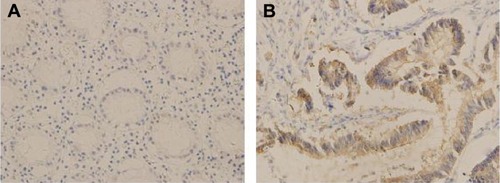
Table 1 Expression comparison of Gal-3 in colorectal cancer and nontumor tissue adjacent to carcinoma
The expression of MUC1 in each group
The positive expression rates of MUC1 in the nontumor tissues adjacent to carcinoma and colorectal cancer were 0.0% (0/20) and 55.6% (25/45), respectively. The positive expression rate of MUC1 in the colorectal cancer was significantly higher than that in the nontumor tissue adjacent to carcinoma, and the expression difference was statistically significant (P<0.005), as shown in and .
Figure 2 Expression of MUC1 in nontumor tissue adjacent to carcinoma (A) and colorectal cancer (B).
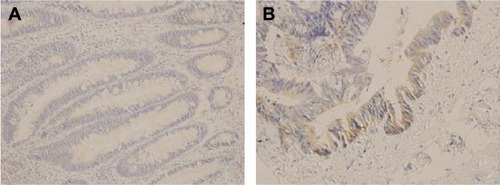
Table 2 Expression of MUC1 in colorectal cancer and nontumor tissue adjacent to carcinoma
The relationship between lymph node metastasis and Gal-3 and MUC1
The positive expression rate of Gal-3 in the patients without lymph node metastasis was 61.5% (16/26), and the positive expression rate of the patients with lymph node metastasis was 89.5% (17/19). The expression difference was statistically significant (P=0.0363). The positive expression rate of MUC1 in the group without lymph node metastasis was 34.6% (9/26), the positive expression rate of the patients with lymph node metastasis was 84.2% (16/19), and the expression difference was statistically significant (P=0.0009), as shown in .
Table 3 Relationship of Gal-3 and MUC1 expression with lymphatic metastasis in colorectal cancer
Discussion
Research has proved that Gal-3 is involved in many pathological processes, such as cell progression, new blood vessel formation, immune escape, tumor and embolism formation, and so on.Citation11–Citation12 Gal-3 is expressed in thyroid cancer, bladder cancer, laryngeal squamous carcinoma, kidney tumor, and so on.Citation12–Citation16 The mechanisms of Gal-3 in the occurrence and development of cancer have been suggested as follows: the sugar affinity of Gal-3 promoted the mitosis of vascular endothelial cells and smooth muscle, and Gal-3 participated in vessel formation during the tumor angiogenesis.Citation17 Gal-3 was the decomposition substrate of MMP-2/-9. High expression of MMP-2/-9 was associated with tumor. Therefore, Gal-3 might play a role in tumor invasion and metastasis.Citation18 Gal-3 had similar antiapoptotic function with sequence structure to that of Bcl-2; namely, the carboxyl end contained an NWGR quaternary structure, so the antiapoptotic mechanisms of Gal-3 may be related to the Bcl-2.Citation19 Gal-3 induced tumor cell cluster, which enabled tumor embolism for blocking capillaries, and benefited the coalescence of tumor cells and vascular endothelial cells, thus increasing the overflow of tumor cells and the potential metastasis of tumor cell.Citation20
MUC1 regulated the interaction between cells, mediated cell signal transduction, and participated in immune regulation and other functions. In recent years, the function of MUC1 in tumor progression and metastasis has attracted much attention. The present study found that the incomplete glycosylation of MUC1 in tumor cells could be used as hapten and induced antitumor CTL immune response; at the same time, it could also suppress the lethal effect of immune activity cells against tumor cells. The patients with high expression of MUC1 had poor prognosis, indicating that MUC1 participated in the regulation of the body’s immune system.Citation21 MUC1 had dual functions of adhesion and anti-adhesion. It could decrease the expression of E-calcium mucins,Citation22 while E-calcium mucins cell adhesion molecules required by calcium ion, mediated intercellular adhesion. Therefore, MUC1 may be involved in anti-adhesion effect. Research suggested that MUC1 formed a high density of filamentous molecules on the surface of cancer cellsas well as MUC1 sugar chain sialic acid, which enabled cells to obtain a negative charge and made cancer cells fall off from their original site, thus facilitating the metastasis of tumor cells.Citation13,Citation23 Other studies showed that MUC1 activated the expression of the antiapoptotic gene of TNF receptor-related factors, then inhibited cell apoptosis, and also induced the formation of vascular endothelial growth factor, in order to promote tumor metastasis and angiogenesis.Citation14,Citation24
The research from Ramasamy et al suggested that MUC1 was the ligand of Gal-3, Gal-3 formed a bridge between MUC1 and epidermal growth factor receptor (EGFR), and Gal-3 was indispensable in the interaction between MUC1 and EGFR, which was mediated by EGFR.Citation25 The study also showed MUC1 C -Gal-3 was one part of control loop of MiR-322, and had its function, while EGFR had a significant role in regulating tumor cell growth, repair, survival, angiogenesis, invasion, and metastasis. Therefore, Gal-3 and MUC1 worked in tumor progression.
At present, at home and abroad, there are no research reports on joint detection of Gal-3 and MUC1 in colorectal cancer. The present experiment applied the immunohistochemical SP method to detect Gal-3 and MUC1 expression in 45 cases of colorectal cancer and colon cancer. The results showed that the expression of Gal-3 in the large intestine cancer was significantly higher than that in the nontumor tissues adjacent to carcinoma, and the difference was statistically significant. Gal-3-positive expression ratio in the patients with lymph node metastasis was significantly higher than without lymph node metastasis. MUC1 expressions in the nontumor tissues adjacent to carcinoma and colorectal cancer were 0.0% (0/20) and 55.6% (25/45), respectively, and the positive expression of MUC1 with lymph node metastasis was 84.2% (16/19), which was higher than that without lymph node metastasis, accounting for 34.6% (9/26). The differences were statistically significant. The research results showed that Gal-3 and MUC1 were important in the occurrence and metastasis of colorectal cancer.
The expression of Gal-3 and MUC1 should be analyzed by other methods in addition to the immunohistochemistry SP method, such as Western blot and reverse transcription polymerase chain reaction to further confirm the findings in the present investigation. However, because it is very hard to prepare the Western blot samples of cancer tissue without inference from of other tissues and cells (when collecting samples during operation), especially blood cells, we think that the Western blot method might not be a good method to explore expression of Gal-3 and MUC1. Furthermore, there were no significant changes in the messenger RNA level of Gal-3 and MUC1 in these tissues.
Therefore, Gal-3 and MUC1 were closely related with the occurrence and metastasis of colorectal cancer. The detection of these two indexes could be applied in the diagnosis of colorectal cancer and as indicators of colorectal cancer prognosis. In addition, there was research on immune and gene therapy of Gal-3 and MUC1, which will provide a new idea to the gene and immune treatment of colorectal cancer.Citation14
Conclusion
In conclusion, the expression of Gal-3 and MUC1 was significantly higher than that in the nontumor tissue adjacent to carcinoma. There was a correlation between Gal-3 and MUC1 expression and lymphatic metastasis. The outcome and future prospects of the patients should be addressed in the future to further investigate the correlation between Gal-3 and MUC1 expression and lymphatic metastasis.
Disclosure
The authors report no conflicts of interest in this work.
References
- MaCCLiPWangLHThe value of single-incision laparoscopic surgery for colorectal cancer: a systematic literature reviewHepatogastroenterology2015621374550
- CloutierASFaronMHonoréCSecond-look surgery plus HIPEC for patients with colorectal cancer at high risk of peritoneal carcinomatosis: Should we resect the initial anastomosis? An observational studyEur J Surg Oncol Epub201543
- KumarARajendranVSethumadhavanRPurohitREvidence of colorectal cancer-associated mutation in MCAK: a computational reportCell Biochem Biophys201367383785123564489
- KumarAPurohitRCancer associated E17K mutation causes rapid conformational drift in AKT1 pleckstrin homology (PH) domainPLoS One201385e6436423741320
- RajendranVSethumadhavanRDrug resistance mechanism of PncA in Mycobacterium tuberculosisJ Biomol Struct Dyn201432220922123383724
- BhaumikPGopalakrishnanCKamarajBPurohitRSingle nucleotide polymorphisms in microRNA binding sites: implications in colorectal cancerScientific World Journal2014201454715425654126
- PurohitRRole of ELA region in auto-activation of mutant KIT receptor: a molecular dynamics simulation insightJ Biomol Struct Dyn20143271033104623782055
- MitchellEDPickwell-SmithBMacleodURisk factors for emergency presentation with lung and colorectal cancers: a systematic reviewBMJ Open201554e006965
- RajendranVPurohitRSethumadhavanRIn silico investigation of molecular mechanism of laminopathy caused by a point mutation (R482W) in lamin A/C proteinAmino Acids201243260361521989830
- KumarAPurohitRUse of long term molecular dynamics simulation in predicting cancer associated SNPsPLoS Comput Biol2014104e100331824722014
- YuLGAndrewsNZhaoQGalectin-3 interaction with Thomsen-Friedenreich disaccharide on cancer-associated MUC1 causes increased cancer cell endothelial adhesionJ Biol Chen20072821773781
- BartolazziABellottiCSciacchitanoSMethodology and technical requirements of the galectin-3 test for the preoperative characterization of thyroid nodulesApplImmunohistochem Mol Morphol201220127
- de MatosLLDel GiglioABMatsubayashiCOde Lima FarahMDel GiglioAda Silva PinhalMAExpression of CK-19, galectin-3 and HBME-1 in the differentiation of thyroid lesions: systematic review and diagnostic meta-analysisDiagn Pathol201279722888980
- SakakiMFukumoriTFukawaTClinical significance of Galectin-3 in clear cell renal cell carcinomaJ Med Invest2010571–215215720299755
- CanesinGGonzalez-PeramatoPPalouJUrrutiaMCordón-CardoCSánchez-CarbayoMGalectin-3 expression is associated with bladder Cancer progression and clinical outcomeTumour Biol201031427728520401558
- Li-huaMAXuanLIUHuaLIUExpression and significance of Galectin-3 protein in laryngeal squamous carcinomaChinese Journal of Laboratory Diagnosis201115914851487
- Nangia-MakkerPBaccariniSRazACarbohydrate-recognition and angiogenesisCancer Metastasis Rev2000191–2515711191063
- FukumoriTTakenakaYYoshiiTCD29 and CD7 mediate galectin-3-induced type II T-cell apoptosisCancer Res200363238302831114678989
- ZuckerSCaoJChenWTCritical appraisal of the use of matrix metalloproteinase inhibitors in cancer treatmentOncogene200019566642665011426650
- InoharaHAkahaniSKothsKRazAInteractions between galectin-3 and Mac-2-binding protein mediate cell-cell adhesionCancer Res19965619453045348813152
- Li-xinZHANGChun-haiLIYa-fenLUMUC1 immune biological function and its application in tumor biological therapyChin J Cancer Biother200073165170 Chinese
- WesselingJvan der ValkSWHilkensJA mechanism for inhibition of E-cadherin-mediated cell-cell adhesion by the membrane-associated mucin episialin/MUC1Mol Biol Cel199674565577
- WesselingJvan der ValkSWVosHLSonnenbergAHilkensJEpisialin (MUC1) overexpression inhibits integrin-mediated cell adhesion to extracellular matrix componentsCell Biol19951291255265
- NickelWThe mystery of nonclassical protein secretion. A current view on cargo proteins and potential export routesEur J Biochem2003270102109211912752430
- RamasamySDuraisamySBarbashovSKawanoTKharbandaSKufeDThe MUC1 and galectin-3 oncoproteins function in a microRNA-dependent regulatory loopMol Cell2007276992100417889671