
Abstract
Background
The protein p27 (p27Kip1) is a member of the cyclin-dependent kinase inhibitor family, which negatively regulates cell cycle progression, and the phosphorylation of p27 has been proven to affect its stability and nuclear export. Clinical studies on the relation between p27 and phosphorylated p27 (p-p27Ser10) in breast invasive ductal carcinomas are still scarce.
Methods
We examined the expression of p27 and p-p27Ser10 using immunohistochemistry in 107 breast invasive ductal carcinomas and analyzed the relationship of these biomarkers and tumor characteristics.
Results
Of the 107 tumor samples, 38.3% (41 of 107) overexpressed p27 and 64.5% (69 of 107) overexpressed p-p27Ser10. Analysis of correlation with clinical characteristics showed that high expression level of p-p27Ser10 was linked to poor differentiation, advanced disease stage, and lymph node metastasis, whereas a contrary trend was observed for p27 (all P<0.05). In addition, the expression of p-p27Ser10 was significantly higher in malignant tumors than in adjacent tissues, while p27 showed the opposite trend. Also, there were different levels of p27 and p-p27Ser10 in different types of breast cancer.
Conclusion
p27 and p-p27Ser10 are involved in the development of invasive ductal carcinoma and are potential indicators to judge the degree of malignancy as well as recurrence and metastasis.
Keywords:
Introduction
Mammalian cell proliferation is strictly controlled by the ordered activation of cyclin-dependent kinases (Cdks).Citation1 The Cdk inhibitor p27 is a tumor inhibitor that regulates G0-to-S phase transitions by binding and inhibiting the activity of Cdks.Citation1,Citation2 The expression of p27 is high in quiescent cells and declines as cells enter the S phase.Citation3 Multiple experiments have shown that p27 is an independent prognostic factor in a variety of human malignancies.Citation4–Citation6 In breast cancer, reduced p27 expression was associated with poor patient outcome.Citation5 During the carcinogenic process, the expression of p27 gradually decreased: normal breast duct epithelia (95%), premalignant atypical ductal hyperplasia (85%), ductal carcinoma in situ (40%), and invasive cancer (34%).Citation7 The activities of p27 are influenced by its intracellular concentration and subcellular localization.Citation8 However, the phosphorylation of p27 can affect its nuclear–cytoplasmic localization.Citation9 It has previously been confirmed that Ser10 was the principal phosphorylation site in p27, contributing to approximately 70% of the total phosphorylation of this protein.Citation10 When p27 was phosphorylated at Ser10 by kinase interacting stathmin, it could form a functional complex by combining with Jun activation domain-binding protein-1 (JAB1) and then migrate from the nucleus to the cytoplasm by recruiting chromosome region maintenance 1 (CRM1).Citation11
Currently, only a small number of studies have focused on the expression of phosphorylated p27 (p-p27Ser10) in human malignancies. It has been proven that p-p27Ser10 overexpression is strongly associated with advanced stage and tumor grade in human epithelial ovarian carcinoma.Citation12 No studies have been reported to investigate the expression of p-p27Ser10 and the correlation between p27 expression and clinical characteristics in breast cancer. We detected the expression of p27 and p-p27Ser10 in 107 breast invasive ductal carcinomas and analyzed their relationships with tumor size, pathologic classification, and lymph node metastasis, as well as the expressions of estrogen receptor α (ERα), progesterone receptor (PR), human epidermal growth factor receptor-2 (Her2), and antigen identified by monoclonal antibody Ki-67 (Ki67). Moreover, the expression of p27 and p-p27Ser10 in malignant tumors and adjacent tissues, as well as in different types of breast cancer, was explored. Our study was designed to demonstrate the clinical significance of p27 and p-p27Ser1 0 in the development of breast cancer and to find new and effective predictors and therapeutic targets.
Materials and methods
Ethical approval
The study was approved by the Ethics Committee of Jinling Hospital, Southern Medical University, People’s Republic of China. All patients provided written informed consent.
Study subjects
We studied 107 breast cancer patients who underwent radical or modified radical mastectomy in the Ganzhou Cancer Hospital, Jiangxi Province, People’s Republic of China, from January 2008 to January 2013. Inclusion criteria of breast cancer patients included a set of clinical symptoms, imaging examinations, and pathological diagnoses. We also obtained 15 paired primary breast carcinomas and corresponding adjacent noncancerous tissues among 107 cases. Immunohistochemistry for ERα, PR Her2, and Ki67 was conducted. ERα and PR were scored as a percentage of cells staining positive, using 10% as a positive cutoff point. The cutoff value for Ki67 index was set at 14% considering the optimum threshold determined by Cheang et al.Citation13 Scoring of Her2 was done on a scale of 0–3 (−, no staining; +, weak or barely perceptible membranous staining in >10% of the tumor cells; ++, moderate membranous staining in >10% of the tumor cells; +++, strong complete membranous staining in >10% of the tumor population). Furthermore, the uncertain Her2 score (++) was confirmed by fluorescence in situ hybridization (FISH). Score of +++ and positive expression by FISH of ++ cases were defined as Her2-positive expression, and the remaining scores were considered negative.Citation14 On the basis of the 2013 St Gallen Consensus, molecular subtypes of breast cancer (Luminal A, Luminal B, Her2-overexpressing, basal-like, and normal breast-like) were categorized according to the immunohistochemical profiles of ERα, PR, Ki67, and Her2.Citation15 Patient information consisted of age, menopausal status, and lymph node metastasis, and the tumor-node-metastasis (TNM) stage was obtained from clinical inquiry, medical records, and relevant inspections. The TNM classification was performed independently by two different physicians in accordance with the TNM Classification of Malignant Tumors, Seventh Edition, published by the Union for International Cancer Control. The present research was approved by the Ethics Committee of Jinling Hospital, Southern Medical University, People’s Republic of China.
Immunohistochemistry of p27 and p-p27ser10
To evaluate the expression of immune cell antigens using immunohistochemistry, 3 μm tumor sections of paraffin-embedded, formalin-fixed tissues were deparaffinized in xylene and rehydrated in a series of graded ethanol. Endogenous peroxidase activity was blocked by immersing sections in 3% H2O2 for 5 minutes. The sections were blocked in 10% fetal bovine serum for 10 minutes at room temperature and then incubated with primary antibodies to p27Kip1 (rabbit polyclonal, ab7961, 1:100; Abcam) and p-p27Ser10 [EP233(2)Y] (rabbit polyclonal, ab62364, 1:200; Abcam) for 60 minutes of staining. Immunostaining was carried out using the MaxVision™ HRP-Polymer anti-Rabbit IHC Kit (KIT-5005; Maxim) for 15 minutes according to the manufacturer’s protocol and finally visualized with diaminobenzidine. In addition, sections were then counterstained with hematoxylin. The testis tissue served as the positive control.
Evaluation of p27 and p-p27ser10 immunoreactivity
Two investigators separately evaluated the staining patterns of the tumor and control tissue samples in a blinded, randomized manner and scored the same. For each slide, five random fields were evaluated; p27 immunoreactivity was scored for the percentage of nucleus-staining tumor cells and p-p27Ser10 immunoreactivity was scored for the percentage of cytoplasm-staining tumor cells (<10%, −; >10%, +).
Statistical analyses
Statistical analyses were conducted with SPSS 19.0 version software (IBM Corporation, Armonk, NY). The Wilcoxon signed-rank test was used for comparison of expression levels of p27 and p-p27Ser10 between breast tumor tissues and the corresponding adjacent normal tissues in 15 pairs of samples. The χ2 test or the Fisher’s exact test was used to compare the expression of each index for different factors. The correlation between indicators and the molecular subtypes of breast cancer associated with Ki67 was analyzed using the χ2 test or the Fisher’s exact test. Kappa test was used to analyze the relevance between the expression of p27 and p-p27Ser10 and the correlation between the expression of p27 and p-p27Ser10 and clinicopathological characteristics in invasive ductal cancers. All statistical tests were two-sided, and the statistical significance threshold was set at P<0.05.
Results
Correlation between clinicopathological features and p27 and p-p27ser10 expression in 107 breast cancer cases
The positive rates of p27 and p-p27Ser10 were 38.3% (41 of 107) and 64.5% (69 of 107) in tumors, respectively. In breast invasive ductal carcinomas, the level of p27 varied in subgroups with different states of tissue differentiation (χ2=16.612, P<0.001), TNM stage (χ2=14.755, P<0.0001), and lymph node metastasis (χ2=23.017, P<0.0001). The p27 protein was higher in tissue differentiation G1 grade, I–II TNM stages, and lymph node-negative group. But p27 expression in various subgroups of age (χ2=0.587, P=0.444), menstrual status (χ2=1.765, P=0.184), and tumor size (χ2=5.75, P=0.058) had no difference in distribution. Expression of p27 protein was related with the level of ERα, Her2, and Ki67. The kappa values were 0.222, −0.281, and −0.300, respectively. The expression of p27 was positively correlated with the expression of ERα (P=0.020), but it was negatively correlated with the expression of Her2 (P=0.002) and Ki67 (P=0.001). No significant difference was found in the expression of p27 and PR (P>0.05) (). The level of p-p27Ser10 in breast invasive ductal cancers was differentially regulated in subgroups with various stages of tissue differentiation (χ2=7.688, P=0.021), TNM (χ2=4.408, P=0.044), and lymph node metastasis (χ2=10.591, P=0.001). The expression of p-p27Ser10 protein in tissue differentiation G2 grade, III–IV TNM stages, and lymph node-positive group was higher. This difference was statistically significant (P<0.05). The expression of p-p27Ser10 protein in the various age (χ2=1.964, P=0.164) and menstrual status (χ2=0.826, P=0.363) subgroups showed no difference in distribution. Moreover, the expressions of p-p27Ser10 protein and ERα (P=0.615), PR (P=0.594), Her2 (P=0.064), and Ki67 (P=0.067) were not related (). Meanwhile, the authors analyzed the correlation between the expression of p27 and p-p27Ser10 in invasive ductal carcinomas, and the results suggested that there was an inverse correlation between p27 and p-p27Ser10 in invasive ductal carcinomas, which was statistically significant (; ) (kappa values =−0.611, P<0.0001).
Figure 1 Representative examples of immunohistochemical analysis of p27 and p-p27ser10 in breast invasive ductal carcinomas and adjacent tissues.
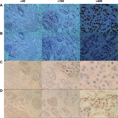
Table 1 Correlation between clinicopathogical features and p27 expression in 107 breast cancer cases
Table 2 Correlation between clinicopathological features and p-p27ser10 expression in 107 breast cancer cases
Table 3 Correlation between the expression levels of p27 and p-p27ser10 in invasive ductal carcinoma
Expression of p27 and p-p27ser10 in 15 breast invasive ductal carcinomas and adjacent tissues
We further analyzed the paired specimens of tumor tissues and adjacent tissues. In 15 tumor tissues and corresponding benign tissues, p27 overexpression was detected in 40.0% (6 of 15) of malignant tumors and 86.7% (13 of 15) of adjacent tissues (). Furthermore, p-p27Ser10 overexpression was found in 73.3% (11 of 15) of malignant tumors and 26.7% (4 of 15) of adjacent tissues (). The expression of p27 was higher in adjacent normal tissues than in tumor tissues (P=0.004), while the expression of p-p27Ser10 was reverse. The expression of p27 and p-p27Ser10 was different between malignant tumors and tumor adjacent tissue (P=0.037). Difference between these groups had statistical significance (P<0.05).
Table 4 Expression of p27in breast invasive ductal carcinomas and adjacent tissues
Table 5 Expression of p-p27ser10 in breast invasive ductal carcinomas and adjacent tissues
The expression of p27 and p-p27ser10 in different subtypes of breast cancer
Among the breast cancer samples, 31 (29.0%) cases were of Luminal A; 24 (22.4%) cases were of Luminal B; 20 (18.7%) cases showed Her2 overexpression; and 32 (29.9%) cases were of the basal-like subtype. The expression of p27 in different subtypes of breast cancers (χ2=14.653, P=0.002) was statistically significant (P<0.05) and the rate of positive p27 expression was lower in breast cancers with Her2 overexpression. The expression of p-p27Ser10 in different subtypes (χ2=6.442, P=0.090) showed no significant difference (P>0.05) ().
Table 6 Expression of p27 and p-p27ser10 in different subtypes of breast cancer
Discussion
p27 is an important component of the cell cycle machinery.Citation16 Quiescent normal epithelia of breast, prostate, ovary, and lung highly express nuclear p27.Citation3,Citation17,Citation18 An overwhelming amount of data showed the inverse correlation between expression of p27 and prognosis in a variety of human neoplasms, including those of the breast, colon, stomach, and prostate.Citation4–Citation6,Citation19 p-p27Ser10 could affect the nuclear–cytoplasmic localization of p27. A recent report provided convincing evidence for a serine 10-dependent regulatory mechanism affecting p27 tumor suppressive activity during epithelial ovarian cancer development, as well as increased p-p27Ser10 expression with enhanced malignancy.Citation12,Citation20 Point mutations in Ser10 and base mutations within the nuclear export sequence damaged p27 nuclear export and indicated that the induction of p27 expression was associated with declined cancer cell proliferation.Citation12,Citation20 In addition, the survival analyses showed that Jab1 and p-p27Ser10 expression is significantly associated with poor prognosis in hepatocellular carcinoma.Citation21
Our research illustrated by immunohistochemistry that there was a correlation between p27 and p-p27Ser10 expression and invasive ductal breast carcinomas. Comparison between malignant tumors and benign tumors showed that the expression levels of p27 and p-p27Ser10 were significantly different. Results showed that p27 and p-p27Ser10 expression is obviously correlated with the degree of differentiation, TNM staging, and lymph node metastasis number. Higher expression of p-p27Ser10 was observed in tumors that showed poor histological differentiation, high TNM staging, and incidence of lymph node metastasis. The negative correlation between the expression of nuclear p27 and cytoplasm p-p27Ser10 suggested that the degradation of nuclear p27, which p-p27Ser10 mediates, might be an important mechanism in breast cancer. The results concur with those of several previous studies in other human tumors.Citation19–Citation21
Currently, the treatment and prognosis of breast cancer are based primarily on molecular subtypes.Citation22 Effective prognostic markers that differentiate patient outcomes when analyzing an intermediate-stage disease are crucially important in making appropriate therapeutic evaluations. In human breast cancer, Her2 amplification is apparently correlated with reduced levels of p27, which is consistent with the results from previous reports.Citation23 Given the facts mentioned herein about the relation between p27 and p-p27Ser10 expression and different molecular subtypes of breast cancer, they can be used to guide the clinical treatment. For example, the correlation between p27 and the ERα and PR duo in invasive ductal carcinoma suggested that p27 was associated with endocrine therapy as a good prognostic factor. Because breast cancers often become chemotherapy resistant, p27 as a predictive factor of response to chemoprevention for malignancies requires further investigation.Citation24 So far, few large prospective randomized studies explicitly indicate that p27 predicts response to certain chemotherapeutic drugs. Further research in this area would be needed to make clear the prognostic value of p27 and p-p27Ser10 for effectiveness of treatment in different contexts.
In conclusion, our research provides evidence that p27 and p-p27Ser10 have correlation with clinicopathological characteristics in breast cancers. p27 and p-p27Ser10 are involved in the development of invasive ductal carcinoma and are potential biomarkers to judge the degree of malignancy as well as recurrence and metastasis. It is demonstrated that p27 and p-p27Ser10 potentially can be used as a clinical prognosis and therapeutic target.
Acknowledgments
This work was supported by the National Natural Science Foundation of China (grant number 81470357) and the Foundation for Clinical Medicine Science and Technology Special Project of the Jiangsu Province, People’s Republic of China (grant number BL2014071 to XG).
Disclosure
The authors report no conflicts of interest in this work.
References
- SherrCJRobertsJMCDK inhibitors: positive and negative regulators of G1-phase progressionGenes Dev199913121501151210385618
- CarranoACEytanEHershkoAPaganoMSKP2 is required for ubiquitin-mediated degradation of the CDK inhibitor p27Nat Cell Biol19991419319910559916
- HengstLReedSITranslational control of p27Kip1 accumulation during the cell cycleScience19962715257186118648596954
- ChenGChengYZhangZMartinkaMLiGPrognostic significance of cytoplasmic p27 expression in human melanomaCancer Epidemiol Biomarkers Prev201120102212222121828232
- FilipitsMRudasMHeinzlHAustrian Breast and Colorectal Cancer Study GroupLow p27 expression predicts early relapse and death in postmenopausal hormone receptor-positive breast cancer patients receiving adjuvant tamoxifen therapyClin Cancer Res200915185888589419723645
- OginoSShimaKNoshoKA cohort study of p27 localization in colon cancer, body mass index, and patient survivalCancer Epidemiol Biomarkers Prev20091861849185819505918
- HanSParkKKimHYLeeMSKimHJKimYDReduced expression of p27Kip1 protein is associated with poor clinical outcome of breast cancer patients treated with systemic chemotherapy and is linked to cell proliferation and differentiationBreast Cancer Res Treat1999552159165
- EkholmSVReedSIRegulation of G 1 cyclin-dependent kinases in the mammalian cell cycleCurr Opin Cell Biol200012667668411063931
- RodierGMontagnoliADi MarcotullioLp27 cytoplasmic localization is regulated by phosphorylation on Ser10 and is not a prerequisite for its proteolysisEMBO J200120236672668211726503
- GuanXDuLChenLChenYWangJVariation of gene expression profile linked to p27 (Kip1) Ser (10) phosphorylation status in MCF-7 cell lineBiomed Pharmacother201165853754121216562
- IshidaNHaraTKamuraTYoshidaMNakayamaKNakayamaKIPhosphorylation of p27Kip1 on serine 10 is required for its binding to CRM1 and nuclear exportJ Biol Chem200227717143551435811889117
- WangYWangYXiangJKnockdown of CRM1 inhibits the nuclear export of p27Kip1 phosphorylated at serine 10 and plays a role in the pathogenesis of epithelial ovarian cancerCancer Lett2014343161324018641
- CheangMCChiaSKVoducDKi67 index, HER2 status, and prognosis of patients with luminal B breast cancerJ Natl Cancer Inst20091011073675019436038
- JacobsTWGownAMYazijiHBarnesMJSchnittSJSpecificity of HercepTest in determining HER-2/neu status of breast cancers using the United States Food and Drug Administration–approved scoring systemJ Clin Oncol19991771983198710561248
- GoldhirschAWinerEPCoatesASPanel MembersPersonalizing the treatment of women with early breast cancer: highlights of the St Gallen International Expert Consensus on the primary therapy of early breast cancer 2013Ann Oncol20132492206222323917950
- YoonMKMitreaDMOuLKriwackiRWCell cycle regulation by the intrinsically disordered proteins p21 and p27Biochem Soc Trans201240598198822988851
- RivardNL’AllemainGBartekJPouyssegurJAbrogation of p27Kip1 by cDNA antisense suppresses quiescence (G0 state) in fibroblastsJ Biol Chem199627118337183418702474
- PaganoMTamSWTheodorasAMRole of ubiquitin-proteasome pathway in regulating abundance of the cyclin-dependent kinase inhibitor p27Science19962696826857624798
- GuoSLPengZYangXmiR-148a promoted cell proliferation by targeting p27 in gastric cancer cellsInt J Biol Sci20117556757421552422
- RaduMSopranoDRSopranoKJS10 phosphorylation of p27 mediates at RA induced growth arrest in ovarian carcinoma cell linesJ Cell Physiol2008217255856818615582
- WangYYuYNSongSJAB1 and phospho-Ser10 p27 expression profile determine human hepatocellular carcinoma prognosisJ Cancer Res Clin Oncol2014140696997824671224
- DawoodSHuRHomesMDDefining breast cancer prognosis based on molecular phenotypes: results from a large cohort studyBreast Cancer Res Treat2011126118519220711652
- YangXYangSMcKimmeyCGenistein induces enhanced growth promotion in ER-positive/erbB-2-overexpressing breast cancers by ER-erbB-2 cross talk and p27/kip1 downregulationCarcinogenesis201031469570220067990
- HolohanCVan SchaeybroeckSLongleyDBJohnstonPGCancer drug resistance: an evolving paradigmNat Rev Cancer2013131071472624060863