Abstract
Background
Endemic/Enzootic maintenance mechanisms like vertical transmission (pathogen passage from infected adults to their offspring) are central in the epidemiology of zoonotic pathogens. In Kenya, Rift Valley fever virus (RVFV) may be maintained by vertical transmission in ground-pool mosquitoes such as Aedes mcintoshi. RVFV can cause serious morbidity and mortality in humans and livestock. Past epidemics/epizootics have occurred in sub-Saharan Africa but, since the late 1970s, RVFV has also appeared in North Africa and the Middle East. Preliminary results revealed RVFV-infected eggs in Ae. mcintoshi after virus injection into the hemocoel after the first of two blood meals, justifying further study.
Methods
Mosquitoes were collected from an artificially flooded water-catching depression along a stream in Kenya, shipped live to the USA, and studied using an immunocytochemical method for RVFV-antigen localization in mosquito sections.
Results and conclusion
After virus injection into the hemocoel, RVFV-infected reproductive tissues were found, particularly follicular epithelia and oocyte/nurse cells. Ovarian infection from the hemocoel is a crucial step in establishing a vertically transmitting mosquito line. Ovarian follicles originate from germarial cells, primordia located distally in each ovariole, and infection of these cells is expected to be requisite for long-term vertical transmission. However, no germarial cell infection was found, so establishing a new line of vertically transmitting mosquitoes may require two generations. The findings support the hypothesis that Ae. mcintoshi is involved in the endemic maintenance of RVFV by vertical transmission. Detection of distinct pathology in infected eggs raises the possibility of virus-laden eggs being deposited among healthy eggs, thereby providing an exogenous source of infection via ingestion by mosquito larvae and other organisms. This has potentially significant epidemiological implications. Possible modes of entry of virus from the hemocoel into the ovaries and routes by which larvae might become infected by ingesting virus are discussed.
Introduction
The mechanisms of endemic/enzootic maintenance are central to understanding the epidemiology of zoonotic pathogens. An example among arthropod-borne disease systems is vertical transmission, the passage of a pathogen from infected adults to their eggs (transovarial transmission), through the larval instars (transstadial transmission), and, ultimately, to the next generation of adults. Among arthropods, transovarial/transstadial pathogen transmission was first discovered in ticks relative to the rickettsia associated with Rocky Mountain spotted feverCitation1 and later in mosquitoes relative to LaCrosse virus.Citation2 Vertical transmission has subsequently been found in several mosquito/arbovirus combinations.Citation3–Citation8
In this paper, evidence for Rift Valley fever virus (RVFV)-infected eggs (ova) in the African floodwater mosquito Aedes mcintoshiCitation9 after intrathoracic (IT) infection between two blood meals is reported. This finding is consistent with the vertical (transovarial) transmission of RVFV by this species.
Several mosquito-borne arboviruses are active in the USA and, given the introduction and rapid spread of West Nile virus beginning in 1999,Citation10,Citation11 it is clear that other arboviruses are likewise capable of importation into the USA, for example RVFV.Citation12,Citation13 RVFV (genus Phlebovirus, family Bunyaviridae) can cause serious human and livestock morbidity and mortality and is listed as a Category A potential bioterrorism agent.Citation14 In the past, RVFV epidemics/epizootics have occurred sporadically throughout much of sub-Saharan Africa, but, from the late 1970s, this virus has appeared in North Africa and the Middle East.Citation15–Citation20 Cases associated with a recent outbreak in South Africa continue to appear in 2011.Citation21 In sub-Saharan Africa, RVFV is thought to be maintained by vertical transmission in ground-pool Aedes species; for example, one of the hypothetical interepizootic maintenance species in Kenya is the African floodwater mosquito, Ae. mcintoshi.Citation22
The authors of this paper have been involved for many years in studies designed to elucidate the interaction between mosquitoes and arboviruses, including the use of in situ methods to locate RVFV antigen and genome segments in sections of mosquito tissues.Citation23–Citation27 In a preliminary study of a Kenyan strain of RVFV in Ae. mcintoshi, a single RVFV antigen-positive chorionated egg was found after a second blood meal in a female that had been intrahemocoelically infected after the first blood meal. It was therefore evident that in this species, the reproductive system, particularly the follicular epithelia and oocyte/nurse cells, can become infected with virus originating in the hemocoel. Ovarian infection from the hemocoel after dissemination of virus from the midgut after a viremic blood meal is logically a critical first step in the establishment of a vertically transmitting line of mosquitoes de novo in a previously nonvertically transmitting strain or species. On this basis, the sequence of events that led to infection of follicular epithelia and of an egg was duplicated in a larger sample of mosquitoes.
While the research reported here was carried out several years ago, the ideas presented have developed from discussions, especially between the first and second authors during a study of western equine encephalitis virus infection in Culex tarsalis in which the same phenomenon was observed. The study of western equine encephalitis virus/Cx. tarsalis will be reported separately, but the results are fully consistent with our findings in RVFV-infected Ae. mcintoshi.
Materials and methods
Mosquito collection
Specimens of Ae. mcintoshi were collected from an artificially flooded dambo (natural water-catching depression along a stream) at Sakari Ranch near Nairobi, Kenya, and shipped live to the US Army Medical Research Institute of Infectious Diseases (USAMRIID), Fort Detrick, Frederick, Maryland.
Mosquito infection and analysis
In a preliminary study at USAMRIID, four mosquitoes that had obtained blood from an unknown host in Kenya were inoculated with RVFV (Kenya strain 21445) by injection into the hemocoel through a soft area of cuticle in the thorax (IT infection/innoculation), held at 26°C for several days, allowed to oviposit, and then given a second blood meal from an uninfected hamster. These mosquitoes were killed and fixed for immunocytochemical examination after being held long enough for vitellogenesis to occur.
Based on preliminary results, a larger sample of adult female mosquitoes that had been collected in Kenya and held at USAMRIID were likewise IT inoculated with the 21445 strain of RVFV. After 3 days incubation, mosquitoes were allowed to ingest, digest, and assimilate a blood meal from an uninfected hamster and provided with an opportunity to oviposit. A second blood meal opportunity was then provided and the resulting 35 engorged females incubated for about 3 days. Gravid females were then killed and fixed in 5% formaldehyde for approximately 5 hours and stored in 70% ethyl alcohol. IT-inoculated mosquitoes and similarly treated uninfected virus-diluent-injected female Culex pipiens mosquitoes (100% infection rate when IT inoculated) were prepared as positive and negative controls, respectively.
Immunocytochemistry
To detect viral antigen in serial paraffin sections of mosquitoes, the avidin-biotin-peroxidase complex immunocytochemical technique for light-level microscopy was used.Citation28,Citation29 The “primary antibody” was a blend of monoclonal antibodies directed against RVFV nucleocapsid protein and two envelope glycoproteins or a monoclonal antibody directed against RVFV nucleocapsid protein, both effective in antigen detection. Dr James Meegan and Dr Jonathan Smith provided these antibodies. Appropriate reagent elimination studies had been carried out earlier.Citation29
Results
In the preliminary study, one of the four specimens had an antigen-positive egg with a distinct positive signal in all serial sections of that egg as well as an antigen-positive follicular epithelium. The results of the 35 RVFV-infected female Ae. mcintoshi study are shown in and . Representative sections from control specimens are shown in .
Figure 1 (A, B) RVFV antigen in chorionated eggs showing pathology and RVFV antigen-negative germarium (B). (C) Chorionated egg with distinct traces of RVFV antigen. (D) Uninfected chorionated egg with antigen-positive ovariolar sheath. (E) Uninfected chorionated eggs with RVFV antigen-positive ovarian sheath. (F) Sagittal section in posterior part of the abdomen showing antigen-positive oviduct epithelium and tissues in proximity to the spermathecae. (G) Antigen-positive small tracheae associated with the ovaries. (B) Section of spermathecae and adjacent antigen-positive tissues, and antigen-negative accessory gland. (I–L) Negative control sections.
Abbreviations: Ag, RVFV antigen; AG, accessory gland; Ch, chorion; CO, common oviduct; FE, follicular epithelium; Ge, germarium; OS, ovary sheath; OvS, ovariole sheath; RVFV, Rift Valley fever virus; Sp, spermatheca; TL, tracheal lumen; Tr, trachea; UE, uninfected egg; UF, uninfected follicle; Yo, yolk.
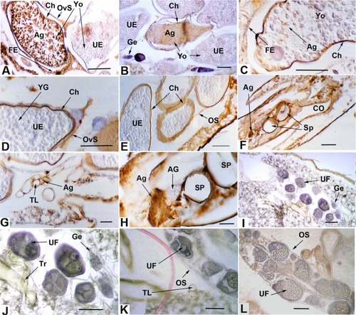
Table 1 RVFV antigen in Aedes mcintoshi female reproductive tissues
Among the ultimate follicles with chorionated eggs, RVFV antigen was seen in a single egg in three different specimens; ie, 3/35 or 8.6%. Antigen could be seen in several serial sections of each infected egg along with distinct pathology characterized by the coalescence of yolk granules having a somewhat clumped, smooth appearance (). This pathology was in contrast to the granular yolk texture observed in uninfected eggs adjacent to the infected eggs () and in the control ().
Both ovarian () and ovariole sheaths () were observed to contain antigen in many individuals, 74.3% and 51.4%, respectively. In no case was the germarium () observed to contain antigen. A single penultimate follicle was observed to be antigen positive. A much higher frequency of antigen presence was observed in the follicular epithelia of ultimate follicles (), 62.9% in the cells associated with the anterior (distal) part of an egg, 55.9% in the cells of the central region of an egg, and 84.4% in the cells associated with the posterior (proximal) region of an egg (including the adjacent pedicel and calyx). The oviducts () displayed high frequencies of antigen presence, as did cells near the spermathecae (). In no case was the accessory gland (), or spermatozoa, when present in the spermathecae, seen to contain antigen. Many instances of antigen in the trachea associated with the ovaries were observed (). In one case a chorionated egg contained distinct traces of RVFV antigen ().
Discussion
The findings of this study are consistent with the hypothesis that Ae. mcintoshi is involved in the endemic maintenance of RVFV by vertical transmission.Citation22 The occurrence of RVFV antigen-positive eggs is evidence that RVFV in Ae. mcintoshi can reach and infect eggs from the hemocoel when there is a disseminated infection (virus in the hemocoel beyond the midgut epithelium). The fact that mosquitoes were IT inoculated, given an uninfected blood meal, allowed to oviposit, and then given a second blood meal may have influenced the receptivity of the ovaries to RVFV infection.
The occurrence of strongly antigen-positive eggs with antigen in virtually every section, along with associated pathology, suggests that eggs heavily infected with RVFV can be deposited in the aquatic habitat along with uninfected viable eggs. The observation of distinct traces of antigen in an otherwise normal-appearing egg () suggests that some eggs could have a mild or even latent viral infection with no pathology and which might remain viable.
The appearance of distinct antigen in the cytoplasm of smaller tracheae suggests that virus entry into the ovaries could possibly occur via ovarian tracheae. It has been suggested that tracheae could be virus escape conduits from the midgut epithelium into the hemolymph (dissemination).Citation25,Citation26 Virus could enter the ovaries by direct infection of the ovarian and ovariolar sheaths and then the follicular epithelia. Because antigen was observed in all of these tissues, it is possible that both tracheal and sheath routes are involved in infection of the ovaries.
Regardless of mode of ingress into an ovary, infection of a developing egg with virus from the hemocoel can logically be expected to be a critical step in the establishment de novo of a line of vertically transmitting mosquitoes. A permanent vertically transmitting line of mosquitoes would ultimately require infection of the germaria, the undifferentiated primordial tissue at the apex of each ovariole from which the oocytes/nurse cells and follicular epithelia originate. Virus in germarial cells would have ample opportunity to infect oocytes and follicular epithelia as they develop. It seems safe to assume that many, if not all, follicles originating from an infected germarium would be infected. However, no evidence of antigen in germarial tissue was observed. How, then, could a permanent vertically transmitting line be established?
The finding of what appears to be a lightly infected egg suggests the possibility of other eggs having undetected or latent infections. If such eggs remained viable, a line of a vertically transmitting mosquito generations with a “stabilized” viral infectionCitation30 could be established. This is because infection would be systemic from the very beginning as opposed to the virus from a viremic blood meal being required to infect several differentiated tissues to spread. Virus that has infected an egg would have opportunity to spread during virtually all of the processes of growth and development, that is, during embryogenesis, during the larval stadia, and during metamorphosis to the adult stage. Virus present during metamorphosis would be in a position to infect any developing adult tissues, including primordial germ cells and adult salivary gland cells. Infected female germ cells would be expected to produce at least some infected germaria and hence ovarioles that would produce infected eggs. Any infected but viable eggs among all eggs deposited would be expected to undergo the same sequence of tissue-infection events during growth and development, thereby leading to infected eggs in the next generation, and so on, a generational repeat of vertical transmission, that is, a stabilized infection.Citation30
Likewise, such a systemic infection in a male egg might produce infected male gametes (spermatozoa), raising the possibility of venereal transmission, a phenomenon first described in male Aedes triseriatus, tree-hole mosquitoes infected with LaCrosse encephalitis virusCitation31 and later in two other mosquito-arbovirus combinations.Citation32,Citation33 Infection of spermatheca-associated tissues could lead to “transovum” transmission, in which case virus might eventually enter chorionated eggs via the micropyle along with spermatozoa as they enter eggs passing along the oviduct.
Though admittedly speculative, another way a permanent vertically transmitting line could be established stems from the fact that heavily infected eggs displayed signs of pathology. This indicates that likely nonviable virus-laden eggs can be deposited along with viable uninfected eggs. Once in the aquatic environment, these eggs could conceivably be a source of viral infection for mosquito larvae or other aquatic organisms. A logical route of egress of virus from an infected egg would be via the micropyle, the opening through which sperm enter and then initiate fertilization. The eventual breakdown of a heavily infected egg could also release virus. Larvae from viable eggs deposited along with one or more infected eggs and larvae from female adults of the same or different species could easily ingest released virus particles. Infected eggs (or larvae) are also likely to be ingested by other animals (vertebrate or invertebrate), offering a putative mechanism for cross-specific viral transmission.Citation34 Such transmission within the larval habitat could play a very significant role in endemic maintenance and could also enhance virus spread and amplification associated with animal and/or human disease outbreaks. Mosquito larval habitats generally teem with other organisms, including arthropods and other invertebrates, Monera, and Protists,Citation35 and it is well known that various vertebrates (fish, tadpoles, frogs, toads, birds, and bats) prey on mosquito larvae and/or adults.
The idea of infected eggs releasing virus, which infect larvae when ingested, is consistent with the demonstration that larval ingestion of particles from homogenized, RVFV-infected hamster livers in the rearing medium lead to transstadial virus transmission and produce adults that transmit virus to new hosts during blood feeding.Citation36 It is unclear how infected hamster livers and infected mosquito eggs compare in regard to the amount of virus present, but the very strong positive reaction of the egg shown in is indicative of widespread infection within the egg and suggests the possibility that the viral titer within an infected egg could be reasonably high.
To establish infection in a mosquito larva, ingested virus has to come into contact with midgut epithelial cell surface-receptor molecules associated with the plasma membranes. However, the larval foregut is lined with an impermeable, noncellular chitinous intimaCitation37,Citation38 and ingested particles in the midgut lumen are completely invested in the cylindrical noncellular peritrophic membrane (PM). The larval PM, while permeable to large molecules such as the mosquitoes’ own proteolytic enzymes,Citation39,Citation40 is not permeable to virus-sized particles. However, the work of Turell et alCitation36 demonstrates that ingested RVFV particles must in fact reach plasma membrane and infect epithelial cells. The question remains regarding how virus-sized particles circumvent the foregut intima and larval PM and initiate infection. Relative to the structure of the mosquito alimentary canal and to developmental changes that happen in the fourth instar larval stage, there are at least two possibilities. First, it seems possible that virus could reach the foregut–midgut junction within the larval proventriculus, immediately anterior to where the larval PM forms but posterior to the cells to which the foregut intima attaches and terminates. There is evidence that RVFV can infect the corresponding region in adult mosquitoes and possibly disseminate from there into the hemocoel.Citation23,Citation27 A second plausible way for ingested virus to reach the plasma membranes of larval midgut epithelial cells is during the pharate pupal stage at the time when the larval PM is destroyed or egested but before the meconial PM forms.Citation41 In this regard, it is noteworthy that Turell et al used fourth instar larvae in their experiments.Citation36
Although four individual eggs infected in four different mosquitoes may seem to be a low frequency of infection, that 4/35 individuals (11.4%) contain any RVFV antigen-positive eggs could be very significant, especially in view of the potentially large populations of floodwater mosquitoes emerging in a short period of time. Arbovirus-infected egg pathology has also been observed in Cx. tarsalis in association with an arbovirus in a different family, that is, the Togaviridae,Citation42 suggesting that our observations of infected eggs associated with apparent pathology are not just random artifacts and may well play a role in the establishment of a line of vertically transmitting mosquitoes.
Disclosure
This research was financially supported by the US Army Research and Development Command (contract number DAMD17-86-C-6133).
The views of the authors do not purport to reflect the positions of the US Department of the Army or the US Department of Defense. Research was conducted in compliance with the Animal Welfare Act and other federal statutes and regulations relating to animals and experiments involving animals and adheres to principles stated in the Guide for the Care and Use of Laboratory Animals, National Research Council, 1996. The facility where this research was conducted is fully accredited by the Association for Assessment and Accreditation of Laboratory Animal Care International.
References
- BurgdorferWVertical transmission of spotted fever group and scrub typhus rickettsiaeHarrisKFCurrent Topics in Vector ResearchSanta Barbara, CAPraeger19847792
- WattsDMPantuwatanaSYuillTMDeFoliartGRThompsonWHHansonRPTransovarial transmission of LaCrosse virus in Aedes triseriatusAnn N Y Acad Sci19752661351431072592
- LeakeCJTransovarial transmission of arboviruses by mosquitoesMayoMAHarrapKAVectors in Virus BiologyLondonAcademic Press19846391
- RosenLOvarian infection and transovarial transmission of viruses in insectsNotkinsALOldstoneMBAConcepts in Viral PathogenesisNew York, NYSpringer-Verlag1984194198
- TeshRBTransovarial transmission of arboviruses in their invertebrate vectorsHarrisKFCurrent Topics in Vector ResearchSanta Barbara, CAPraeger19845776
- TurellMJHorizontal and vertical transmission of viruses by insect and tick vectorsMonathTPThe Arboviruses: Epidemiology and EcologyBoca Raton, FLCRC Press1988128152
- WilsonMLRift Valley fever virus ecology and the epidemiology of disease emergenceAnn N Y Acad Sci19947401691807840448
- HiggsSInfluences of arthropod vectors on encephalic arbovirusesShoskesReiss CNeurotropic Viral InfectionsCambridge, UKCambridge University Press2008362381
- HuangY-MA new African species of Aedes (Diptera: Culicidae)Mosquito Systematics198517108120 Available from: http://www.mosquitocatalog.org/files/pdfs/MS17N02P108.pdfAccessed August 18, 2011
- CalisherCHWest Nile virus in the New World: appearance, persistence, and adaptation to a new econiche – an opportunity takenViral Immunol200013441141411192287
- HayesEBKomarNNasciRSMontgomerySPO’LearyDRCampbellGLEpidemiology and transmission dynamics of West Nile virus diseaseEmerg Infect Dis20051181167117316102302
- GarganTP2ndClarkGGDohmDJTurellMJBaileyCLVector potential of selected North American mosquito species for Rift Valley fever virusAm J Trop Med Hyg19883824404462895591
- TurellMJDohmDJMoresCNPotential for North American mosquitoes to transmit Rift Valley fever virusJ Am Mosq Control Assoc200824450250719181056
- Centers for Disease Control and PreventionBioterrorism Agents/DiseasesAtlanta, GACenters for Disease Control and Prevention2011 Available from: http://emergency.cdc.gov/agent/agentlist-category.aspAccessed July 20, 2011
- ArthurRRel-SharkawyMSCopeSERecurrence of Rift Valley fever in EgyptLancet19933428880114911507901480
- MeeganJMBaileyCLRift Valley feverMonathTPArboviruses: Epidemiology and EcologyBoca Raton, FLCRC Press19895176
- MeeganJMThe Rift Valley fever epizootic in Egypt 1977–1978. Description of the virological studiesTrans R Soc Trop Med Hyg1979736618623
- MeeganJMKhalilGMHoogstraalHAdhamFKExperimental transmission and field isolation studies implicating Culex pipiens as vector of Rift Valley fever in EgyptAm J Trop Med Hyg1980296140514107446827
- FagboSFThe evolving transmission pattern of Rift Valley fever in the Arabian PeninsulaAnn N Y Acad Sci200296920120412381591
- Abdo-SalemSGerbierGBonnetPDescriptive and spatial epidemiology of Rift valley fever outbreak in Yemen 2000–2001Ann N Y Acad Sci2006108124024217135517
- The Outbreak Response Unit, Special Pathogens Unit, and South African Field Epidemiology and Laboratory Training Programme (SAFELTP), National Institute for Communicable Diseases (NICD) of the National Health Laboratory Service (NHLS)Rift Valley Fever Outbreak: Interim Report on the 2011 Rift Valley Fever (RFV) Outbreak in South Africa2011 [update Mar 22]. Available from: http://www.samedical.org/assets/files/doctors-corner/outbreaks/rift-valley-fever/Rift%20Valley%20Fever%20Outbreak%2022%20March.pdfAccessed July 20, 2011
- LinthicumKJDaviesFGKairoABaileyCLRift Valley fever virus (family Bunyaviridae, genus Phlebovirus). Isolations from Diptera collected during an inter-epizootic period in KenyaJ Hyg (Lond)19859511972092862206
- RomoserWSFaranMEBaileyCLA newly recognized route of arbovirus dissemination from the mosquito midgutJ Med Entomol19872444314323305948
- RomoserWSFaranMEBaileyCLLerdthusneeKAn immunocytochemical study of the distribution of Rift Valley fever virus in the mosquito Culex pipiensAm J Trop Med Hyg19924644895011575297
- RomoserWSWasieloskiLPJrPushkoPEvidence for arbovirus dissemination conduits from the mosquito midgutJ Med Entomol200441346747515185952
- RomoserWSTurellMJLerdthusneeKPathogenesis of Rift Valley fever virus in mosquitoes – tracheal conduits and the basal lamina as an extra-cellular barrierArch Virol Suppl20051989100
- LerdthusneeKRomoserWSFaranMEDohmDJRift Valley fever virus in the cardia of Culex pipiens: an immunocytochemical and ultrastructural studyAm J Trop Med Hyg19955343313377485683
- HsuSMRaineLFangerHUse of avidin-biotin-peroxidase complex (ABC) in immunoperoxidase techniques: a comparison between ABC and unlabeled antibody (PAP) proceduresJ Histochem Cytochem19812945775806166661
- FaranMERomoserWSRoutierRGBaileyCLUse of the avidin-biotin-peroxidase complex immunocytochemical procedure for detection of Rift Valley fever virus in paraffin sections of mosquitoesAm J Trop Med Hyg1986355106110673532843
- TurellMJHardyJLReevesWCStabilized infection of California encephalitis virus in Aedes dorsalis, and its implications for viral maintenance in natureAm J Trop Med Hyg1982316125212597149111
- ThompsonWHBeatyBJVenereal transmission of La Crosse (California encephalitis) arbovirus in Aedes triseriatus mosquitoesScience19771964289530531850794
- ShroyerDAVenereal transmission of St. Louis encephalitis virus by Culex quinquefasciatus males (Diptera: Culidicae)J Med Entomol19902733343372159075
- MavaleMParasharDSudeepAVenereal transmission of chikungunya virus by Aedes aegypti mosquitoes (Diptera: Culicidae)Am J Trop Med Hyg20108361242124421118928
- SbranaETonryJHXiaoSYda RosaAPHiggsSTeshRBOral transmission of West Nile virus in a hamster modelAm J Trop Med Hyg200572332532915772330
- LairdMThe Natural History of Larval Mosquito HabitatsOxfordAcademic Press1988
- TurellMJLinthicumKJBeamanJRTransmission of Rift Valley fever virus by adult mosquitoes after ingestion of virus as larvaeAm J Trop Med Hyg19904366776802267972
- RomoserWSVenardCEThe development of the ventral oesophageal diverticulurn in Aedes triseriatus (Diptera: Culicidae)Ann Entomol Soc Am1967593484489
- WalkerMCRomoserWSThe origin and movement of gas during adult emergence in Aedes aegypti: an hypothesisJ Am Mosq Control Assoc1987334294323504927
- DetraRLRomoserWSPermeability of Aedes aegypti (L.) (Diptera: Culicidae) larval peritrophic membrane to proteolytic enzymeMosquito News1979393582585
- EdwardsMJJacobs-LorenaMPermeability and disruption of the peritrophic matrix and caecal membrane from Aedes aegypti and Anopheles gambiae mosquito larvaeJ Insect Physiol20004691313132010844150
- MoncayoACLerdthusneeKLeonRRobichRMRomoserWSMeconial peritrophic membrane structure, formation and meconial degeneration in mosquito pupae/pharate adults: histological and ultrastructural aspectsJ Med Entomol200542693994416465731
- Neira OviedoMVRomoserWSJamesCBLMahmoodFReisenWKInfection dynamics of western equine encephalomyelitis virus (Togaviridae: Alphavirus) in four strains of Culex tarsalis (Diptera: Culicidae): an immunocytochemical studyRes Rep Trop Med201120112657722629118