
Abstract
Background
An arm supported robotic drill has been recently demonstrated for preparing cochleostomies in a pilot research clinical trial. In this paper, a hand-guided robotic drill is presented and tested on human cadaver trials.
Methods
The innovative smart tactile approach can automatically detect drilling mediums and decided when to stop drilling to prevent penetrating the endosteum. The smart sensing scheme has been implemented in a concept of a hand guided robotic drill.
Results
Experiments were carried out on two adult cadaveric human bodies for verifying the drilling process and successfully finished cochleostomy on three cochlea. The advantage over a system supported by a mechanical arm includes the flexibility in adjusting the trajectory to initiate cutting without slipping. Using the same concept as a conventional drilling device, the user will also be benefit from the lower setup time and cost, and lower training overhead.
Conclusion
The hand-guided robotic drill was recently developed for testing on human cadavers. The robotic drill successfully prepared cochleostomies in all three cases.
Introduction
Over the last 30 years, robotic surgery has made its mark as a precise mean of tool deployment in surgical procedures.Citation1,Citation2 It has demonstrated consistent resultsCitation3–Citation5 for certain procedures, such as laparoscopic surgery, with reduced length of stay and blood loss.Citation6,Citation7 For many other procedures, the upfront cost, consumable costs, surgeon training overhead, and maintenance of a large system cannot be justified.Citation8 At the meanwhile, a number of hand-guided robotic systems, which are smaller and intuitive to use, have been developed, for example, assisting gripping tissues (laparoscopy), guiding hand-held instruments, and cutting applications (knee joint replacement surgery).Citation8–Citation12 Hand-held robots have the advantage of being compact and easily integrated into routine surgical practice. These devices have a physically smaller footprint, make use of much of the surgeon’s existing dexterity, and are typically lower in cost with minimal setup time and lower training overhead.Citation13 The development of such devices faces the crucial challenge of achieving successful results within a less-structured working environment such as deforming tissue, and they need the robustness to accomplish this with disturbances both induced by surgeon and patient motions. Sensing systems, protocol, and configuration need to address this challenge.
An innovative tactile method to automatically discriminate mediums and structures ahead on a cutting tool trajectory has been demonstrated successfully in surgery to produce precise cochleostomies.Citation14 The method enables preservation of fine tissue structures by simultaneous determining of the state of the process and automatically stopping the drilling if undesired drilling medium is detected. Most importantly, this is used to achieve high tissue preservation and low tissue trauma in surgery.Citation15–Citation17 This tactile tissue-guided sensing approach enables extension of an arm-supported robotic drill explored as a hand-guided unit. It relies on an innovative method for tactile sensing to determine and respond to the state of both the tissue being drilled and the tissue about to be drilled.
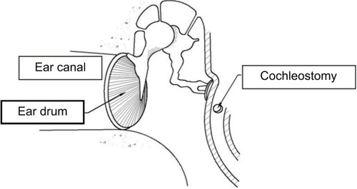
A cochlear implant is a surgically implanted device that allows rehabilitation of hearing in patients with severe-to-profound hearing loss. It represents the gold standard treatment for patients who derive limited or no benefit from conventional hearing aids. The anatomy of ear is shown in with indication of the position of cochleostomy. The cochlear implant is inserted inside cochlear through the cochleostomy hole.
Figure 1 Diagram illustrating the anatomy of the ear and location of a cochleostomy.
Residual hearing preservation has attracted increasing attention in recent years. Poor preservation of tissue could cause poor hearing preservation during the implantation process. Although it is an ongoing debate about the optimal procedure for opening cochlear through cochleostomy or round window, sometimes cochleostomies cannot be avoided if the round window is difficult to access. Among different stages in the surgical procedure of cochlear implantation, cochleostomy is considered crucial to hearing preservation. The reasons are twofold, the considerable chance of inadvertent perforation being the first. Inadvertent perforation is destructive as it exposes the cochlea to perilymph contamination – by bone dust and exotic fluid such as blood, and the risk of drill bit entering scala vestibuli and potentially damaging the basilar membrane where sensory cells are located. Second, the action of drilling on the delicate central sensing organ can cause acoustic mechanical trauma – inner ear trauma resulted from excessive acoustic stimuli or in general mechanical disturbance. Drill-induced mechanical trauma is proven to be severe in middle ear surgery especially if the ossicular chain is drilled unintentionally. Using a robotic device to perform cochleostomy could help to improve the consistency and accuracy. Several robotic devices have been developed for minimally invasive cochlear implantation.Citation18–Citation20 Such robotic devices require high-resolution computed tomography (CT) images for the operator to preplan the drilling pathCitation18,Citation19 or calibrate the robot.Citation20 During the surgery, image navigation system is used to track the movement of the robotic arm relative to the patient. Primarily, such robotic device development is focused on creating access tunnel to cochlea avoiding facial nerve during the drilling process. In contrast, the present research is focusing on the opening of cochlear. The presented robotic device is in the format of a hand-guided device for easy setup and handling. The device does not require preplanned trajectory and works similar to the conventional drill. The advantage is that it can automatically decide when to stop the drilling before entering undesired layer of the structure, ie, endosteum. The unique smart sensing algorithm uses information of the interaction between the tool and the drilling medium to discriminate the drilling stage. In this article, a human cadaver trial for a hand-guided robotic drill is presented to evaluate the setup and performance of the device in a clinical environment.
Methods
Hand-guided robotic drill
The concept of a hand-guided robotic drill has been inspired by an automated, mechanical arm-supported, robotic drill recently applied in clinical practice to produce cochleostomies.Citation17 The smart sensing algorithm uses information derived from coupled force and torque transient discriminating tissue boundaries/structures ahead on the drilling path. This valuable approach robustly detects and preserves the endosteum underlying bone tissue of the cochlear capsule to produce a membrane window of correct diameter ready for electrode insertion into the cochlea. The process achieves precise feed characteristics with micron-level accuracy to deform tissue boundaries. Earlier successful clinical trials demonstrated reduced disturbances in tissues, thus reducing trauma to the inner ear. Novel methods of measurement indicate that the technique reduces peak-to-peak amplitude of intracochlear disturbances to 1% of manual drilling.Citation17
A hand-held drill is more convenient to use than a device constrained by a mechanical support arm. From the perspective of surgeons, who are used to deploy tools by hand, it is likely to appear more intuitive to use. Previous research has proved that the flexibility in the drilling trajectory will help the control of drilling into the basal turn of the cochlea. Initial cutting without slip is achieved more readily when the drilling trajectory is perpendicular to the surface.Citation20,Citation21
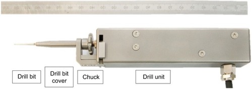
The hand-guided drilling system contains three units, such as a drill unit, a hard-wired control unit, and an output screen. shows the system containing all the three units, and shows the drill unit. The drill unit uses standard drill bit driven by a servo motor. The design of the chuck helps to change the drill bit easily and transfer the pushing force to the sensor inside the unit. The hardwired control unit contains two microcontrollers. One is to provide servo control of the drill unit, and the other is to control the information communication to the output screen through ethernet. There are also LED bars on the control unit showing the pushing force during drilling. It is important to maintain the pushing force in the range between 0.5 and 1.5 N shown as green area on the LED bars. If pushing too hard or too light, the LED bar will display red. On the output screen, a user interface is displayed to show information such as pushing force, rotation torque, and rotation speed. The system has been tested on a variety of phantoms such as raw eggs and porcine cochlear.Citation21 The feasibility results demonstrated the consistency and robustness when drilling on variety phantoms.
Figure 2 The experimental hand-guided surgical robot drill system.
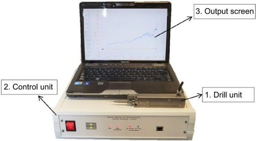
Figure 3 The hand-guided robotic drill unit.
Cadaver experiments
The cadaver trials were carried out on two adult cadaveric human bodies bequeathed for medical education and research purposes. Specimens were obtained within 120 h of death and frozen at −20°C. Experiments were carried out within 3 months of death. Otoscopy and tympanometry were carried out prior to temporal bone drilling. To achieve easy access to the promontory and the basal turn of the cochlea, a wide cortical mastoidectomy and posterior tympanotomy were performed on each side of the head of each specimen. Care was taken to retain the ear canal wall intact throughout the whole experimental procedure to make sure that middle ear transfer function can be measured at different stages. The ossicular chain and the inner ear were examined carefully, and no abnormality was found. Although the purpose of experiments was not to investigate middle ear mechanism, the tympanic membrane, ossicular chain, and all ligaments and tendons were preserved throughout the whole experimental process. This was to eliminate any effect of an incomplete sound conducting system on the cochlear dynamics.
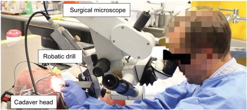
The drilling was performed by an ENT surgeon for both preparing the access to cochlea and then drilling the cochleostomy. Written informed consent was provided by the person in to have the image published. The drilling process is shown in . A total of 1 mm diameter diamond burrs were used during the trials. The robotic drill was held by the surgeon’s left hand resting on the armed chair to avoid too much movement. In theater, one would use a shoulder bolster next to the patients’ head to allow direct wrist support and minimization of tremor.Citation23,Citation24 The drilling processes were performed under a surgical microscope. The drill bit was first applied to the desired position before the drilling process started. The drill process started by pressing the start button on the control user interface. After starting, the operating surgeon guided the drill unit forward to perform the drilling. Pressure applied throughout the robotic drilling process was monitored and kept constant – the surgeon was able to correct the force applied according to a real-time signal. The drill process will automatically stop when a cochleostomy was created before penetrating the endosteum. The drilling can be stopped by the operator at any time of the drilling process for checking or cleaning if needed. The operator can continue drilling by following the same process of start drilling to finish the cochleostomy.
Figure 4 Drilling cochleostomy using hand-guided robotic drill on cadaver.
Ethic approval
This work was approved by University Research Ethics Committee of the Brunel University London with a reference number 3129-TISS-Jun/2016-3192-1.
Results
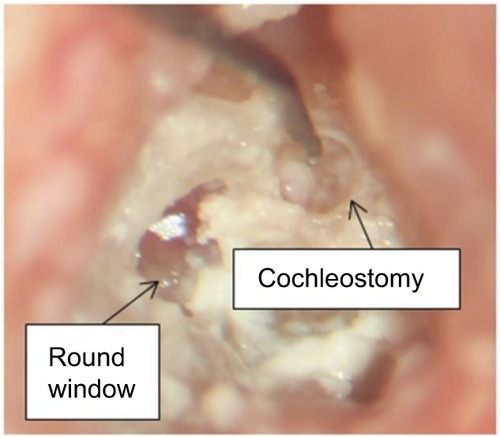
Four cases of cochleostomies were performed. The first cochlea was primarily used for the surgeon to practice the use of robotic drill on – mitigating the gap in surgeon’s experience with using conventional drilling. The other three cases of cochleostomy were successfully finished with intact underlying endosteal membrane on two cadaver heads. One finished result of a cochleostomy is shown in . The underlying membrane remained successfully intact.
Figure 5 The finished cochleostomy using both hand-guided robotic drill and conventional surgical drill with endosteal membrane.
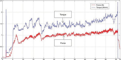
The correlated coupled force and torque transients are shown in . The force level during drilling was maintained at ~1 N over the range from 0.6 to 1.3 N. The operator begins by increasing the feed force to ensure that the drill is cutting and is stable on the surface. The result is an initial force building transient. Following this period, the fluctuating force amplitude is primarily due to unsteady motion imparted by the operator. This could be due to the unusual posture to support the drill, which is in need of improvement as simply indicated in the earlier section. At the end of the drilling process (56 s), a rapid increase in the torque and dropping of the force can be observed. These coupled characteristics together are indicative of completion of the cochleostomy. Although significant disturbances induced by operator’s hand tremor and movement are present in the signals, the automated discrimination of completion of the cochleostomy is not interrupted and the robotic drilling process successfully completes the cochleostomy as required.
Figure 6 A typical force and torque transient during the drilling.
Discussion
In this article, hand-guided robotic drilling is shown to be a beneficial process over that of conventional drilling for avoiding inadvertent penetration of the delicate endosteum. The sensing technique enables control of drilling to produce accurate and consistent results relative to tissue interfaces. The robot is in the same form as conventional surgical drills, such that it can be easily integrated into existing surgical procedures without any significant training time. The robotic device is also similar in the size and setting up time compared to conventional surgical drill. The familiar form of the device, weight, and balance, with that of a conventional drill, enables ready application by the operator. Compared to other robotic systems for cochlear implantation discussed in the studies by Caversaccio et al,Citation18 Majdani et al,Citation19 and Nguyen et al,Citation20 the presented robotic drill does not require high-resolution CT scan information or preplanned trajectory. The robotic drill is hand guided by the operating surgeon so that no further optical tracking system is needed. Instead of simply following the preplanned path, the robotic drill can automatically discriminate the drilling stage and make decision when to stop drilling before the penetration of endosteum. At the meanwhile, the system can feedback and record the drilling information, ie, applied force and the rotating torque, to the operator for monitoring purpose.
The presented system is focused on the procedure for opening cochlea during the cochlear implantation. It is an ongoing debate regarding the best route for cochlear insertion, whether directly through the round window or after the creation of a cochleostomy. However, an accurately placed cochleostomy may provide a better insertion angle compared to a round window approach, as shown in Meshik et al,Citation25 subsequently reducing the likelihood of trauma later in the insertion process. Although it does not provide the function for creating a minimally invasive tunnel to cochlea, the unique sensing technology can be integrated into a robot for such purpose, as discussed in Williamson et al.Citation26 The combination of the smart drill with the robotic system could enable a more fully integrated minimally invasive procedure spanning minimally invasive access through the facial recess and atraumatic cochleostomy.
The former mechanical arm-supported version of the robotic drill has been used in the operating room and has shown significant reduction in intracochlear disturbances induced while drilling a cochleostomy.Citation17 There is anticipated benefit in the reduction of tissue trauma as a result. Further investigation will be required to contrast the reduction in disturbances induced using the robotic hand-guided drilling solution over that of conventional devices, such that the beneficial contribution toward reducing trauma is known.
Conclusion
The robotic microdrilling method applied to surgery can discern tissue interfaces ahead on a drill path. This enables tools to cut up to delicate tissue interfaces without penetration automatically. Applied to a cochlear implantation procedure, the process can be deployed to advantageously maximize tissue preservation and minimize trauma during surgery. This article presented the trial for creating cochleostomy on two human cadaver heads using the hand-guided drill. This helps to verify the performance of the device in a clinical setup and environment. The endosteum was remained induct in three cases of drilling while one cochlea was used for training the surgeon to operate the robotic device. The hand-guided robotic drill produces consistent outcomes and augments surgeon control and skill. The advantage over an arm-supported system is that it offers flexibility in adjusting the trajectory. This can be important to initiate cutting without slipping and then to proceed on the desired trajectory.
Disclosure
The authors report no conflicts of interest in this work.
References
- DrakeJMJoyMGoldenbergAKreindlerDComputer and robotic assisted resection of brain tumoursProceedings of the Fifth International Conference on Advanced RoboticsPisaICAR1991888892
- TaylorRHPaulHAMittelstadtBDAn image based robotic system for hip replacement surgeryJ Robot Soc Jpn199085111116
- GuthartGSSalisburyJKThe intuitive telesurgery system: overview and applicationIEEE International Conference on Robotics and Automation (ICRA ‘00)San Francisco, California12000618621
- JakopecMRodriguez y BaenaFHarrisSJThe hands-on orthopaedic robot ‘acrobot’: early clinical trials of total knee replacement surgeryIEEE Trans Robot Autom2003195902911
- LonnerJHJohnTKCondittMARobotic arm-assisted UKA improves tibial component alignment: a pilot studyClin Orthop Relat Res2010468114114619593669
- HuJCGuXLipsitzSRComparative effectiveness of minimally invasive vs open radical prostatectomyJAMA2009302141557156419826025
- RamsayCPickardRRobertsonCSystematic review and economic modelling of the relative clinical benefit and cost-effectiveness of laparoscopic surgery and robotic surgery for removal of the prostate in men with localised prostate cancerHealth Technol Assess201216411313
- LaskarisJReganKSoft Tissue Robotics – The Next Generation (VII)MD Buyline2014
- JessHLGlennJKRobotically assisted unicompartmental knee arthroplastyOper Tech Orthop2012224182188
- JaramazANikouCSimoneANavioPFS for unicondylar knee replacement: early cadaver validationBone Jt J201395-BSuppl 2873
- SchullerBRigollGCanSFeussnerHEmotion sensitive speech control for human-robot interaction in minimal invasive surgeryRO-MAN 2008. The 17th IEEE International Symposium on Robot and Human Interactive CommunicationMunich, Germany4582008453
- NelsonCAZhangXShahBCGoedeMROleynikovDMultipurpose surgical robot as a laparoscope assistantSurg Endosc20102471528153220039068
- PayneCJYangGZHand-held medical robotsAnn Biomed Eng20144281594160524927713
- TaylorRPDuXProopsDWReidAPCoulsonCBrettPNA sensory-guided surgical micro-drillProc Inst Mech Eng C J Mech Eng Sci2010224715311537
- JamesCAlbeggerKBattmerRPreservation of residual hearing with cochlear implantation: how and whyActa Otolaryngol2005125548149116092537
- ZouJBretlauPPyykköIStarckJToppilaESensorineural hearing loss after vibration: an animal model for evaluating prevention and treatment of inner ear hearing lossActa Otolaryngol2001121214314811349766
- CoulsonCJZoka AssadiMTaylorRPA smart micro-drill for cochleostomy formation: a comparison of cochlear disturbances with manual drilling and a human trialCochlear Implants Int20131429810622333534
- CaversaccioMGavaghanKWimmerWRobotic cochlear implantation: surgical procedure and first clinical experienceActa Otolaryngol2017137444745428145157
- MajdaniORauTSBaronSA robot-guided minimally invasive approach for cochlear implant surgery: preliminary results of a temporal bone studyInt J Comput Assist Radiol Surg20094547548620033531
- NguyenYMiroirMVellinJFMinimally invasive computer-assisted approach for cochlear implantation: a human temporal bone studySurg Innov201118325926721502203
- DuXAssadiMZJowittFRobustness analysis of a smart surgical drill for cochleostomyInt J Med Robot20139111912623081742
- BrettPDuXZoka-AssadiMCoulsonCReidAProopsDFeasibility study of a hand guided robotic drill for cochleostomyBiomed Res Int201420147
- HildmannHSudhoffHMiddle Ear SurgerySpringer Science & Business Media2006Berlin, Germany
- CoulsonCJSlackPSMaXThe effect on wrist rest on hand tremorMicrosurgery201030756556820853330
- MeshikXHoldenTACholeRAHullarTEOptimal cochlear implant insertion vectorsOtol Neurotol20103115819707168
- WilliamsonTDuXBellBMechatronic feasibility of minimally invasive, atraumatic cochleostomyBiomed Res Int2014201418162425110661