
Abstract
Intraoral removal of a submandibular gland stone is less invasive than submandibulectomy, with no life-threatening airway complications reported until now. We report a case involving airway obstruction caused by pharyngolaryngeal swelling after intraoral removal of a submandibular gland stone. A 31-year-old man with a left submandibular gland stone underwent intraoral removal of the stone under general anesthesia and was discharged 1 day after surgery. That night, he was transported back to our hospital by ambulance for dyspnea. Because computed tomography showed pharyngolaryngeal swelling, he was intubated and managed by a ventilator. Four days later, the pharyngolaryngeal swelling had mostly resolved and he was extubated and discharged. Surgeons should be vigilant of the potential risk of airway complications following an intraoral approach for submandibular gland stone removal.
Introduction
Salivary gland stones are commonly located in the submandibular gland,Citation1,Citation2 with around 50% of such stones found in the proximal/hilar region.Citation3 Small stones in this region can now be removed with minimal invasiveness by sialendoscopy;Citation4–Citation6 large stones are removed through an intraoral incision.Citation6–Citation8 Although intraoral removal of proximal/hilar or intraglandular stones is technically difficult, intraoral removal has several advantages over submandibular sialoadenectomy, including no visible scarring, no facial nerve injury, shorter operation time, and better gland function.Citation1,Citation9 The complications reported for intraoral removal are mainly temporary lingual nerve paralysis, postoperative infection or bleeding, development of traumatic ranula, and ductal stricture. No life-threatening airway complications have been reported to date. We report here such a case, involving airway obstruction caused by pharyngolaryngeal swelling after intraoral removal of a submandibular gland stone.
Case report
A 31-year-old man with no allergies attended another otolaryngology clinic for a left submandibular swelling and was diagnosed with submandibular gland stone. Intraoral removal of the stone was attempted at under local anesthesia, but the stone migrated deeply and could not be removed. The stone remained in its deep location over 4 months of follow-up. Therefore, he was referred to our department for sialendoscopy. Upon presentation to us, he had no past medical history except for obesity (body mass index: 26). Computed tomography (CT) showed a stone (4.4×4.0×3.9 mm) located in hilar region of left submandibular gland (). We performed outpatient sialendoscopy of the palpable stone under local anesthesia, but the sialendoscope could not reach the stone due to a ductal stricture. Therefore, several weeks later, we performed intraoral removal of the stone under general anesthesia. After retraction of the tongue using a tongue retractor, a 3-cm mucosal incision was made on the lingual aspect of the retromolar region. The exposed sublingual gland was retracted laterally, and Wharton’s duct and the lingual nerve were identified. After careful dissection along Wharton’s duct to the hilum of the submandibular gland, the submandibular gland was raised with extraoral finger pressure and the medial aspect of the submandibular gland was exposed. An incision was made on the hilum after palpation of the stone, and the stone was removed. The hilum was sutured to the oral mucosa, and an orifice was made for salivary flow. Operation time was 28 minutes and bleeding was negligible. The patient was discharged 1 day after surgery without complications; he had no lingual nerve paralysis nor dyspnea. However, that night he experienced dyspnea when supine and was transported by ambulance back to our hospital. The left sub-mandibular region was moderately swollen, the oral floor was slightly swollen, and there was no bleeding from the wound. CT showed pharyngolaryngeal edema from the left submandibular gland to the epiglottis (). The fiberscopic image showed edematous swelling from the epiglottis to the left lateral arytenoid cartilage (). After discussion with anesthesiologists and otolaryngologists, we intubated and mechanically ventilated the patient and admitted him to the intensive care unit. He was given antibiotics and steroids. Although the wound was released by suture removal after intubation, there was no bleeding or hematoma. Follow-up CT and fiberoptic nasal endoscopy 4 days later showed that the pharyngolaryngeal swelling had mostly resolved, and he was extubated and discharged.
Figure 1 Preoperative CT.
Abbreviation: CT, computed tomography.
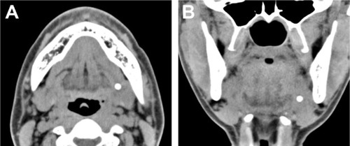
Figure 2 CT before tracheal intubation showing pharyngolaryngeal edema from the left submandibular gland to the epiglottis.
Abbreviation: CT, computed tomography.
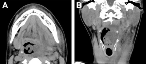
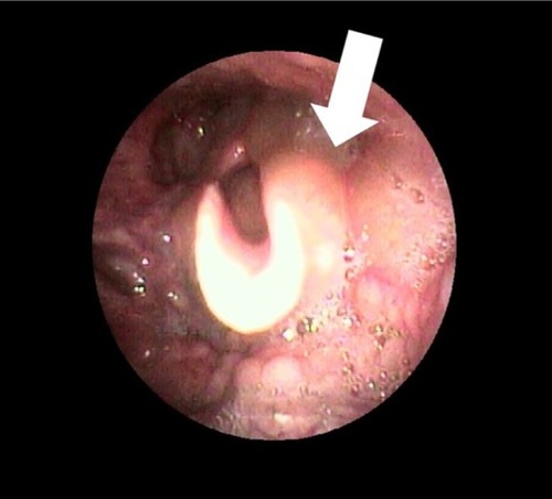
Figure 3 Fiberscopic image showing edema (arrow) from the epiglottis to the left lateral arytenoid cartilage.
Discussion
Proximal/hilar submandibular gland stones are traditionally treated with sialoadenectomy, with potential risks of facial nerve injury and visible scarring. Therefore, a minimally invasive approach has been favored in recent years, with the application of intraoral removal, endoscopic removal, or a combination of both for gland preservation.Citation1–Citation9 Although small stones in this region can be removed by sialendoscopy, intraoral removal of large proximal/hilar or intraglandular stones is challenging because the surgical field is limited.Citation2,Citation9 However, intraoral removal of deep submandibular gland stones has several advantages over submandibular sialoadenectomy, including shorter operation time and better gland function.Citation1,Citation9 To overcome the technical difficulties, we have developed a novel tongue retractor.Citation8
The potential complications of intraoral removal of sub-mandibular gland stones are lingual nerve paralysis, ranula and ductal stenosis, and edema of soft tissues. Although swelling of the oral floor after intraoral removal is observed in 75%–100% cases,Citation6 to our knowledge, there are no previous reports of life-threatening airway complications after conventional intraoral stone removal. However, airway obstruction was reported as a complication of sialendoscopy, which is a minimally invasive procedure.Citation5 In that case, the airway complication was caused by extraductal leaking of saline injected to spread the duct after the duct perforation.
Our patient was obese (with a body mass index >25) and snored during sleep. A retrospective review of preoperative CT images revealed ipsilateral tonsillar hypertrophy. Consequently, he had high risk of postoperative airway complication. Based on our experience in this case, we now routinely evaluate pharyngolaryngeal swelling on CT images 1 day after simultaneous intraoral removal of bilateral proximal/hilar submandibular gland stones. We commonly see pharyngeal edema that does not involve the larynx, where the soft tissue swelling is likely caused by release of the suprahyoid muscles from the medial aspect of the submandibular gland. Postoperative pharyngeal pain during swallowing may be due to this pharyngeal swelling.
Conclusion
Surgeons should be vigilant of the potential risk of airway complications following an intraoral approach for submandibular gland stone removal. We recommend close observation of patients with tonsillar hypertrophy, obstructive sleep apnea, obesity, or bilateral submandibular gland stones as these may be risk factors for airway complications.
Ethics statement
Written informed consent was obtained from the patient, and the review and approval of the institutional review board was not required. The patient agreed for his photos, images, and information to be applied to this research and allowed them to be published in this journal.
Disclosure
The authors report no conflicts of interest in this work.
References
- CombesJKaravidasKMcGurkMIntraoral removal of proximal submandibular stones – an alternative to sialadenectomy?Int J Oral Maxillofac Surg200938881381619398191
- RohJLParkCITransoral removal of submandibular hilar stone and sialodochoplastyOtolaryngol Head Neck Surg2008139223523918656721
- SigismundPEZenkJKochMSchapherMRudesMIroHNearly 3,000 salivary stones: some clinical and epidemiologic aspectsLaryngoscope201512581879188225994240
- SuCHTsengHLeeKSTsengTMHungSHExperiences in the treatment of obstructive sialoadenitis with sialendoscopyB-ENT201612319920629727124
- BaptistaPGimenoCVSalvinelliFRinaldiVCasaleMAcute upper airway obstruction caused by massive oedema of the tongue: unusual complication of sialoendoscopyJ Laryngol Otol2009123121402140319470191
- CapaccioPClementeIAMcGurkMBossiAPignataroLTransoral removal of hiloparenchymal submandibular calculi: a long-term clinical experienceEur Arch Otorhinolaryngol201126871081108621298387
- McGurkMSurgical release of a stone from the hilum of the submandibular gland: a technique noteInt J Oral Maxillofac Surg200534220821015695053
- OguriSIwaiTUedaJTohnaiIUse of a novel tongue retractor for intraoral removal of proximal/hilar submandibular gland stoneJ Oral Maxillofac Surg Med Pathol2017294306308
- EunYGChungDHKwonKHAdvantages of intraoral removal over submandibular gland resection for proximal submandibular stones: a prospective randomized studyLaryngoscope2010120112189219220938953