
Abstract
Descemet’s membrane detachment is an important reason for corneal endothelial decompensation after intraocular surgery. During cataract surgery, it is an unusual complication. We report a case of Descemet’s membrane detachment in which approximately 60% of Descemet’s membrane (DM) involving approximately the upper two-thirds of the cornea was torn out during a routine phacoemulsification. It caused diffuse corneal edema and blurred vision in the 2 months following the surgery. Topical prednisolone acetate (1%) and sodium chloride (5%) had been used for treatment, with slow improvement in the patient’s symptoms and vision. Interestingly, the cornea regained clarity 2 months after surgery without further surgical treatment.
Introduction
Descemet’s membrane detachment (DMD) is often attributable to cataract surgery.Citation1–Citation3 Various types of DMD can cause corneal endothelial dysfunction. In 1928, DMD was first described by Samuels;Citation4 he reported three patients with DMD after iridectomy. Different from the findings of Samuels,Citation4 ScheieCitation5 realized the serious surgical complication in his report on three patients who developed DMD after cataract extraction. Since the publication of these initial reports, DMD has also appeared after a wide variety of intraocular surgeries, including iridectomy,Citation6 penetrating keratoplasty,Citation7 lamellar keratoplasty,Citation8 pars plana vitrectomy,Citation9 and viscocanalostomy.Citation10 Few cases of Descemet’s membrane (DM) tears were reported, and the cornea regained clarity 2 months after an uncomplicated phacoemulsification without surgical intervention.
To our knowledge, this is the first report of DMD and tears involving approximately the upper two-thirds of the cornea without surgical intervention and the cornea spontaneously regaining clarity.
Case report
All procedures conformed to the Declaration of Helsinki, and written informed consent was acquired from the participant. The study was approved by the Ethics Committee of Affiliated Hospital of Nantong University. An 83-year-old Chinese man presented with complaints of diminished vision in both eyes, with a best-corrected visual acuity of 20/125 in his right eye and 20/160 in his left eye. Slit-lamp exam (Topcon SL-2D, Japan) of both eyes revealed that he had nuclear type cataract in both eyes (right eye: nuclear opalescence [NO]4, nuclear color [NC]4 and left eye [NO5, NC5]) according to the Lens Opacities Classification System III.Citation11 Intraocular pressures were 14 mmHg in both eyes by noncontact tonometer (Canon TX-20, Japan). Cornea was clear and the anterior chamber depth in the left eye was found to be 2.20 mm. Specular endothelial microscopy and cell count showed no significant difference between the two eyes (2,527 cells/mm2 oculus dexter, 2,420 cells/mm2 oculus sinister; Topcon-SP3000).
After topical anesthesia, a 3.0 mm corneoscleral tunnel incision and one side port were made at 11 and 2 o’clock in his left eye. A peristaltic pump-based (LAUREATE®, Alcon, INFINITI®, USA) phacoemulsification was done. DuoVisc® (Alcon) was used during the procedure. Low ultrasound energy (5%) was used with an effective phacoemulsification time of 5 seconds. While using the irrigation/aspiration device to remove the lens cortex, we found that the DM was torn, involving approximately the upper two-thirds of the cornea in his left eye. Then a foldable posterior chamber intraocular lens (AcrySof® 1-piece, Alcon, USA) was implanted.
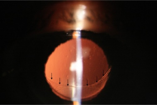
On day 1 after the surgery, the patient came with vision of counting fingers at approximately 10 cm with diffuse corneal edema in the left eye. The intraocular pressure was 18 mmHg. He was started on 1% prednisolone acetate and 5% sodium chloride three and six times a day, respectively, for 7 days. Seven days after the surgery, his vision was 20/200 and the remainder of the DM was completely reattached. On the subsequent follow-up, transparency of the cornea was gradually restored. The vision improved to 20/40 2 months after the surgery and the cornea regained clarity without corneal opacification and scar (). One year after the surgery, the best-corrected visual acuity in the left eye was 20/50, and the cornea remained clear ().
Figure 1 Slit-lamp color photographs at presentation 2 months after phacoemulsification.
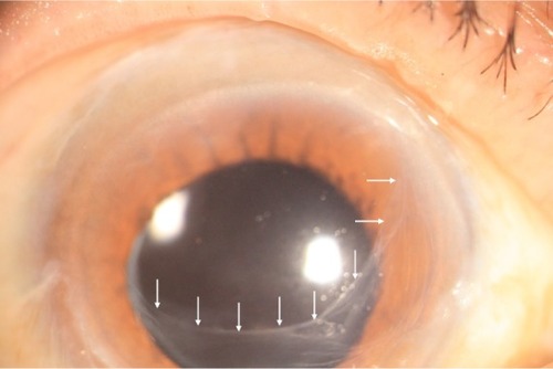
Figure 2 Slit-lamp color photographs at presentation 1 year after phacoemulsification.
Discussion
A study reported the incidence of DMD to be 2.6% for extracapsular cataract extraction and 0.5% for phacoemulsification.Citation12 DMD has also been reported in Descemet membrane endothelial keratoplasty and deep anterior lamellar keratoplasty.Citation13,Citation14 Several mechanisms have been implicated in the etiology of DMD, such as a shallow anterior chamber,Citation1 use of blunt microkeratomes,Citation15 shelved and/or anteriorly placed incisions,Citation16,Citation17 engagement of the DM during intraocular lens implantation,Citation18 the inadvertent injection of saline into the space between the deep stroma and the DM.Citation12
A study explored the possibility of an underlying anatomic predisposition for the development of DMD, possibly explained by an abnormality in the fibrillary stromal adhesion to DM.Citation2 Some patients may have an abnormal attachment between the stroma and DM caused by dysfunction of the anchoring protein βig-h3.Citation8,Citation19 We reported the case of DMD and tears during irrigation/aspiration. Many reasons have been attributed to the occurrence of detachments after cataract surgery. However, the scarce literature discusses reasons responsible for DMD when they were torn. Why had this patient developed DM tears during cataract surgery? A logical explanation for this unusual presentation is that the surgeon was inexperienced and using blunt keratomes. During surgery, a tight main incision can not fit the phaco probe. This is the another reason for DMD. The surgeon did not find the DMD before removing the lens cortex until the membrane was torn and removed by irrigation/aspiration device. Interestingly, endothelial dysfunction and bullous keratopathy did not occur in this case. The remainder of corneal endothelium staying viable is the reason the cornea regained clarity. This factor has been attributed to corneal endothelial cell hypertrophy, migration, redistribution, and/or repopulation.
Some similar cases have been reported, but this case report is different. This case of DMD involved approximately two-thirds of the cornea, and recovery without surgical intervention is very rare. There are few manuscripts reporting the detachment and tearing of DM during routine phacoemulsification.
Conclusion
It is vital to be aware of the potential causes of DMD during cataract surgery, especially when the operation is being performed by an inexperienced surgeon. Before initiating a complex surgical procedure such as a keratoplasty for this kind of case, which requires good postoperative care and regular follow-up, proper medication for DM tears is definitely a worthwhile attempt.
Disclosure
The authors report no conflicts of interest in this work.
References
- StewartCMLiFMcAlisterJCLate-onset persistent Descemet’s membrane detachment following uncomplicated clear corneal incision cataract surgeryClin Experiment Ophthalmol201139217117421401844
- KansalSSugarJConsecutive Descemet membrane detachment after successive phacoemulsificationCornea200120667067111473175
- DatarSKelkarAJainAKRepeat Descemetopexy after Descemet’s membrane detachment following phacoemulsificationCase Rep Ophthalmol20145220320625126074
- SamuelsBDetachment of Descemet’s membraneTrans Am Ophthalmol Soc19282642743716692811
- ScheieHGStripping of Descemet’s membrane in cataract extractionTrans Am Ophthalmol Soc19646214015214269887
- MackoolRJHoltzSJDescemet membrane detachmentArch Ophthalmol1977953459463843278
- WiggintonSAJungschafferDALeeDAPostoperative Descemet membrane detachment with maintenance of corneal clarity after trabeculectomyJ Glaucoma20009220020210782634
- HiranoKKojimaTNakamuraMHottaYTriple anterior chamber after full-thickness lamellar keratoplasty for lattice corneal dystrophyCornea200120553053311413412
- JonesMRRepair of Descemet’s membrane detachment with perfluoropropane (C3F8)Cornea19981744579676924
- UnluKAksungerADescemet membrane detachment after viscocanalostomyAm J Ophthalmol2000130683383411124308
- ChylackLTJrWolfeJKSingerDMThe lens opacities classification system III. The Longitudinal Study of Cataract Study GroupArch Ophthalmol199311168318368512486
- MulhernMBarryPCondonPA case of Descemet’s membrane detachment during phacoemulsification surgeryBr J Ophthalmol19968021851868814754
- HeindlLMRissSBachmannBOLaaserKKruseFECursiefenCSplit cornea transplantation for 2 recipients: a new strategy to reduce corneal tissue cost and shortageOphthalmology2011118229430120723996
- VenkatramanASpontaneous resolution of double anterior chamber with perforation of Descemet’s membrane in deep anterior lamellar keratoplastyOman J Ophthalmol20125211211422993468
- MackoolRJHoltzJSDull knives and Descemet’s membrane detachmentsArch Ophthalmol1978963542
- ZusmanNBWaringGO3rdNajarianLVWilsonLASulfur hexafluoride gas in the repair of intractable Descemet’s membrane detachmentAm J Ophthalmol198710466606623688108
- JohnMENoblittRLBoleynKLRaananMGDeLucaMEffect of a superficial and a deep scleral pocket incision on the incidence of hyphemaJ Cataract Refract Surg19921854954991403755
- MakleyTAJrKeatesRHDetachment of Descemet’s membrane with insertion of an intraocular lensOphthalmic Surg19801184924946999409
- StreetenBWQiYKlintworthGKEagleRCJrStraussJABennettKImmunolocalization of beta ig-h3 protein in 5q31-linked corneal dystrophies and normal corneasArch Ophthalmol1999117167759930162