
Abstract
Dysplasia epiphysealis hemimelica, also termed Trevor disease, is a rare disorder that, although benign in nature, can be locally aggressive, particularly when affecting the ankle joint, which is the joint most frequently affected, followed by the knee. The female:male ratio is 1:3, and it is generally diagnosed between 2 and 14 years of age. Surgical treatment with complete resection is recommended before irreversible joint damage and deformity occurs. We presented a case in which dysplasia epiphysealis hemimelica was diagnosed on the medial aspect of a right ankle joint.
Introduction
Dysplasia epiphysealis hemimelica (DEH), also known as Trevor disease, is a rare disorder with unknown etiology. Its estimated incidence is 1:1,000,000.Citation1–Citation4 The ankle joint is most frequently affected, followed by the knee. The female:male ratio is 1:3, and DEH is generally diagnosed between 2 and 14 years of age.Citation1–Citation4
DEH was first described by Mouchet and BelotCitation5 as “tarsomegalie” in 1926. In 1950, TrevorCitation6 reported a case series, including ten patients and proposed the term “tarso-epiphysial aclasis”. In 1956, FairbankCitation1 reported a case series of 14 patients and used the term “dysplasia epiphysealis hemimelica”. DEH is considered a benign, asymmetrical, intra-articular mass lesion confined by epiphysis, resulting from an abnormal proliferation of cartilage tissue.Citation3,Citation4 We presented a case in which DEH was diagnosed at the medial aspect of the right ankle joint.
Case report
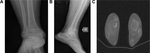
A 9-year-old girl was presented with swelling of the right ankle and pain during walking, for >3 months. The patient had no history of trauma, and there was pain during palpation at the medial aspect of the ankle. An osteocartilaginous mass (2.5 cm ×2.0 cm ×2.0 cm in size), localized at the distal end of the tibia and extending from the vicinity of the medial malleolus to the medial aspect of the talus, was observed using plain radiograph and computerized tomography (CT) ().
Figure 1 Ankle images of the patient.
Abbreviations: AP, anterior–posterior; CT, computerized tomography.
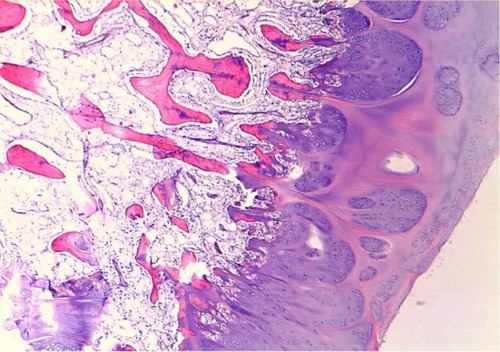
During surgical intervention, a medial incision at the medial aspect of the medial malleolus was made, and subcutaneous tissues were explored. The lesion, of osteocartilaginous appearance and originating from the distal tibial epiphysis at medial malleolus level and extending to the medial aspect of the talus, was exposed, which destructed the medial cortex of the talus. Four large osteocartilaginous lesions were removed from the talus and ankle joint (). Histological examination revealed that the lesion was mostly hyperplastic cartilage with little osseous tissue, and these features were identical to that of osteochondroma (). However, the patient was diagnosed with DEH when the radiological and gross appearance of the lesion was compared with similar reports in the literature.Citation1–Citation9
Figure 2 Intraoperative view of patient.
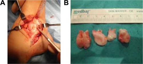
Figure 3 Histopathological findings in Trevor disease.
During the 14-month follow-up, the patient returned to normal activities. Range of motion at the ankle was normal without pain or disability. This study was conducted in accordance with the ethical guidelines of the Declaration of Helsinki. The patient provided written informed consent before participation. The study protocol was approved by the Kayseri Training and Research Hospital, Kayseri, Turkey.
Discussion
DEH is a rare, osseous developmental disorder manifesting with epiphyseal involvement.Citation3,Citation9 The localized form of the disease generally involves bones in the ankle and hindfoot. DEH is benign and its prognosis is favorable; no malignant transformation has been reported.Citation4,Citation7–Citation9 The etiology of DEH is uncertain. Potential causes include a congenital error affecting the limb bud during early fetal life or the presence of abnormal chondrocytes that continue to proliferate in an unregulated manner.Citation9
In Trevor disease, histopathological diagnosis is not pathognomonic.Citation3 However, EXT1 and EXT2 gene expressions can be studied by additional molecular assays.Citation9 Gene expressions are within normal ranges in DEH, whereas they are lower in osteochondroma owing to a mutation. These tests are costly; hence, clinical and radiological findings are important diagnostic tools.Citation7 CT and magnetic resonance imaging can confirm the diagnosis, aid surgical planning, and define the relationship of the mass to local structures.Citation3,Citation4,Citation9,Citation10 Specifically, CT can help define the anatomical relationship between the mass and the host bone, and magnetic resonance imaging can show the extent of epiphyseal involvement, joint deformity, and the status of the articular surface.Citation1,Citation3,Citation4,Citation9 Radiographs usually demonstrate a partially ossified, lobulated, cartilaginous mass arising unilaterally from the affected epiphysis with or without an osseous connection.Citation3,Citation4,Citation9,Citation10
DEH was classified by Azouz et alCitation11 as having three distinct presentations: localized, classic, and generalized. In Trevor disease, histopathological findings are similar to those in benign osteochondroma,Citation3,Citation4,Citation9,Citation10 and it is also difficult to discriminate DEH from benign osteochondroma clinically and pathologically.Citation4,Citation10 Both diseases are specific to the developmental period. Thus, radiological findings are important in differential diagnosis. Irregular bone growth originating from epiphysis and a distinct ossification focus that can be seen in radiological evaluation are the most important discriminative features of Trevor disease.Citation2–Citation4,Citation9,Citation10 In addition to osteochondroma, intra-articular loose body, myositis ossificans, or synovial chondromatosis can be considered in differential diagnosis.Citation3,Citation4,Citation9
Management options for Trevor disease include observation and surgical excision. Asymptomatic lesions may be treated nonoperatively,Citation3,Citation4,Citation7,Citation8 but surgery should be considered when the lesion causes pain, loss of function, limitation of movement, and deformity.Citation3,Citation4,Citation7,Citation8
Conclusion
In conclusion, DEH is an uncommon, benign disease. Although histopathological findings in Trevor disease are similar to those in osteochondroma, it has differential features that can be distinguished clinically and radiologically. However, molecular analysis has to be done for the definitive diagnosis. Early diagnosis and treatment are necessary to prevent articular dysfunction.
Disclosure
The authors report no conflicts of interest in this work.
References
- FairbankTJDysplasia epiphysialis hemimelica (tarso-ephiphysial aclasis)J Bone Joint Surg Br195638123725713295331
- YurdogluCSahlanSOkluTHemimelic epiphyseal dysplasia (a case report)Acta Orthop Traumatol Turc2004265348349
- KuoRSBellemoreMCMonsellFPFrawleyKKozlowskiKDysplasia epiphysealis hemimelica: clinical features and managementJ Pediatr Orthop19981845435489661870
- NowickiPDBordersHDysplasia epiphysealis hemimelica (Trevor disease) of the ankleOrthopedics2015384269271
- MouchetABelotJTarsomegalieJ Radiol Electrol192610289293
- TrevorDTarso-epiphysial aclasis: a congenital error of epiphysial developmentJ Bone Joint Surg Br195032220421315422019
- GokkusKAydinATUyanACengizMDysplasia epiphysealis hemimelica of the ankle joint: a case reportJ Orthop Surg (Hong Kong)201119225425621857058
- OuyangZXuMLiXPengDDysplasia epiphysealis hemimelica with involvement of the distal tibial epiphysis and talus: recurrence of a case and literature reviewJ Foot Ankle Surg201453219920224412001
- TylerPARajeswaranGSaifuddinAImaging of dysplasia epiphysealis hemimelica (Trevor’s disease)Clin Radiol201368441542123040212
- StruijsPAKerkhoffsGMBesselaarPPTreatment of dysplasia epiphysealis hemimelica: a systematic review of published reports and a report of seven patientsJ Foot Ankle Surg201251562062622819617
- AzouzRMSlomicAMMartonDRigaultPFinidoriGThe variable manifestations of dysplasia epiphysealis hemimelicaPediatr Radiol198515144493969295