
Abstract
Objectives
Behçet’s disease (BD) is a chronic, recurring vasculitis of unknown etiology. Patients with BD may use a lot of medications associated with the clinical symptoms. Drugs that are used in the treatment of BD may cause bone loss. The aims of the current study were to compare the bone mineral density (BMD) values between BD and healthy volunteers and describe the effect of disease duration on mandibular BMD.
Materials and methods
The study comprised 30 healthy volunteers (15 males and 15 females, mean age 35.50±6.80 years) and 45 patients with BD (24 males and 21 females, mean age 38.93±8.93 years). The BD group was subdivided according to disease duration (0–5, 6–10, and >10 years). The BMD value of the mandibular body was determined by the dual energy X-ray absorptiometry technique.
Results
The mean mandibular body BMD values were 1.294±0.21 g/cm2 in the control group and 1.216±0.22 g/cm2 in the BD patients, although there was no statistically significant difference. The BMD was observed to decrease with increased disease duration but not to a statistically significant degree.
Conclusion
The results of this study showed that although the BMD value decreased as the duration of the disease increased, no statistically significant difference was found between the BD patients and the healthy control group.
Introduction
Behçet’s disease (BD) is an idiopathic, chronic, relapsing, multisystemic, vasculitic inflammatory disorder presenting with triple-symptom complex of recurrent oral aphthous ulcers, genital ulcers, and uveitis. Gastrointestinal, pulmonary, musculoskeletal, cardiovascular, and neurological systems can be involved.Citation1,Citation2 BD is generally seen in East Asia and the Mediterranean rather than in Western countries. The disease affects both sexes equally and the age of onset is in the third or fourth decade of life.Citation3
The etiopathogenesis of BD remains unclear because of its rarity and corresponding lack of clinical evidence, but the most credited hypothesis suggests a complex interaction among immune mechanisms, genetic factors (especially, in Turks and Japanese patients, HLA-B51 gene may be responsible for genetic susceptibility), and environmental factors, such as microbial agents (Saccharomyces cerevisiae, Mycobacteria, Borrelia burgdorferi, Helicobacter pylori, Escherichia coli, Staphylococcus aureus, Mycoplasma fermentans, Streptococcus sanguinis, herpes simplex virus).Citation4–Citation6 Chronicity, inflammation, and the drugs used for treatment may lead to bone loss in BD.
Chronic inflammation may cause the development of osteoporosis (OP), which is a significant skeletal disorder characterized by low bone mass. Reduced bone mineral density (BMD) and OP are major risk factors for bone fractures and are frequently observed in several rheumatological disorders such as BD, rheumatoid arthritis (RA), ankylosing spondylitis, familial Mediterranean fever (FMF), and systemic lupus erythematosus (SLE).Citation7–Citation9 Besides the chronic inflammation, the drugs used for treatment of the various symptoms of BD such as anticonvulsants, corticosteroids, disease-modifying antirheumatic drugs, hormone replacement therapy, bisphosphonates, vitamin D, xuoride, calcitonin, and diuretics may affect bone mineral status.Citation10,Citation11 Some studies have suggested that long-term drug use may reduce BMD,Citation12,Citation13 although evidence of the real effect remains controversial.Citation10
BMD is seen as an important factor in treatment planning by oral surgeons and affects the success of dentomaxillofacial operations such as bone grafting, periodontal procedures, and especially dental implants.Citation14–Citation17 In the success of dental implant surgery, osteointegration is influenced by bone volume and quality.Citation18 Therefore, systemic inflammatory diseases that affect BMD become a highly significant and attractive issue for oral surgeons.
In the evaluation of BMD, dual energy X-ray absorptiometry (DXA) is a valid, noninvasive technique. Some studies have reported investigations of lumbar and femoral BMD in patients with BD.Citation3,Citation10 However, to the best of our knowledge, the effect of BD on mandibular bone density has not yet been investigated. The first aim of the present study was to evaluate mandibular BMD in BD patients with DXA. The second aim was to compare the BMD values of BD patients and healthy subjects using DXA. The third aim was to describe the effect of long-term disease duration on BMD.
Materials and methods
Patients
Approval for the study was granted by the Ethics Committee of Bezmialem Vakif University Faculty of Medicine. DXA examinations were then applied in the Bone Densitometry Unit of the Department of Physical Medicine and Rehabilitation of Afyon Kocatepe University.
The study included 45 adults with BD (male/female: 24/21) and 30 volunteer healthy adults (male/female: 15/15) as a control group. All patients gave informed written consent. A questionnaire was used to collect information on age, sex, disease history, drug status, menopausal status, weight, and height. The control group was randomly chosen from patients in the Department of Oral and Maxillofacial Surgery, Faculty of Dentistry, Afyon Kocatepe University. All BD patients were receiving 1–1.5 mg/day of colchicines therapy. The patients were divided into four groups according to the disease duration: healthy controls (group 1); patients with BD between 0 and 5 years disease duration (group 2); patients with BD between 6 and 10 years disease duration (group 3); and patients with BD more than 10 years disease duration (group 4).
Exclusion criteria
Patients who had been previously diagnosed with any systemic disease other than BD or who had taken any drugs in the last 6 months which could affect BMD (including steroids, anticonvulsants, corticosteroids, disease-modifying antirheumatic drugs, hormone replacement therapy, bisphosphonates, vitamin D, xuoride, calcitonin, and diuretics) were excluded from the study.
Patients with considerable infection or bone pathology (cysts and tumours), absence of all premolars and molars in the lower jaw, and edentulous patients were also excluded.
BMD measurement
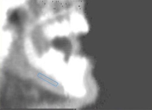
Scans of the mandible were performed and BMD (g/cm2) was calculated using the DXA device (GE Lunar DPX-NT, LUNAR Corp., Madison, WI, USA). Scanning procedures and BMD measurements were performed on the body of the mandible as previously described ().Citation19–Citation21 The BMD of the mandible was measured and analyzed by a single-blind operator.
Figure 1 Measurement of mandibular BMD by dual-energy X-ray absorptiometry.
Statistical analysis
Statistical analysis was performed with the Statistical Package for the Social Science Program (version 20.0, IBM Corporation, Armonk, NY, USA). The normality and homogeneity of the sample were confirmed by the Shapiro–Wilk tests, after which the one-way analysis of variance/Tukey honest significant difference tests were used to determine any intergroup differences. Differences in individual parameters between the whole BD group and the control group were tested using the Student’s t-test. A value of P<0.05 was considered statistically significant.
Results
Evaluation was made of the data of 75 patients, comprising 36 females (48%) and 39 males (52%), with a mean age of 37.56±8.2 years (range: 22–63 years) ( and ). There was only a significant difference between group 4 and the other groups regarding age (P<0.05) (47.27±7.5 years [range: 34–63 years]) ().
Table 1 Characteristics of patients (all groups) according to age, BMI, and BMD
Table 2 Characteristics of patients (all Behçet’s patients and controls) according to age, BMI, and BMD
The characteristics of the study participants are shown (mean ± SD) in and . There was no statistically significant difference between the study and control groups with respect to sex and body mass index (BMI) variables (P>0.05).
There was found to be no statistically significant difference between the three study groups and the control group in respect of the mandibular body BMD (P>0.05). However, the mandibular body BMD was highest in the control group patients (1.294±0.21 g/cm2) and gradually reduced through the study subgroups (group 2: 1.251±0.22 g/cm2, group 3: 1.249±0.20 g/cm2, group 4: 1.147±0.24 g/cm2) (). No statistically significant differences were determined (P>0.05) ().
Discussion
This study with a relatively small sample size group examined the comparison of mandibular BMD between BD patients and healthy individuals and showed that although there was no statistically significant difference, bone loss can occur in BD patients. To the best of our knowledge, this is the first study in the literature to investigate mandibular body BMD in BD patients.
The findings of the study showed the mean mandibular body BMD value of 1.294±0.21 g/cm2 in the control group and 1.247±0.22 g/cm2 in all the participants in our study which is similar to the findings of Buyukkaplan et alCitation20 (1.10±0.31 g/cm2), Pluskiewicz et alCitation22 (1.221±0.3 g/cm2), Devlin and HornerCitation23 (1.15±0.26 g/cm2), Drage et alCitation24 (1.38±0.39 g/cm2), and Esfahanizadeh et alCitation21 (1.386±0.320 g/cm2). Different BMD values in studies may be associated with “region of interest (ROI)” or dentulous/edentulous patients.
OP is a systemic skeletal disorder characterized by bone loss and structural deterioration of bone tissue, which may lead to bone fragility and an increased susceptibility to fractures. Recently, there have been many studies on OP in several rheumatological and chronic inflammatory disorders such as BD, RA, ankylosing spondylitis, FMF, and SLE.Citation7–Citation9 In these diseases, multiple factors may be responsible for bone mass reduction, especially the inflammatory process and the treatment drugs.
BD is a polysymptomatic and recurrent systemic vasculitis with a chronic course and unknown cause. In the inflammatory process, proinflammatory cytokines, such as tumor necrosis factor-α, interleukin- (IL-) 1β, IL-6, and IL-17 can upregulate and may cause increased bone resorption and decreased BMD.Citation3,Citation25 According to the degree of the disease, various drugs are used to control the signs and symptoms of BD. The main goal of drug therapy is to reduce symptoms, reduce inflammation, control the immune system, maintain remission, and improve the patient’s quality of life. Although there has been no consensus on a treatment program, many medications have been used to reduce inflammation or modify the immune system, including colchicine, corticosteroids, antitumor necrosis factor agents (infliximab and etanercept), thalidomide, and cyclosporine.Citation1 Colchicine and corticosteroids are suspected to reduce BMD.
Due to the inflammatory processes and long-term drug use, BMD may be affected by the disease duration and activity. Laan et alCitation12 reported that BMD can be reduced with a long-term disease duration in patients with RA. However, Biçer et alCitation10 reported no association between disease duration and BMD. In the present study, a reduction in BMD values was determined in patients with long-term disease duration, although it was not statistically significant.
In oral and maxillofacial surgery, decreased BMD can affect the decision-making for placement, success/survival and osseointegration of dental implants, success of bone regeneration therapies (osteotomies, bone augmentations, guided-bone-regeneration, and bone grafting), and bone loss in periodontitis.Citation26–Citation29 Studies have shown a positive correlation between BMD and bone quality.Citation30–Citation32 Pommer et alCitation33 found that radiographic bone density significantly impacted implant stability values and local jawbone density was reported to be the major determinant of primary implant stability in the maxillary sinus floor of shortened height. Türkyılmaz et alCitation34 reported significant correlations between bone quality and implant stability parameters. Therefore, all diseases and conditions that affect BMD are closely related to oral surgery.
Esfahanizadeh et alCitation21 reported that since there was a significant correlation between the densities of skeletal and jaw bones and the presence of OP or osteopenia, the maxilla and mandible may reflect the same situation. Pluskiewicz et alCitation22 also reported that mandibular BMD may be an appropriate measurement site for assessment of OP. However, Gulsahi et alCitation35 suggested that the BMD of the jaws was not correlated with femoral BMD. It was explained that these different results may have arisen from different sizes of ROI selection and the measurement of specific areas (anterior, premolar and molar regions of maxilla and mandible).
Mandibular BMD can be measured in the body, symphysis, and ramus regions. Horner et alCitation36 suggested that the mandibular body has greater sensitivity and specificity compared with the ramus and symphysis regions in the detection of OP. Therefore, in the present study, the BMD measurements were performed on the body of the mandible.
DXA is the gold standard for measurement of BMD. Due to its rapid, highly reproducible, very precise, low cost, and radiation dosage properties, DXA is the most widely accepted and utilized technology.Citation37
Limitations
Limitations of the present study include the small sample size and that there was no information about drug dosages and types and dentolous/edentulous patients. Further studies are needed of larger study groups.
Conclusion
The results of this study have shown that long-term disease duration may cause decreased mandibular BMD and BD patients had lower BMD than control group, although it was not statistically important.
Disclosure
The authors report no conflicts of interest in this work.
References
- SalehZArayssiTUpdate on the therapy of Behcet diseaseTher Adv Chronic Dis20145311213424790727
- RiganteDThe fresco of autoinflammatory diseases from the pediatric perspectiveAutoimmun Rev201211534835622024500
- TekinNSOzdolapSSarikayaSEsturkEGumustasSBone mineral density and bone turnover markers of patients with Behcet’s diseaseJ Eur Acad Dermatol Venereol2007211252917207163
- YurdakulSYaziciHTuzunYThe arthritis of Behcet’s disease: a prospective studyAnn Rheum Dis19834255055156625699
- DireskeneliHBehcet’s disease: infectious aetiology, new autoantigens, and HLA-B51Ann Rheum Dis20016011996100211602462
- GaleoneMColucciRD’ErmeAMMorettiSLottiTPotential infectious etiology of Behcet’s diseasePatholog Res Int2012201259538022254152
- MartinJCMunroRCampbellMKReidDMEffects of disease and corticosteroids on appendicular bone mass in postmenopausal women with rheumatoid arthritis: comparison with axial measurementsBr J Rheumatol199736143499117173
- JuanolaXMateoLNollaJMRoig-VilasecaDCampoyERoig-EscofetDBone mineral density in women with ankylosing spondylitisJ Rheumatol20002741028103110782832
- GilboeIMKvienTKHaugebergGHusbyGBone mineral density in systemic lupus erythematosus: comparison with rheumatoid arthritis and healthy controlsAnn Rheum Dis200059211011510666165
- BicerATursenUKayaTIBone mineral density in patients with Behcet’s diseaseRheumatol Int200424635535814556035
- SkefWHamiltonMJArayssiTGastrointestinal Behcet’s disease: a reviewWorld J Gastroenterol201521133801381225852265
- LaanRFBuijsWCVerbeekALBone mineral density in patients with recent onset rheumatoid arthritis: influence of disease activity and functional capacityAnn Rheum Dis199352121268427509
- GoughAKLilleyJEyreSHolderRLEmeryPGeneralised bone loss in patients with early rheumatoid arthritisLancet1994344891423277912297
- JemtTLekholmUImplant treatment in edentulous maxillae: a 5-year follow-up report on patients with different degrees of jaw resorptionInt J Oral Maxillofac Implants19951033033117615326
- JaffinRABermanCLThe excessive loss of Branemark fixtures in type IV bone: a 5-year analysisJ Periodontol1991621242002427
- KlemettiECollinHLForssHMarkkanenHLassilaVMineral status of skeleton and advanced periodontal diseaseJ Clin Periodontol19942131841888157771
- TezalMWactawski-WendeJGrossiSGHoAWDunfordRGencoRJThe relationship between bone mineral density and periodontitis in postmenopausal womenJ Periodontol20007191492149811022780
- van SteenbergheDQuirynenMMollyLJacobsRImpact of systemic diseases and medication on osseointegrationPeriodontol 200020033316317112950849
- BuyukkaplanUSGuldagMUYildizMGumusBAComparison of mandibular bone mineral density in osteoporotic, osteopenic and normal elderly edentulous subjects measured by the dual-energy X-ray absorptiometry techniqueGerodontology2012292e1098e110222288568
- BuyukkaplanUSTongucMOGuldagMUYildizMGumusBAComparison of mandibular bone mineral densities in dentate and edentulous patientsJ Prosthodont2013221232722946895
- EsfahanizadehNDavaieSRoknARCorrelation between bone mineral density of jaws and skeletal sites in an Iranian population using dual X-ray energy absorptiometryDent Res J (Isfahan)201310446046624130580
- PluskiewiczWTarnawskaBDrozdzowskaBMandibular bone mineral density measured using dual-energy X-ray absorptiometry: relationship to hip bone mineral density and quantitative ultrasound at calcaneus and hand phalangesBr J Radiol20007386728829210817045
- DevlinHHornerKA study to assess the relative influence of age and edentulousness upon mandibular bone mineral density in female subjectsOral Surg Oral Med Oral Pathol Oral Radiol Endod2007104111712117577551
- DrageNAPalmerRMBlakeGWilsonRCraneFFogelmanIA comparison of bone mineral density in the spine, hip and jaws of edentulous subjectsClin Oral Implants Res200718449650017517057
- LaneNERehmanQOsteoporosis in the rheumatic disease patientLupus2002111067567912413067
- BlomqvistJEAlberiusPIsakssonSLindeAHanssonBGFactors in implant integration failure after bone grafting: an osteometric and endocrinologic matched analysisInt J Oral Maxillofac Surg199625163688833304
- AugustMChungKChangYGlowackiJInfluence of estrogen status on endosseous implant osseointegrationJ Oral Maxillofac Surg2001591112851289 discussion 1290–128111688027
- GondimVAunJFukudaCTSevere loss of clinical attachment level: an independent association with low hip bone mineral density in postmenopausal femalesJ Periodontol201384335235922548585
- PassosJSViannaMIGomes-FilhoISOsteoporosis/osteopenia as an independent factor associated with periodontitis in postmenopausal women: a case-control studyOsteoporos Int20132441275128323001114
- Fuster-TorresMAPenarrocha-DiagoMPenarrocha-OltraDPenarrocha-DiagoMRelationships between bone density values from cone beam computed tomography, maximum insertion torque, and resonance frequency analysis at implant placement: a pilot studyInt J Oral Maxillofac Implants20112651051105622010089
- KidoHSchulzEEKakuraKYamamotoKMorinagaKMatsuuraMHuman mandibular trabecular bone density correlation with mechanical strength: implications for implant dentistryImplant Dent201120432332621778888
- MunakataMTachikawaNHondaEShiotaMKasugaiSInfluence of menopause on mandibular bone quantity and quality in Japanese women receiving dental implantsArch Osteoporos20116515722207877
- PommerBHofMFadlerAGahleitnerAWatzekGWatzakGPrimary implant stability in the atrophic sinus floor of human cadaver maxillae: impact of residual ridge height, bone density, and implant diameterClin Oral Implants Res2014252e109e11323167282
- TurkyilmazIMcGlumphyEAInfluence of bone density on implant stability parameters and implant success: a retrospective clinical studyBMC Oral Health200883219025637
- GulsahiAPaksoyCSOzdenSKucukNOCebeciARGencYAssessment of bone mineral density in the jaws and its relationship to radiomorphometric indicesDentomaxillofac Radiol201039528428920587652
- HornerKDevlinHAlsopCWHodgkinsonIMAdamsJEMandibular bone mineral density as a predictor of skeletal osteoporosisBr J Radiol199669827101910258958019
- MaricicMUse of DXA-based technology for detection and assessment of risk of vertebral fracture in rheumatology practiceCurr Rheumatol Rep201416843624938441