Abstract
Background
The aim was to evaluate factor XIII activity (FXIIIa) and monocyte-derived microparticles (MDMPs) in cancer patients.
Methods
In total, 138 cancer patients (31 malignant lymphomas, 39 multiple myelomas, and 68 lung cancers) were analyzed. We measured various biomarkers including FXIIIa and MDMPs.
Results
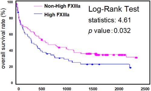
The values of endothelial activation markers, monocyte chemoattractant peptide (MCP)-1, soluble (s)CD14, and MDMPs were higher in cancer patients than in non-cancerous controls. MCP-1, sCD14, and MDMPs were significantly correlated with FXIIIa in multivariate analysis in cancer patients. In addition, MCP-1, sCD14, and MDMP levels were significantly increased in the high FXIIIa group of patients. Finally, the survival rate of the high FXIIIa group was significantly poor in the Kaplan–Meier analysis.
Conclusion
These results suggest that abnormal levels of FXIIIa and MDMPs may offer promise as poor prognostic factors in cancer patients.
Introduction
In recent years, oncocardiology has emerged as a new area of academic research, mainly in Europe and the United States.Citation1,Citation2 Progression in cancer treatment including the development of molecular-targeted drugs has been made against this background.Citation3 While survival has improved, new cardiotoxicity or cardiovascular toxicity events due to such new treatments have been recorded. As a result, there is a growing need for cardiovascular treatment in parallel with cancer therapy.Citation4 The cardiotoxicity of chemotherapies is classified as heart failure, coronary heart disease, hypertension, thromboembolism, and arrhythmia.Citation5 Venous thromboembolism (VTE) is particularly important,Citation6 and came to be described as cancer-associated thrombosis (CAT) when VTE is related to cancer.Citation7 Cancer cells or the inflammatory region around cancer cells release substances that function in thrombosis such as tissue factor (TF), coagulation factor, or cytokines.Citation8 Vascular disorders, associated with chemotherapy or enhancement of the coagulation system, are important as factors of VTE.Citation9
Factor XIII (FXIII) is present in the blood as a heterotetramer that is formed by FXIII-A and FXIII-B subunit polymers.Citation10 FXIII-A is present mainly in platelets, megakaryocytes, monocytes, and macrophages.Citation11,Citation12 In contrast, FXIII-B is generated and secreted by liver cells, and binds to FXIII-A in peripheral blood.Citation10–Citation12 FXIII is activated by thrombin which is produced in the final stages of coagulation, in a process called FXIII activity (FXIIIa).Citation11 FXIIIa forms covalent bonds between fibrins to eventually stabilize them.Citation11,Citation12 Therefore, FXIII has an essential role in normal hemostasis, in which it contributes to the regulation of fibrinolysis.Citation13 Specifically, this factor contributes to clot strength in the final step of coagulation with platelets and fibrinogen. Additionally, functional cellular FXIII becomes exposed on the surface of stimulated platelets.Citation14 These observations suggest that a close relationship exists between activated platelets and FXIII. However, it has been reported that FXIII has various functions.Citation15–Citation17 For example, high FXIIIa is associated with the risk of developing cardiovascular events.Citation18 Furthermore, abnormal FXIII expression levels or activity are found in some cancer patients.Citation19,Citation20
Activated monocytes and monocyte-derived microparticles (MDMPs) might be markers for vasculopathies in patients with lifestyle-related diseases.Citation21–Citation23 In addition, monocyte chemotactic peptide (MCP)-1 is unregulated in some patients along with endothelial cell dysfunction markers such as soluble (s) E-selectin and soluble vascular cell adhesion molecule-1 (sVCAM-1).Citation24,Citation25 However, little is known about FXIIIa and MDMPs in cancer patients. We investigated FXIIIa, MDMPs, and various biomarkers in cancer patients.
Materials and Methods
Subjects
Cancer patients and healthy volunteers were recruited from Kansai Medical Hospital and Kansai Medical University (Osaka, Japan) between August 2014 and September 2018. In total, 138 cancer patients were analyzed, and 14 had thrombotic complications within 6 months after their first examination. The types of cancer studied were malignant lymphoma (ML; n = 31), multiple myeloma (MM; n = 39), and lung cancer (LC; n = 68). The disease type of ML was classified using World Health Organization (WHO) classification 2017.Citation26 ML was classified into four stage (I, II, III and IV) by the Lugano classification.Citation27 The staging of MM was classified using the revised international staging system,Citation28 and LC stage was classified using the TNM classification of the international association for the study of LC.Citation29 This study was conducted in accordance with the Declaration of Helsinki and was performed with approval from the Institutional Review Board of Kansai Medical University. Written informed consent was obtained from all participants.
Measurement of FXIIIa
Patient blood samples (1.8 mL) were collected in 0.2 mL sodium citrate-containing tubes to a total volume of 2 mL. Citrated plasma was isolated by centrifugation for 15 min at 1500 ×g at 4°C. Plasma was divided into aliquots and FXIIIa was measured by photometric assay.Citation30 In a one-step procedure, FXIII was activated by thrombin, and Ca2+ and cross-linked glycine-ethyl ester to a specific glutamine-containing peptide substrate. The released ammonia was incorporated into alpha-ketoglutarate by glutamate dehydrogenase, and the NADH consumption of this reaction was measured photometrically at 340 nm. NADH-consumption was directly proportional to FXIIIa.
Measurement MDMPs
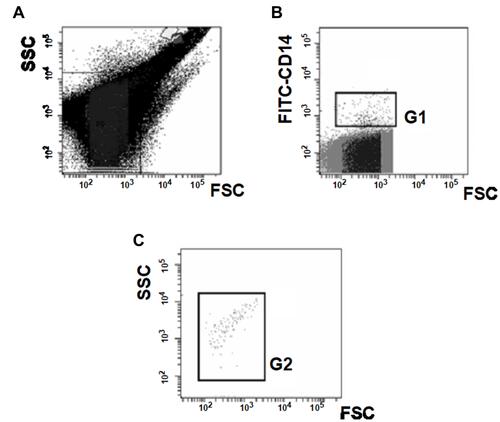
Blood samples were collected using a 21-gauge needle from a peripheral vein into vacutainers containing ethylenediaminetetraacetic acid (NIPRO Co. Ltd., Osaka, Japan) to minimize platelet activation. The samples were handled as described in the manufacturer’s protocol. Briefly, the samples were gently mixed by inverting the tube once or twice, stored at room temperature for 2–3 h, and centrifuged at 8000 ×g for 5 min at room temperature. Storing samples at room temperature for 2–3 h did not affect MDMP levels. MDMPs were analyzed using an FACS Cant II flow cytometer (BD Biosciences, Franklin Lakes, NJ, USA). All flow cytometry data of forward light scatter (FSC), side scatter (SSC), and fluorescence intensity (FL) were analyzed in log space. MDMPs were identified and quantified based on their FSC/SSC characteristics according to their size and reactivity to the monocyte-specific monoclonal antibody (CD14). The lower detection limit was placed at a threshold above the electronic background noise of the flow cytometer, and the upper threshold for FSC (1μm) was set with the use of standard beads (Megamix, BioCytex, Tokyo, Japan) (). To identify positively stained events, thresholds were set based on FITC-CD14 (G1 gate) (). Finally, events in the G1 gate were expanded to FSC/SSC (G2 gate) (). The density of MDMPs in the G2 gate was set to less than 10 events/μL using blood samples from healthy volunteers.
Measurement of MCP-1, sE-selectin, sVCAM-1, sCD14 and PAI-1
Patient blood samples were collected in empty or sodium citrate-containing tubes and left at room temperature for a minimum of 1 h. Serum and citrated plasma were isolated by centrifugation for 20 min at 1000 ×g at 4°C. Serum was divided into aliquots and frozen at −30°C until use. Recombinant products and standard solutions provided with commercial kits served as positive controls. Plasma concentrations of MCP-1, sE-selectin, sVCAM-1 and PAI-1 were measured using monoclonal antibody-based ELISA kits (Invitrogen, Carlsbad, CA, USA). Plasma sCD14 levels were measured by ELISA (R&D Systems Inc., Minneapolis, MN, USA). All kits were used according to the manufacturers’ instructions.
Statistical Analysis
Data are expressed as mean ± standard deviation (SD) and were analyzed using multiple regression (stepwise method), as appropriate. A receiver operating characteristics (ROC) curve analysis was used to estimate the value of each biomarker. Between-group comparisons were made using the Newman–Keuls test and Scheffe’s test. Overall survival (OS) was defined as the time from initial diagnosis to the time of death from any cause or the date the patient was last known to be alive. Univariate analyses of OS were performed using the Kaplan–Meier product-limit method with the Log-rank test and the Cox proportional hazards model. All statistical analyses were performed using StatFlex (v7) software, with P < 0.05 being considered statistically significant.
Results
Cell Type and Staging of Cancers of Cancer Patients
The types of ML examined in this study included 19 mature B cell lymphomas, 10 mature T/natural killer (NK) cell lymphomas, and two Hodgkin lymphomas. The frequency of stage III and IV in ML was 34%. All patients with MM were symptomatic myeloma,Citation31 and the frequency of stage III was 45%. The cell type of LC was 60 non-small cell and eight small cell LC. The frequency of stage IIIb and IV was 56%. The mortality rate during the observation period was 32% (ML, 5/31; MM, 10/39; and LC, 32/68).
Clinical Characteristics of Cancer Patients
Patient demographic and clinical characteristics are shown in . We confirmed that all biomarker values were distributed normally, and we estimated the value of each biomarker using a ROC curve. Age, body mass index (BMI), white blood cells and platelets were similar in the cancer and the non-cancer controls. Hb in cancer patients was significantly lower than the non-cancerous controls. However, the values of red blood cell distribution width (RDW)-SD, mean platelet volume (MPV), FXIIIa, sE-selectin, sVCAM-1, PAI-1, MCP-1, sCD14, and MDMPs were higher in cancer patients than the healthy controls.
Table 1 Demographic and Clinical Characteristics of Cancer Patients and Healthy Controls
Univariate and Multivariate Regression Analyses
We investigated the associations between 14 variables and FXIIIa in cancer patients (). Univariate analysis showed that RDW, platelets, MPV, sE-selectin, sVCAM-1, PAI-1, MCP-1, sCD14, and MDMPs were factors significantly associated with FXIIIa, whereas sVCAM-1, PAI-1, MCP-1, sCD14, and MDMPs were significantly correlated with FXIIIa in multivariate analysis.
Table 2 Multiregression Analysis of FXIIIa in Cancer Patients
Comparison of All Markers of High FXIIIa or Non-High FXIIIa Groups in Cancer Patients
We calculated the cutoff value for FXIIIa using ROC curve analysis. A cutoff value of 108.23 was found to be an identifying value for patients with cancers. We divided the patients with cancer into two groups according to the cutoff value of 108.23 for FXIIIa. There were no significant differences in age, BMI, WBC, Hb, RDW-SD, platelets and MPV between the non-high FXIIIa and high FXIIIa groups (). However, sE-selectin, sVCAM-1, PAI-1, MCP-1, sCD14 and MDMP levels were significantly increased in the high FXIIIa group ().
Table 3 Plasma Levels of Various Biomarkers in the Non-High FXIIIa and High FXIIIa Groups
Kaplan–Meier Curves of Non-High FXIIIa and High FXIIIa Patient Groups
shows the Kaplan–Meier curves for OS of the cancer patients with or without high FXIIIa. The log-rank P-value at 2000 days between patients with or without high FXIIIa was 0.032.
Discussion
It has been known for some time that cancer is accompanied by the risk of thrombosis such as Trousseau’s syndrome.Citation32,Citation33 It is necessary to consider the thrombosis associated with the treatment for cancer separately from the original risk of cancer itself.Citation9 In addition, even if the cancer treatment is successful, we have to consider the other factors or side effects of anticancer drugs. Therefore, predicting CAT is an important proposition for cancer in clinical practice. Several biomarkers have been reported in terms of CAT predictions thus far.Citation34–Citation39 Zöller et alCitation34 reported that RDW-SD is associated with long-term incidence of the first event of VTE among middle-aged subjects, although not necessarily cancer. Other reports suggest that MPV is associated with VTE risk and survival in cancer patients.Citation35–Citation37 In the present study, we observed the high level of RDW-SD, MPV, sE-selectin, sVCAM-1, PAI-1, MCP-1 and sCD14 in cancer patients, although we could not recognize the significance of these markers in CAT. However, an interesting marker was MDMP, because MPs could associate with CAT.Citation38–Citation43
MDMPs contribute to the development of thrombosis at sites of vascular injury, since they express TF and enhance the exogenous tract of coagulation.Citation44 The binding of these MDMPs to activated endothelial cells may promote the accumulation of tissue factor and localize thrombin generation, resulting in the development of thrombosis.Citation34,Citation45 Some previous reports demonstrated the importance of tumor-derived MPs in CAT or the increased levels of TF-positive MPs in cancer patients with VTE compared with cancer patients without VTE.Citation39–Citation43 Furthermore, prospective studies suggested that high levels of TF-positive MPs activity may predict VTE in cancer patients.Citation37–Citation39 A meta-analysis by Cui et alCitation38 demonstrated the increased presence of TF-positive MPs that represented an increased risk of CAT with overall odds of 1.76. In addition, the role of TF-positive MPs originating from tumor cells in CAT development has been clearly shown in human pancreatic BxPc-3 cells orthotopically grown in nude mice.Citation46,Citation47 Thus, TF-positive MPs including MDMPs have an important role in CAT. Interestingly, in the present study, MDMP significantly increased in cancer patients compared with healthy controls. However, CAT appears to be involved in the mechanism of TF-independent MDMP.Citation48 This suggests that it is necessary to consider another role of MDMP other than coagulofibrinolytic imbalance in CAT as a future potential therapeutic target.
Abnormal FXIIIa appears to cause thrombotic states such as cardiovascular diseases.Citation18,Citation49,Citation50 The same abnormal activity has been reported in cancer patients.Citation17,Citation20 A study in non-small cell lung cancer found that FXIIIa levels in advanced-stage patients are higher than those in healthy controls. These patients indicated that FXIIIa expression could potentially be used as a predictive marker of advanced non-small cell lung cancer.Citation20 In the present study, FXIIIa was significantly increased in cancer patients compared with healthy controls. In the present study, the frequency of patient with advanced cancer was 34% in ML, 45% in MM and 56% in LC. Unfortunately, we could not confirm a significant elevation of FXIIIa in these advanced cases. Therefore, the expression of FXIIIa by tumor-infiltrating macrophages was not determined. In the present study, MCP-1, sCD14 and MDMPs were higher in cancer patients than in non-cancerous controls. In addition, the levels of these markers were significantly increased in the high-FXIIIa group. These findings suggest that the association of monocytes and FXIIIa occurs in cancer patients, resulting in the poor prognosis of the high-FXIIIa cancer patients in our Kaplan–Meier analysis. However, the role of FXIII in CAT is still unclear, and the relationship between FXIII and cancer needs to be further elucidated.
Conclusions
In conclusion, RDW-SD, MPV, FXIIIa, sE-selectin, sVCAM-1, PAI-1, MCP-1, sCD14, and MDMP levels were higher in cancer patients than in non-cancerous controls. sVCAM-1, PAI-1, MCP-1, sCD14, and MDMPs were significantly correlated with FXIIIa by multivariate analysis in cancer patients. In addition, sE-selectin, sVCAM-1, PAI-1, MCP-1, sCD14, and MDMP levels were increased significantly in the high FXIIIa group. Finally, the survival rate of the high-FXIIIa group was significantly poor in the Kaplan–Meier analysis. Therefore, abnormal levels of FXIIIa and MDMPs may offer promise as poor prognostic factors in cancer patients.
Nevertheless, this study has some limitations. First, we could not confirm a direct relationship of FXIIIa or MDMPs in our thrombotic patients. Second, we did not attempt a more accurate method than flow cytometry to detect MDMPs. For example, antigenic detection of TF-positive MDMPs by scanning confocal microscopy may be appropriate.Citation48,Citation51,Citation52 These limitations are issues to be considered in the future.
Abbreviations
RDW-SD, red blood cell distribution width-standard deviation; MPV, mean platelet volume; FXIIIa, factor XIII activity; MCP-1, monocyte chemotactic peptide-1; sCD14, soluble CD14; BMI, body mass index; TF, tissue factor; sE-selectin, soluble E-selectin; sVCAM-1, soluble vascular cell adhison molecule-1; PAI-1, plasminogen activator inhibitor-1; MDMP, monocyte-derived microparticle; LC, lung cancer; ML, malignant lymphoma; MM, multiple myeloma; CAT, cancer-associated thrombosis; VTE, venous thromboembolism.
Author Contributions
All authors contributed to data analysis, drafting and revising the article, gave final approval of the version to be published, and agree to be accountable for all aspects of the work.
Acknowledgment
We thank H. Nikki March, PhD, from Edanz Group for editing a draft of this manuscript.
Disclosure
The authors declare that there are no conflicts of interest.
References
- Koene RJ, Prizment AE, Blaes A, Konety SH. Shared risk factors in cardiovascular disease and cancer. Circulation. 2016;133(11):1104–1114. doi:10.1161/CIRCULATIONAHA.115.020406
- Bonnie KY. Cardio-oncology. In: Braunwald E, editor. Heart Disease. 11thed. Philadelphia: Saunders; 2018:1641–1650.
- Chang HM, Okwuosa TM, Scarabelli T, Moudgil R, Yeh ETH. Cardiovascular complications of cancer therapy: best practices in diagnosis, prevention, and management-part 2. J Am Coll Cardiol. 2017;70(20):2552–2565. doi:10.1016/j.jacc.2017.09.1095
- Okwuosa TM, Prabhu N, Patel H, et al. The cardiologist and the cancer patient: challenges to cardio-oncology (or onco-cardiology) and call to action. J Am Coll Cardiol. 2018;72(2):228–232. doi:10.1016/j.jacc.2018.04.043
- Edwards BK, Noone AM, Mariotto AB, et al. Annual report to the nation on the status of cancer, 1975-2010, featuring prevalence of comorbidity and impact on survival among persons with lung, colorectal, breast, or prostate cancer. Cancer. 2014;120(9):1290–1314. doi:10.1002/cncr.28509
- Lyman GH, Khorana AA, Falanga A, et al. American Society of Clinical Oncology guideline: recommendations for venous thromboembolism prophylaxis and treatment in patients with cancer. J Clin Oncol. 2007;25(34):5490–5505. doi:10.1200/JCO.2007.14.1283
- Khorana AA, Francis CW, Culakova E, Kuderer NM, Lyman GH. Thromboembolism is a leading cause of death in cancer patients receiving outpatient chemotherapy. J Thromb Haemost. 2007;5(3):632–634. doi:10.1111/j.1538-7836.2007.02374.x
- Khorana AA. Venous thromboembolism and prognosis in cancer. Thromb Res. 2010;125(6):490–493. doi:10.1016/j.thromres.2009.12.023
- Mukai M, Komori K, Oka T. Mechanism and management of cancer chemotherapy-induced atherosclerosis. J Atheroscler Thromb. 2018;25(10):994–1002. doi:10.5551/jat.RV17027
- Muszbek L, Bereczky Z, Bagoly Z, et al. Factor XIII: a coagulation factor with multiple plasmatic and cellular functions. Physiol Rev. 2011;91(3):931–972.
- Ichinose A. Factor XIII is a key molecule at the intersection of coagulation and fibrinolysis as well as inflammation and infection control. Int J Hematol. 2012;95(4):362–370. doi:10.1007/s12185-012-1064-3
- Smith KA, Pease RJ, Avery CA, et al. The activation peptide cleft exposed by thrombin cleavage of FXIII-A2 contains a recognition site for the fibrinogen α chain. Blood. 2013;121(11):2117–2126. doi:10.1182/blood-2012-07-446393
- Fraser SR, Booth NA, Mutch NJ. The antifibrinolytic function of factor XIII is exclusively expressed through 2-antiplasmin cross-linking. Blood. 2011;117(23):6371–6374.
- Mitchell JL, Lionikiene AS, Fraser SR, et al. Functional factor XIII-A is exposed on the stimulated platelet surface. Blood. 2014;124(26):3982–3990. doi:10.1182/blood-2014-06-583070
- Schroeder V, Kohler HP. New developments in the area of factor XIII. J Thromb Haemost. 2013;11(2):234–244. doi:10.1111/jth.12074
- Tahlan A, Ahluwalia J. Factor XIII: congenital deficiency factor XIII, acquired deficiency, factor XIII A-subunit, and factor XIII B-subunit. Arch Pathol Lab Med. 2014;138(2):278–281. doi:10.5858/arpa.2012-0639-RS
- Shi DY, Wang SJ. Advances of coagulation factor XIII. Chin Med J. 2017;130(2):219–223. doi:10.4103/0366-6999.198007
- Kreutz RP, Schmeisser G, Schaffter A, et al. Prediction of ischemic events after percutaneous coronary intervention: thrombelastography profiles and factor XIIIa activity. TH Open. 2018;2(2):e173–181. doi:10.1055/s-0038-1645876
- Gonçalves E. Lopes da Silva R, Varandas J, Diniz MJ. Acute promyelocytic leukaemia associated factor XIII deficiency presenting as retro-bulbar haematoma. Thromb Res. 2012;129(6):810–811. doi:10.1016/j.thromres.2012.03.008
- Lee SH, Suh IB, Lee EJ, et al. Relationships of coagulation factor XIII activity with cell-type and stage of non-small cell lung cancer. Yonsei Med J. 2013;54(6):1394–1399. doi:10.3349/ymj.2013.54.6.1394
- Omoto S, Nomura S, Shouzu A, et al. Detection of monocyte-derived microparticles in patients with type II diabetes mellitus. Diabetologia. 2002;45(4):550–555. doi:10.1007/s00125-001-0772-7
- Matsumoto N, Nomura S, Kamihata H, et al. Increased level of oxidized LDL-dependent monocyte-derived microparticles in acute coronary syndrome. Thromb Haemost. 2004;91(1):146–154. doi:10.1160/TH03-04-0247
- Nomura S, Shouzu A, Omoto S, et al. Effect of valsartan on monocyte/endothelial cell activation markers and adiponectin in hypertensive patients with type 2 diabetes mellitus. Thromb Res. 2006;117(4):385–392. doi:10.1016/j.thromres.2005.04.008
- Colotta F, Borre A, Wang JM, et al. Expression of monocyte chemotactic cytokine by human mononuclear phagocytes. J Immunol. 1992;148(3):760–765.
- Fries JWU, Williams AJ, Atkins RC, et al. Expression of VCAM-1 and E-selectin an in vivo model of endothelial activation. Am J Pathol. 1993;143(3):725–737.
- Jsffe ES. Introduction and over view of the classification of lymphoid neoplasmas. In: Swerdlow SH, editor. WHO Classification of Tumors of Haematopoietic and Lymphoid Tissues. Lyon: IARC; 2017:pp190–198.
- Cheson BD, Fisher RI, Barrington SF, et al. Recommendations for initial evaluation, staging, and response assessment of Hodgkin and non-Hodgkin lymphoma: the Lugano classification. J Clin Oncol. 2014;32(27):3059–3068. doi:10.1200/JCO.2013.54.8800
- Greipp PR, San Miguel J, Durie BG, et al. International staging system for multiple myeloma. J Clin Oncol. 2005;23(15):3412–3420. doi:10.1200/JCO.2005.04.242
- Rami-Porta R, Bolejack V, Giroux DJ, et al. The IASLC lung cancer staging project: the new database to inform the eighth edition of the TNM classification of lung cancer. J Thorac Oncol. 2014;9(11):1618–1624. doi:10.1097/JTO.0000000000000334
- Fickenscher K, Aab A, Stuber W. A photometric assay for blood coagulation factor XIII. Thromb Haemost. 1991;65(5):535–540. doi:10.1055/s-0038-1648185
- Rajkumar SV, Dimopoulos MA, Palumbo A, et al. International Myeloma Working Group updated criteria for the diagnosis of multiple myeloma. Lancet Oncol. 2014;15(12):e538–548. doi:10.1016/S1470-2045(14)70442-5
- Varki A. Trousseau’s syndrome: multiple definition and multiple mechanisms. Blood. 2007;110(6):1723–1729. doi:10.1182/blood-2006-10-053736
- Ikushima S, Ono R, Fukuda K, et al. Trousseau’s syndrome: cancer-associated thrombosis. Jpn J Clin Oncol. 2016;46(3):204–208. doi:10.1093/jjco/hyv165
- Zöller B, Melander O, Svensson P, Engström G. Red cell distribution width and risk for venous thromboembolism: a population-based cohort study. Thromb Res. 2014;133(3):334–339. doi:10.1016/j.thromres.2013.12.013
- Riedl J, Kaider A, Reitter EM, et al. Association of mean platelet volume with risk of venous thromboembolism and mortality in patients with cancer. Results from the Vienna Cancer and Thrombosis Study (CATS). Thromb Haemost. 2014;111(4):670–678. doi:10.1160/TH13-07-0603
- Díaz JM, Boietti BR, Vazquez FJ, et al. Mean platelet volume as a prognostic factor for venous thromboembolic disease. Rev Med Chil. 2019;147(2):145–152. doi:10.4067/s0034-98872019000200145
- Inagaki N, Kibata K, Tamaki T, Shimizu T, Nomura S. Prognostic impact of the mean platelet volume/platelet count ratio in terms of survival in advanced non-small cell lung cancer. Lung Cancer. 2015;83(1):97–101. doi:10.1016/j.lungcan.2013.08.020
- Cui CJ, Wang GJ, Yang S, Huang SK, Qiao R, Cui W. Tissue factor-bearing MPs and the risk of venous thrombosis in cancer patients: a meta-analysis. Sci Rep. 2018;8(1):1675. doi:10.1038/s41598-018-19889-8
- Tesselaar ME, Romijin FP, Van Der Linden IK, Prins FA, Bertina RM, Osanto S. Microparticle-associated tissue factor activity: a link between cancer and thrombosis? J Thromb Haemost. 2007;5(3):520–527. doi:10.1111/j.1538-7836.2007.02369.x
- Zwicker JI, Liebman HA, Neuberg D, et al. Tumor-derived tissue factor-bearing microparticles are associated with venous thromboembolic events in malignancy. Clin Cancer Res. 2009;15(22):6830–6840. doi:10.1158/1078-0432.CCR-09-0371
- Manly DA, Wang J, Glover SL, et al. Increased microparticle tissue factor activity in cancer patients with venous thromboembolism. Thromb Res. 2010;125(6):511–512. doi:10.1016/j.thromres.2009.09.019
- Campello E, Spiezial L, Radu CM, et al. Endothelial, platelet, and tissue factor-bearing microparticles in cancer patients with and without venous thromboembolism. Thromb Res. 2011;127(5):473–477. doi:10.1016/j.thromres.2011.01.002
- Geddings JE, Mackman N. Tumor-derived tissue factor-positive microparticles and venous thrombosis in cancer patients. Blood. 2013;122(11):1873–1880. doi:10.1182/blood-2013-04-460139
- Falati S, Liu Q, Gross P, et al. Accumulation of tissue factor into developing thrombi in vivo is dependent upon microparticle P-selectin glycoprotein ligand 1 and platelet P-selectin. J Exp Med. 2003;197(11):1585–1598. doi:10.1084/jem.20021868
- Del Conde I, Shrimpton CN, Thiagarajan P, López JA. Tissue-factor-bearing microvesicles arise from lipid rafts and fuse with activated platelets to initiate coagulation. Blood. 2005;106(5):1604–1611. doi:10.1182/blood-2004-03-1095
- Pang W, Su J, Wand Y, et al. Pancreatic cancer-secreted miR-155 implicates in the conversion from normal fibroblasts to cancer-associated fibroblasts. Cancer Sci. 2015;106(10):1362–1369. doi:10.1111/cas.12747
- Geddings JE, Hisada Y, Boulaftali Y, et al. Tissue factor-positive tumor microvesicles activate platelets and enhance thrombosis in mice. J Thromb Haemost. 2016;14(1):153–166. doi:10.1111/jth.13181
- Zarà M, Guidetti GF, Camera M, et al. Biology and role of extracellular vesicle (EVs) in the pathogenesis of thrombosis. Int J Mol Sci. 2019;20(11):E2840. doi:10.3390/ijms20112840
- Bereczky Z, Balogh E, Katona E, Czuriga I, Edes I, Muszbek L. Elevated factor XIII level and the risk of myocardial infarction in women. Haematologica. 2007;92(2):287–288. doi:10.3324/haematol.10647
- Ambroziak M, Kurylowicz A, Budaj A. Increased coagulation factor XIII activity but not genetic variants of coagulation factors is associated with myocardial infarction in young patients. J Thromb Thrombolys. 2019;48(3):519–527. doi:10.1007/s11239-019-01856-3
- Owens AP 3rd, Mackman N. Microparticles in hemostasis and thrombosis. Cir Res. 2011;108(10):1284–1297. doi:10.1161/CIRCRESAHA.110.233056
- Weiss R, Gröger M, Rauscher S, et al. Differential interaction of platelet-derived extracellular vesicles with leukocyte subsets in human whole blood. Sci Rep. 2018;8(1):6598. doi:10.1038/s41598-018-25047-x


