
Abstract
Cardiac angiosarcomas are a rare form of malignancy. The majority of cases arise from the right atrium as mural masses. These tumors have extremely aggressive behavior, with early clinical symptoms that vary depending on location, size, and extent of the tumor. Most of these patients have a very short survival time. Surgical therapy is considered the best choice of therapy approach in cardiac angiosarcoma patients with nonmetastatic disease, even though the disease is rarely cured. Advanced diagnostic techniques facilitate accurate, noninvasive assessments of cardiac sarcomas. We report a case of a 62-year-old man with cardiac angiosarcoma who had multiple distant metastases that were revealed by [18F]fluorodeoxyglucose positron emission tomography–computed tomography imaging.
Introduction
Sarcoma is the most common primary malignancy of the heart and pericardium. Primary cardiac sarcomas are rare entities,Citation1–Citation3 with an autopsy prevalence of 0.001%–0.28%.Citation3 Pathologic subtypes of primary cardiac sarcoma are angiosarcoma fibrosarcoma, rhabdomyosarcoma, malignant schwannoma, and mesothelioma. Frequency rate decreases in adults.
Angiosarcoma frequency is less than 1% among sarcomas. Of all angiosarcomas, a primary angiosarcoma of the heart and the great vessels accounts for only 3%.Citation4 Most of cardiac angiosarcomas (80%) originate from right atrium as mural masses. Extension to the pericardium and encasement of the heart may be seen, and usually fills the entire cardiac chamber. The aggressive behavior of cardiac angiosarcomas causes early symptoms and a very short survival rate. Clinical presentation of sarcomas varies depending on the location, size, and extent of the tumor. Advanced diagnostic techniques facilitate accurate, noninvasive assessment of cardiac sarcomas.Citation5
Diagnostic workup usually includes standard echo contrast-enhanced computed tomography (CT), magnetic resonance imaging, and angiography. In the literature, a few case reports have demonstrated the diagnostic contribution of [18F]fluorodeoxyglucose (FDG) positron emission tomography (PET)-CT in the evaluation of the malignant nature of disease and staging before deciding on the therapy aproach.Citation6,Citation7
Even though cardiac angiosarcoma is rarely cured, surgical therapy is considered the best choice of therapy in nonmetastatic disease.Citation8 Complete or partial surgical resection of primary or metastatic cardiac sarcoma can provide significant hemodynamic improvement, successful palliation, and prolonged survival.Citation9,Citation10 Chemotherapy and postsurgical adjuvant radiotherapy have not proven consistently beneficial. However, all treatment choices after surgical resection can ameliorate symptoms with a varied response.Citation11
Case report
Herein, we report the case of a 62-year-old man who had previously undergone tumor resection for primary high-grade cardiac angiosarcoma () at a university hospital 4 months previously. The patient was referred to the Nuclear Medicine Department for 18F-FDG PET-CT imaging to assess residual and/or metastatic disease.
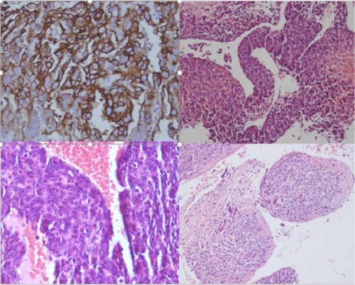
Figure 1 Histopathology findings.
The tumor was surgically approached by medial sternotomy, but complete tumor resection was not possible because of the extensive tumor infiltration. Palliative intervention consisting of partial excision of the tumor mass was aimed at releasing the right ventricular outflow tract.
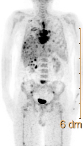
PET-CT scans revealed a residual or relapsed mass in the right atrium, with significantly increased uptake of 18F-FDG (maximum standardized uptake value of 12). The mass extended to the mitral valve, septum, and left ventricle and infiltrated to the pericardium and adjacent mediastinum (). The mass encased and almost completely replaced the atrial wall and nearly filled the entire cardiac chamber. Increased activity in the sternum was concomitant with sternotomy. PET-CT images also revealed multiple distant metastasis in the lung, liver, and bones (). Despite receiving subsequent aggressive therapy, including radiotherapy, the patient died 8 months later.
Figure 2 18F-FDG PET-CT image showing the tumor mass in the right atrium.
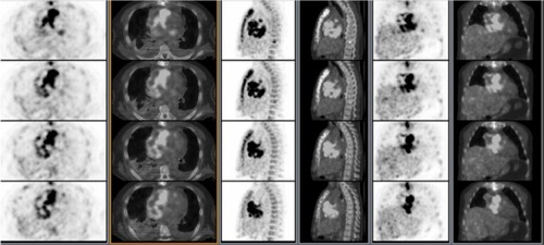
Figure 3 Maximum intensity projection image 18F-FDG PET-CT shows multiple distant metastasis in the lung, liver and bones.
Conclusion
Angiosarcoma is a very aggressive malignant neoplasm with a short fatal clinical process, despite new, cutting edge, treatment modalities, such as extensive surgery, radiotherapy, and chemotherapy.Citation12 Nowadays, through advanced diagnostic technology, such as high-resolution CT, dynamic magnetic resonance imaging, and PET-CT, clinicians are able to diagnose in a shorter time, which allows for more effective radical surgical resection. 18F-FDG PET-CT seems to be an effective and feasible imaging modality to evaluate primary or postsurgical residue and/or relapse and metastases of the tumor.
Disclosure
The authors report no conflicts of interest in this work.
References
- ReynenKFrequency of primary tumors of the heartAm J Cardiol1996771078540447
- SilvermanNAPrimary cardiac tumorsAnn Surg19801911271387362282
- BarreiroMRenillaAJimenezJMPrimary cardiac tumors: 32 years of experience from a Spanish tertiary surgical centerCardiovasc Pathol20132242442723727543
- LantzDADoughertyTHLuccaMJPrimary angiosarcoma of the heart causing cardiac ruptureAm Heart J19891181861882741790
- YuanSMShinfeldALaveeJKupersteinRHaizlerRRaananiEImaging morphology of cardiac tumoursCardiol J200916263519130413
- HoriYFunabashiNMiyauchiHAngiosarcoma in the right atria demonstrated by fusion images of multislice computed tomography and positron emission tomography using F-18 fluoro-deoxyglucoseInt J Cardiol2007123e15e1717316847
- HigashiyamaSKawabeJHayashiTEffectiveness of preoperative PET examination of huge angiosarcoma of the heartClin Nucl Med2009349910219352263
- TruongPTJonesSOMartensBTreatment and outcomes in adult patients with primary cardiac sarcoma: the British Columbia Cancer Agency experienceAnn Surg Oncol2009163358336519830494
- ReardonMJWalkesJCBenjaminRTherapy insight: malignant primary cardiac tumorsNat Clin Pract Cardiovasc Med2006354855316990840
- FurukawaNGummertJBörgermannJComplete resection of undifferentiated cardiac sarcoma and reconstruction of the atria and the superior vena cava: case reportJ Cardiothorac Surg201279623013671
- KakizakiSTakagiHHosakaYCardiac angiosarcoma responding to multidisciplinary treatmentInt J Cardiol1997622732759476688
- Look HongNJPandalaiPKHornickJLCardiac angiosarcoma management and outcomes: 20-year single-institution experienceAnn Surg Oncol20121982707271522476752