
Abstract
Canine hip dysplasia (CHD) is a polygenic and multifactorial developmental disorder characterized by coxofemoral (hip) joint laxity, degeneration, and osteoarthritis (OA). Current diagnostic techniques are largely subjective measures of joint conformation performed at different stages of development. Recently, measures on three-dimensional images generated from computed tomography scans predicted the development of OA associated with CHD. Continued refinement of similar imaging methods may improve diagnostic imaging techniques to identify dogs predisposed to degenerative hip joint changes. By current consensus, joint changes consistent with CHD are influenced by genetic predisposition as well as environmental and biomechanical factors; however, despite decades of work, the relative contributions of each to the development and extent of CHD signs remain elusive. Similarly, despite considerable effort to decipher the genetic underpinnings of CHD for selective breeding programs, relevant genetic loci remain equivocal. As such, prevention of CHD within domestic canine populations is marginally successful. Conservative management is often employed to manage signs of CHD, with lifelong maintenance of body mass as one of the most promising methods. Surgical intervention is often employed to prevent joint changes or restore joint function, but there are no gold standards for either goal. To date, all CHD phenotypes are considered as a single entity in spite of recognized differences in expression and response to environmental conditions and treatment. Identification of distinct CHD phenotypes and targeting evidence-based conservative and invasive treatments for each may significantly advance prevention and management of a prevalent, debilitating condition in canine companions.
Introduction
Canine hip dysplasia (CHD) is a complex developmental disorder characterized by joint laxity and osteoarthritis (OA) in one or both coxofemoral (hip) joints ().Citation1 The polygenic, multifactorial etiologyCitation2 of CHD has challenged veterinarians and researchers since the condition was described in the 1930s.Citation3 Joint changes characteristic of CHD are also associated with environmental factors such as nutrition,Citation4–Citation6 exercise,Citation7 and the process of skeletal ossification.Citation8,Citation9 The condition affects essentially all breeds, with an estimated prevalence ranging from 1% to 80% according to the Orthopedic Foundation for Animals. It appears to occur at a relatively high rate in large-bodied and brachycephalic dogs as well as those with high body length to height ratios.Citation10,Citation11 The periodic appearance of OA in joints other than the coxofemoral jointCitation12,Citation13 has led some to propose systemic contributions to CHD expression.Citation1 These complexities, among others, complicate attempts to manage the CHD by selective breeding despite strict reporting and guidelines.Citation14
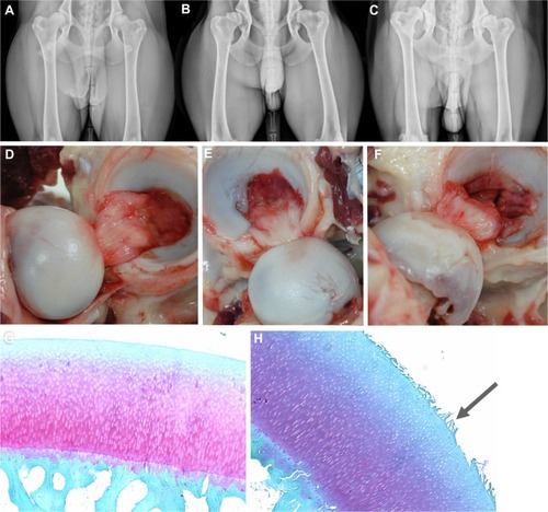
Figure 1 Anatomy of canine hip dysplasia.
There are many theories to explain CHD joint degeneration, but joint laxity and irregular or delayed endochondral ossification are among the most popular. The conditions are not mutually exclusive, and their phenotypic expression is variable within and among breeds.Citation15 Partially ossified hip structures may become distorted during development due to mechanical stresses in joints with delayed endochondral ossification.Citation8,Citation16 Joint components may be more vulnerable to deformation and damage from normal joint kinetics before they are fully ossified.Citation8,Citation9,Citation17 Abnormal and delayed endochondral ossification in the coxofemoral joint has been identified in 15-day-old dogs that developed CHD by the time they were 12 months old,Citation8,Citation9,Citation18 and in Great Danes with experimentally induced hip dysplasia.Citation19 In contrast, comparably earlier joint ossification appears to occur in Greyhounds, a breed with one of the lowest incidences of CHD. While it is clear that variation in the process of endochondral ossification may play a role in the development of CHD, the exact relationships between ossification patterns, abnormal joint structure, and development of OA remain unclear.Citation20
Affected joints usually develop varying degrees of synovial inflammation, articular cartilage damage (), osteophytes, and subchondral bone sclerosis and remodeling.Citation21–Citation23 While there is no single, overarching description of the sequence of events in the process, there are changes that occur in many forms of dysplasia. Recently, the dorsal acetabular rim angle (a measure of the dorsal slope [angle] of the subchondral articular acetabular surface relative to horizontal) was reported to be significantly larger (less femoral head coverage by the acetabulum) in dogs with coxofemoral joint laxity versus normal dogs as early as 1 week of age.Citation17 Subluxation of the femoral head and delays in ossification of the craniodorsal acetabular margin are often visible by 8 weeks, and, in many cases, subluxation of the femoral head increases by around 12 weeks of age.Citation18 Degeneration and microfractures of the articular cartilage, and thickening, inflammation, and deterioration of the joint capsule, tendinous insertions, and ligaments are often apparent by 5 months of age.Citation18 Despite the presence of these degenerative traits in many dogs with degenerative coxofemoral joint changes, clinical signs are variable.Citation21
A direct relationship between joint capsular collagen composition and mechanical properties was proposed over 30 years ago.Citation24 Altered capsular collagen composition has been identified in children with congenitally dislocated hipsCitation25 and dogs with hip joint laxity.Citation26 Joint capsular collagen fibrils were found to be more heterogeneous in 8-month-old Labrador Retriever puppies with severe coxofemoral joint laxity than those with normal joints.Citation15 Abnormal collagen composition is thought to contribute to reduced joint capsule stiffness, which contributes to excess femoral head motion and abnormal mechanical stresses on the femoral greater trochanter and acetabular margins and cartilage.Citation15 Over time, the abnormal forces are thought to result in deformation of the articulating structures and an incongruous joint.Citation18
Despite almost a century of work, many aspects of the development and progression of joint changes and OA associated with CHD remain elusive. This makes establishment of a gold standard for treatment a challenge. The lack of a single, predictable pattern of joint degeneration is likely a reflection of natural variability, including individual responses to external environmental influences. However, ambiguity in disease progression may also reflect distinct disease processes that have yet to be recognized. Continued efforts to identify and characterize patterns in joint changes may lead to identification of CHD phenotypes, which will, in turn, contribute to earlier disease identification and more effective targeted treatments.
Diagnosis
Despite some recognized patterns of joint degeneration characteristic of CHD, there is significant variability in the progression and ultimate severity of the disease as well as inconsistent relationships between gross and radiographic joint changes and clinical signs.Citation21 There are, however, two general behaviors often attributed to CHD, including lameness in young dogs (under 1 year), that increases with activity or trauma, and gait abnormalities and hind limb muscle atrophy in older dogs.Citation27 Notably, hind limb lameness can be due to reasons other than CHD joint changes, including pelvic, distal hind limb, and neurological pathologies, metabolic bone disease, ligament rupture, patellar luxation, and spine disorders.Citation27 Hence, a thorough, comprehensive assessment is paramount to identification of the source of discomfort.
Subjective laxity examinations
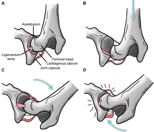
The Ortolani test is a subjective evaluation of coxofemoral joint laxity originally designed for diagnosis of human congenital hip dislocation in the 1930s.Citation28,Citation29 The test is also used as a CHD screening test.Citation28 Dogs are placed in lateral recumbency; one hand of the examiner is used to apply force along the length of the femur from the stifle toward the pelvis as the other braces the back just above the sacrum ().Citation27 This maneuver is intended to displace the femoral head. The stifle is then slowly abducted to reduce the joint.Citation29 An audible or palpable pop as the femur slips back into the acetabulum is considered a positive Ortolani sign indicative of joint laxity. Lack of an Ortolani sign does not necessarily mean that the hip is normal. Joint changes associated with dysplasia, like thickening of the joint capsule and joint tissue, may interfere with the displacement required for a positive sign.Citation28,Citation30 Bardens’ test,Citation31 an examination technique designed to evaluate the hips of babies (aged younger than 6 months), is thought to be more sensitive for detecting coxofemoral joint laxity and/or shallow acetabula in puppies 6–8 weeks of age.Citation29 With the dog in lateral recumbency, the proximal femur is elevated laterally from the body. With the femur elevated, the index finger of the other hand is used to push the femur away from the joint in a dorsal direction with pressure on the greater trochanter. More than 2 mm of displacement is considered a positive sign.Citation31 In general, these and other palpation techniques may be used as part of a comprehensive examination on puppies or dogs suspected to have excessive joint laxity characteristic of CHD. However, the tests alone are not sufficient for diagnosis of CHD.
Figure 2 Schematic illustration of the Ortolani test.
Radiography
Radiography has long been the gold standard to assess and quantify joint changes associated with CHD joint remodeling.Citation32,Citation33 Worldwide, there are five popular, standardized evaluation systems with distinct metrics that are used to grade canine radiographic coxofemoral joint conformation and degenerative changes.
Orthopedic Foundation for Animals
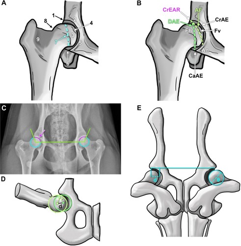
The Orthopedic Foundation for Animals evaluation is performed on hip-extended radiographs performed under heavy sedation or general anesthesia by three independent board-certified radiologists.Citation10 Based on subjective assessment of nine joint parameters (), conformation is categorized as excellent, good, fair, borderline, mild, moderate, or severe. The first three categories are considered to be normal while the last three are dysplastic.Citation10 Metrics are largely subjective assessments of hip conformation and evidence of degenerative joint disease.
Figure 3 Representations of anatomical landmarks and evaluation mechanisms to assess canine hip dysplasia.
Abbreviations: AF, acetabular fossa; An, acetabular notch; CaAE, caudal acetabular edge; CrAE, cranial acetabular edge; CrEAR, cranial effective acetabular rim; DAE, dorsal acetabular edge; Fh, femoral head; Fv, foveal defect.
British Veterinary Association/Kennel Club
The British Veterinary Association/Kennel Club maintains a “pass/fail” evaluation system that was instituted in 1965 and updated in 1984.Citation34 For scoring, dogs must be at least 1 year of age, microchipped (or tattooed), and, if registered with the Kennel Club, the registration number must be included on the radiograph.Citation35 Each dog has one opportunity to be scored by the system. Joints are individually scored on nine criteria from 0 to 5 or from 0 to 6 on hip-extended radiographs by two qualified radiologists on a British Veterinary Association/Kennel Club panel, with 0 being the best score and 106 the worst (53 possible points for each hip).Citation34 The nine criteria () include the Norberg angle () and subjective assessments including subluxation, dorsal acetabular edge, cranial acetabular edge, cranial effective acetabular rim, acetabular fossa, femoral head recontouring, and femoral head and neck exostosis.Citation34,Citation35 An average score for each individual dog breed is published, ie, the breed mean score, and it is recommended that only animals with total scores well below the breed mean be used for breeding purposes.Citation35
Fédération Cynologique Internationale
The Fédération Cynologique Internationale (FCI)Citation36 is one of the largest canine organizations in the world and includes kennel clubs from across Europe, Asia, Africa, and South America. Extended hip and abducted hind limb radiographs performed at 1 year of age (18 months for large breed dogs) are scored according to the official FCI system by radiologists approved by breed-specific kennel clubs.Citation36 Scoring includes the Norberg angle, formed by a horizontal line connecting the centers of the right and left femoral heads and a line connecting each center to the cranial margin of the corresponding acetabulum ()Citation37 as well as subjective hip conformation parameters. Each joint is assigned a grade of A–E, with A representing healthy and E representing severe dysplasia. The more dysplastic of the two joint scores is considered the final score for the individual dog. The same grading scale can also be applied to computed tomography (CT) scans.
Pennsylvania Hip Improvement Program
University of Pennsylvania researchers developed a quantitative method to evaluate canine hip conformationCitation38–Citation40 that was implemented in 1994.Citation41 The primary distinction of the Penn-sylvania Hip Improvement Program (PennHIP) method is that passive hip joint laxity is measured in addition to subjective radiographic conformation.Citation38–Citation40 Three radiographic views are evaluated by PennHIP-certified radiologists: a standard hip-extended view for evidence of degenerative joint disease; a compression view for congruity between the femoral head and acetabulum; and a distraction view, for joint laxity. The distraction index is the ratio of the distance between the centers of the femoral head and acetabulum (d) and the radius of the femoral head (r), as shown in . The closer the score is to 0, the better the fit, ie, minimal femoral distraction, but a score of 1 indicates severe laxity and associated femoral distraction.Citation41 Recently, the PennHIP distraction index and OA scores were found to have strong correlations with microstructural changes in the articular cartilage,Citation42 potentially indicating a relationship between joint laxity measured by this technique and articular surface degeneration.
Dorsolateral subluxation
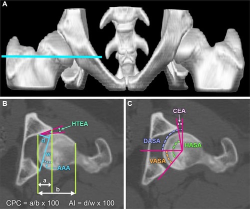
Dorsolateral subluxation is used to quantify joint laxity in a position to simulate weight-bearing (). During general anesthesia, pressure is applied to the femur at the level of the stifle while imaging the dog in ventral recumbency.Citation43 Joints with less than 45% coverage of the femoral head by the lateral aspect of the cranial acetabular rim have an increased chance of developing joint changes and OA over time compared with those with a higher percentage (>55%) of coverage.Citation43
Subjective radiographic evaluations are limited by the inherent variability associated with examiners, image quality, and differences between animals including periarticular soft tissue changes and muscle atrophy. Variation in the degree of muscle relaxation associated with sedation or anesthesia during imaging can influence the ability to identify joint abnormalities by as much as 50%.Citation44 Further, each evaluation system is distinct, so results are based on slightly different criteria. Recently, the Orthopedic Foundation for Animals score was reported to underestimate the likelihood of developing coxofemoral joint OA compared to the PennHIP distraction index.Citation45 Reporting mechanisms also vary widely in public access to individual scores for reproduction decisions. As with any measure, radiographic hip scores should not be used in isolation to evaluate and predict current and future joint structure and function. It is possible that the presence of OA at a young age may be indicative of rapidly progressive joint disease, and, given recognition of the genetic basis for the disease, consideration of the presence and extent of CHD signs in related individuals is likely warranted. Based on this information, it is clear that continued efforts to identify mechanisms for early and accurate CHD diagnosis are of utmost importance. Adaptation of knowledge from decades of research to emerging imaging modalities will, no doubt, continue to improve upon current standards.
Computed tomography
CT technology for pelvic imaging has improved considerably over the past few decades. While radiographs remain the primary method used to image canine coxofemoral joints, CT is becoming increasingly popular. Using three-dimensional CT models, a recent longitudinal study showed that volumetric changes in the acetabulum and proximal femur occurred in a predictable manner during skeletal growth in a colony of dogs with coxofemoral joint laxity.Citation46 Another study demonstrated that two-dimensional CT images and three-dimensional models created from CT images can be used to predict the presence of joint OA at maturity.Citation47 Two-dimensional CT measures included the percent femoral head coverage, acetabular index, and the following angles: acetabular anteversion, ventral, dorsal, and horizontal acetabular sector, center edge, and horizontal toit externe (). Measures on three-dimensional models included femoral head and neck volumes, femoral head and neck radii and femoral neck angle (). The 16-week distraction index and center edge angle combination was the best predictor of mature OA, whereas the 32-week dorsal acetabular sector angle and Norberg angle combination was the most effective predictor of the presence of OA at maturity. Hence, combined measures were the best mechanism for predicting development of OA, and the combinations varied with age. In a separate study, numerous measures were performed on pelvic CT scans of beagles and mixed breed dogs at various time points between the ages of 2 months and 1 year to assess the relationship of the measures with joint laxity.Citation17 The dorsal acetabular rim angle and center distance index (the distance between the femoral head and the center of the acetabulum, divided by the radius of the femoral head, or the PennHIPCitation41 distraction index) were found to be good indicators of joint laxity and dysplastic changes.Citation17 Magnetic resonance imaging is used to evaluate the three-dimensional structure of human articular soft tissues, and relatively recently, canine articular soft tissues,Citation48 but CT is best for bone structure,Citation49 and the cost of magnetic resonance imaging for screening may be prohibitive. As technology advances, and CT and magnetic resonance imaging become more readily available and affordable, use of three-dimensional imaging methodologies will likely become an integral part of diagnosis and assessment of CHD.
Figure 4 Measurements and three-dimensional models for evaluating the dysplastic canine hip.
Abbreviations: AAA, acetabular anteversion angle; AI, acetabular index; CEA, center edge angle; CPC, percentage of femoral head coverage; DASA, dorsal acetabular sector angle (dorsal coverage of the femoral head); HASA, horizontal acetabular sector angle (total acetabular coverage of the femoral head); HTEA, horizontal toit externe angle (orientation of the acetabulum); VASA, ventral acetabular sector angle (ventral coverage of the femoral head).
Therapeutic management and intervention
Conservative management
There are numerous descriptions of multifactorial systems, with numeric, visual analog, and descriptive scales to reproducibly evaluate joint pain associated with CHD.Citation50 Many of the assessments within the systems are subjective evaluations of individual behavior or responses, and there is no single gold standard with which to quantify hip pain in the dog.Citation50 While efforts continue to establish a uniform, standard evaluation system for canine hip joint pain, those systems that include multiple subjective and objective assessments by individuals who are not aware of specific treatments or conditions, are often informative.
Conservative management of CHD generally consists of a combination of mechanisms to reduce progression of joint damage and alleviate discomfort.Citation51 Nonsteroidal anti-inflammatory drugs are commonly used for pain associated with severely arthritic joints in dogs.Citation52 Numerous studies indicate that achieving and maintaining a healthy body weight contributes to delayed onset and reduced clinical signs associated with hip joint pain.Citation4,Citation5,Citation53 Various food supplements reported to alleviate signs of coxofemoral joint pain from OA range from green-lipped mussels (Perna canaliculus)Citation54 to fish oil.Citation55 Polysulfated glycosaminoglycan supplements and injections have been recommended for prevention and treatment of OA in dogs and other mammals.Citation56–Citation59 IntramuscularCitation57 and intra-articular administration has also been reported,Citation56 but responses vary.Citation51 Alternative methods that have also been investigated for the treatment of painful CHD joints include acupuncture and gold bead implantation, among others. The implantation of gold beads at acupuncture points was developed in the USA in the 1970s and implemented to a limited degree in veterinary medicine in the 1990s for degenerative joint disease pain.Citation60,Citation61 Results are mixed, with some studies showing clinical improvementCitation61,Citation62 and others showing no discernible effect.Citation63
Maintenance of optimum body weight has long been considered one of the most effective methods for reducing the signs associated with dysplasia and OA.Citation4 A lifelong dietary restriction of 25% delayed the appearance of OA as well as the intensity of clinical signs in Labrador Retrievers compared with feeding ad libitum.Citation53 Weight loss in conjunction with physiotherapy that included transcutaneous electrical nerve stimulation improved the clinical outcome for obese dogs with radiographic signs of OA.Citation64 Recently, intra-articular botulinum toxin A was reported to reduce the pain associated with OA based on improvements in limb use (ie, gait patterns) measured with a force platform.Citation65 At present, there are few reports of long-term studies concerning the efficacy of nonsurgical or conservative treatment of CHD joint changes. These studies can be limited by the challenges of consistent monitoring and reporting by multiple and individual owners, as well as a wide range of disease severity and canine personalities.Citation66 However, a recent retrospective report indicates that conservative and nonsurgical management (ie, weight control, reduced exercise, and analgesics) of 74 dogs over the span of 13 years did not improve quality of life as anticipated from previous reports.Citation66
There is significant interest in the use of regenerative medicine to treat signs of CHD and OA; however, much of the information reported is subjective in nature. Currently, numerous controlled, preclinical and clinical trials are underway that may provide some perspective on the value of this emerging technology. Intra-articular injection of adipose-derived stem cells has been found to be a safe therapeutic approach for the treatment of symptoms associated with OA.Citation67 Preliminary studies show that injection of adipose-derived stem cells into affected joints may reduce clinical signs of hip pain (ie, lameness) based on subjective clinical evaluationsCitation67 and force platform gait analysis.Citation68,Citation69 A randomized comparison between a single intra-articular injection of adipose-derived stem cells or plasma rich in growth factors showed that both treatments reduced behavior associated with pain, but that the adipose-derived stem cells appeared to be more effective for up to 6 months post-treatment based upon owner assessments.Citation70 This information clearly demonstrates that there is more work to be done on the efficacy of conservative and alternative methods to manage signs of CHD.
Surgery
Despite the prevalence of CHD, a gold standard surgical procedure has yet to be identified.Citation71 As such, there are numerous surgeries to prevent progression of degenerative joint changes or alleviate pain and restore joint function.
Some surgical procedures designed to prevent onset of OA in hips identified as being predisposed to development of OACitation72 include double and triple pelvic osteotomy, acetabular shelf and excision arthroplasty, femoral osteotomy, and juvenile pubic symphysiodesis.Citation73 Both juvenile pubic symphysiodesis and triple pelvic osteotomy are designed to increase femoral head coverage by ventrolateral rotation of the acetabulum. The juvenile pubic symphysiodesis procedure involves premature closure of the pubic symphysis.Citation72,Citation74 Resulting reduction in the pelvic inlet width causes ventrolateral rotation of the acetabulum during pelvic growth, and is thought to result in a 40%–46% improvement in acetabular and dorsal acetabular rim angles compared to no treatment.Citation73–Citation75 Juvenile pubic symphysiodesis appears to have the best outcomes when performed in puppies that are 12–16 weeks old.Citation73 Triple pelvic osteotomy is a much more extensive procedure, and involves osteotomies of the ilium, pubis, and ischium to allow manual rotation of the acetabulum for better femoral head coverage.Citation76 The ilial osteotomy is stabilized with bone plates customized to accommodate the rotation.Citation77 This procedure is generally recommended for young dogs without irreversible (or with mild) degeneration of the coxofemoral joint.Citation77,Citation78 Information about long-term outcomes of the various surgical treatments is limited. Preliminary reports indicate that juvenile pubic symphysiodesis and triple pelvic osteotomy minimally affect laxity and femoral head coverage when performed at 5 months or olderCitation72 compared with earlier reports indicating that the procedure performed slightly earlier (15 weeks) improved acetabular coverage of the femur.Citation75 Mechanisms to reduce the OA characteristic of CHD will undoubtedly improve quality of life for affected dogs. Long-term outcomes will help identify treatments toward this end.
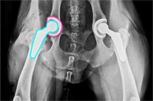
Total hip replacement () is often applied in advanced cases of joint degeneration and is considered a salvage procedure.Citation79 There are no clear guidelines for the best time to implement total hip replacement, but the average time between onset of signs and surgery is 10 months.Citation80 Total hip replacement procedures in dogs began in the 1970s, and a modular system was introduced in the mid 1990s that coupled a fixed monobloc cobalt-chromium alloy femoral implant with an acetabular cup for cemented fixation.Citation81 Further refinements to total hip replacement implants have contributed to a high clinical recovery rate, with loosening of the acetabular cup and cup wear reported as some of the most common complications.Citation81 More recently, cementless fixation has been developed, and is reported to have positive results.Citation79 As the name implies, femoral and acetabular implants are cemented to bone for cemented total hip replacement. In contrast, cementless fixation or uncemented implants are designed so that the bone grows into or onto the prosthesis without the need for cement at the bone–implant interface. Implant loosening is reported to be less than for cemented implants.Citation79,Citation82 A primary concern associated with total hip replacement is the potential for an inflammatory response to implant particulate wear debris from aseptic implant loosening.Citation83 Another consideration is that the persistence of joint laxity may influence the outcomes of total hip replacement.Citation84 Efforts continue to improve upon available total hip replacement implants for dogs. A hybrid system of a cementless acetabular cup and a cemented femoral implant has been successfully applied in dogs relatively recently.Citation85 Hip resurfacing to replace only joint surfaces versus the entire jointCitation86 in dogs is under development, but clinical trials have yet to be reported.Citation81 Unfortunately, large-scale, prospective randomized studies have yet to be conducted for comparison of long-term outcomes for various surgical procedures and nonsurgical management. Hence, individualized care remains largely based on clinician preference and experience.Citation71
Figure 5 Radiograph illustrating a bilateral total hip replacement.
Genetics
While diagnosis and treatment of CHD are central to individual patient care, prevention by selective breeding will help obviate the presence of a debilitating condition in canine companions. With this in mind, there has been significant effort focused on identifying specific genes, genetic mutations, and quantitative trait loci (regions of chromosomes containing DNA for a specific trait), to use in conjunction with standard imaging methods for identification of CHD carriers.Citation87,Citation88 Genetic screening programs are complicated by the polygenic nature of CHD and related OA, as well as environmental influences on phenotypic expression. A few promising quantitative trait loci for OA associated with CHDCitation89 and the CHD phenotype in German shepherdsCitation90 have been identified relatively recently. Additionally, several chromosomal markers for CHD have been reported for a population of cross-bred Labrador Retriever–Greyhounds.Citation91 Notably, several specific single nucleotide polymorphisms and positional candidate genes in dogs with CHD have been found to correlate with genes associated with the expression of OA and developmental dysplasia of the hip in humans.Citation87 While the genomic underpinnings of CHD remain largely elusive, significant progress has been made and will continue, with expanding knowledge of the canine genome and interactions among genes that influence their expression.
Future directions
Despite almost a century of research, the complex etiology and optimal treatment paradigm for CHD remain elusive. As originally proposed by Schnelle at a meeting of the Veterinary Medical Society of New York City in the 1930s,Citation92 CHD is not likely a single affliction but an array of heritable and environmentally induced degenerative disorders that differentially affect the morphology and function of the canine hip. The variable phenotypic expression of CHD makes development and implementation of standard identification procedures difficult. It may be possible to identify specific phenotypes within the broad spectrum of CHD similar to those of the human hip, like acetabular rim syndrome, acetabular retroversion, and femoral head necrosis. The relationship between articular damage in the human hip with morphology suggests a need to evaluate similar relationships in the dog.Citation93 Identification and characterization of CHD phenotypes at the genetic, microstructural, and macrostructural levels will likely contribute to early detection and informed breeding decisions. Another area that will continue to promote progress in both imaging and treatment is development of novel measures on images obtained with contemporary imaging modalities like CT and magnetic resonance imaging. As evidence-based assessments of CHD prevention and treatment strategies become available, their selection and implementation will improve and facilitate the development of novel clinical approaches and surgical procedures. Incremental advances in the diagnosis, treatment, and prevention of the joint degeneration and pain associated with CHD through focused research and clinical evidence will continue to progress toward diminishing and eradicating CHD from our canine companions.
Disclosure
The authors report no conflicts of interest in this work.
References
- LustGAn overview of the pathogenesis of canine hip dysplasiaJ Am Vet Med Assoc199721014431445
- ZhangZWZhuLSandlerJEstimation of heritabilities, genetic correlations, and breeding values of four traits that collectively define hip dysplasia in dogsAm J Vet Res20097048349219335104
- SchnelleGBBilateral congenital subluxation of the coxo-femoral joints in a dogUniversity of Pennsylvania Bulletin School of Veterinary Medicine Veterinary Extension Quarterly1937371516
- ImpellizeriJATetrickMAMuirPEffect of weight reduction on clinical signs of lameness in dogs with hip osteoarthritisJ Am Vet Med Assoc200121610891091
- KealyRDLawlerDFBallamJMEvaluation of the effect of limited food consumption on radiographic evidence of osteoarthritis in dogsJ Am Vet Med Assoc20002171678168011110459
- SteinetzBGWilliamsAJLustGSchwabeCBüllesbachEEGoldsmithLTTransmission of relaxin and estrogens to suckling canine pups via milk and possible association with hip joint laxityAm J Vet Res200869596718167088
- GreeneLMMarcellin-LittleDJLascellesBDAssociations among exercise duration, lameness severity, and hip joint range of motion in Labrador retrievers with hip dysplasiaJ Am Vet Med Assoc20132421528153323683017
- MadsenJSReimannISvalastogaEDelayed ossification of the femoral head in dogs with hip dysplasiaJ Small Anim Pract199132351354
- TodhunterRJZachosTAGilbertROOnset of epiphyseal mineralization and growth plate closure in radiographically normal and dysplastic Labrador retrieversJ Am Vet Med Assoc1997210145814629154197
- The Orthopedic Foundation for Animals [webpage on the Internet]The OFA’s Hip Radiograph Procedures Available from: http://www.offa.org/hd_procedures.htmlAccessed January 10, 2015
- RobertsTMcGreevyPDSelection for breed-specific long-bodied phenotypes is associated with increased expression of canine hip dysplasiaVet J201018326627219959383
- FarquharTBertramJTodhunterRJBurton-WarsterNLustGVariations in composition of cartilage from shoulder joints of young dogs at risk for developing canine hip dysplasiaJ Am Vet Med Assoc1997210148314859154202
- OlsewskiJMLustGRendanoVTSummersBADegenerative joint disease: multiple joint involvement in young and mature dogsAm J Vet Res198344130013086881668
- LeppänenMMäkiKJugaJSaloniemiHFactors affecting hip dysplasia in German shepherd dogs in Finland: efficacy of the current improvement programmeJ Small Anim Pract200041192310713978
- TodhunterRJLustGHip dysplasia: pathogenesisSlatterEDTextbook of Small Animal SurgeryPhiladelphia, PA, USASaunders2003
- Vanden Berg-FoelsWSTodhunterRJSchwagerSJReevesAPEffect of early postnatal body weight on femoral head ossification onset and hip osteoarthritis in a canine model of developmental dysplasia of the hipPediatr Res20066054955416988183
- FujikiMKurimaYYamanokuchiKMisumiKSakamotoHComputed tomographic evaluation of growth-related changes in the hip joints of young dogsAm J Vet Res20076873073417605608
- RiserWHThe dysplastic hip joint: radiologic and histologic developmentVet Pathol197512279305
- WuFMHedhammarÅKrookLOvernutrition and skeletal disease. An experimental study in growing Great Dane dogs. IX. The long bonesCornell Vet19746483114
- ChalmersHJDykesNLLustGAssessment of bone mineral density of the femoral head in dogs with early osteoarthritisAm J Vet Res20066779680016649912
- BarrARSBennyHRGibbsCClinical hip dysplasia in growing dogs: the long-term results of conservative managementJ Small Anim Pract198728243252
- BijlsmaJWBerenbaumFLafeberFPOsteoarthritis: an update with relevance for clinical practiceLancet20113772115212621684382
- BuckwalterJAMartinJAOsteoarthritisAdv Drug Deliv Rev20065815016716530881
- GrantMEProckopDJThe biosynthesis of collagenN Engl J Med19722861941994257078
- SkirvingAPSimsTJBaileyAJCongenital dislocation of the hip: a possible inborn error of collagen metabolismJ Inherit Metab Dis1984727316429438
- MadsenJSThe joint capsule and joint laxity in dogs with hip dysplasiaJ Am Vet Med Assoc1997210146314659154198
- FryTRClarkDMCanine hip dysplasia: clinical signs and physical diagnosisVet Clin North Am Small Anim Pract1992225515581604772
- ChalmanJAButlerHCCoxofemoral joint laxity and the Ortolani signJ Am Anim Hosp Assoc198521671676
- GinjaMMDSilvestreAMGonzalo-OrdenJMFerreiraAJDiagnosis, genetic control and preventive management of canine hip dysplasia: a reviewVet J201018426927619428274
- PuertoDASmithGKGregorTPRelationship between results of the Ortolani method of hip joint palpation and distraction index, Norberg angle, and hip score in dogsJ Am Vet Med Assoc199921449750110029850
- BardensJWHardwickHNew observations on the diagnosis and cause of hip dysplasiaVet Med Small Anim Clin1968632382455183841
- RislerAKlauerJMKeulerNSAdamsWMPuppy line, metaphyseal sclerosis, and caudolateral curvilinear and circumferential femoral head osteophytes in early detection of canine hip dysplasiaVet Radiol Ultrasound20095015716619400461
- SzaboSDBieryDNLawlerDFEvaluation of a circumferential femoral head osteophyte as an early indicator of osteoarthritis characteristic of canine hip dysplasia in dogsJ Am Vet Med Assoc200723188989217867972
- GibbsCThe BVA/KC scoring scheme for control of hip dysplasia: interpretation of criteriaVet Rec19971412752849316245
- DennisRInterpretation and use of BVA/KC hip scores in dogsIn Practice201234178194
- Fédération Cynologique Internationale [webpage on the Internet]FCI Scientific Commission Available from: http://www.fci.be/en/FCI-Scientific-Commission-71.htmlAccessed January 10, 2015
- ComhaireFHCrielACCDassyCAAGuévarPGJSnapsFRPrecision, reproducibility, and clinical usefulness of measuring the Norberg angle by means of computerized image analysisAm J Vet Res20097022823519231956
- SmithGKGregorTPRhodesWHBieryDNCoxofemoral joint laxity from distraction radiography and its contemporaneous and prospective correlation with laxity, subjective score and evidence of degenerative joint disease from conventional hip-extended radiographyAm J Vet Res199354102110428368595
- SmithGKBieryDNGregorTPNew concepts of coxofemoral joint stability and development of a clinical stress-radiographic method for quantitating hip joint laxity in the dogJ Am Vet Med Assoc199019659702295555
- LustGWilliamsABurton-WarsterNJoint laxity and its association with hip dysplasia in Labrador retrieversAm J Vet Res199354199019998116927
- PennHIP [webpage on the Internet]PennHIP Method: Measuring Hip Joint Laxity http://info.antechimagingservices.com/pennhip/navigation/penn-HIP-method/distraction-index-measuring-laxity.htmlAccessed January 10, 2015
- LopezMJLewisBPSwaabMEMarkelMDRelationships among measurements obtained by use of computed tomography and radiography and scores of cartilage and microdamage in hip joints with moderate to severe joint laxity of adult dogsAm J Vet Res20086936237018312135
- FareseJPTodhunterRJLustGWilliamsAJDykesNLDorsolateral subluxation of hip joints in dogs measured in a weight-bearing position with radiography and computed tomographyVet Surg1998273934059749508
- MalmSStrandbergEDanellBAudellLSwensonLHedhammarÅImpact of sedation method on the diagnosis of hip and elbow dysplasia in Swedish dogsPrev Vet Med20077819620917112612
- PowersMYKarbeGTGregorTPEvaluation of the relationship between Orthopedic Foundation for Animals’ hip joint scores and PennHIP distraction index values in dogsJ Am Vet Med Assoc201023753254120807130
- D’AmicoLLXieLAbellLKBrownKTLopezMJRelationships of hip joint volume ratios with degrees of joint laxity and degenerative disease from youth to maturity in a canine population predisposed to hip joint osteoarthritisAm J Vet Res20117237638321355741
- AndronescuAAKellyLKearneyMTLopezMJAssociations between early radiographic and computed tomographic measures and canine hip joint osteoarthritis at maturityAm J Vet Res201576192725535657
- GinjaMMDFerreiraAJJesusSSComparison of clinical, radiographic, computed tomographic and magnetic resonance imaging methods for early prediction of canine hip laxity and dysplasiaVet Radiol Ultrasound20095013514319400458
- PreidlerKWResnickDImaging of osteoarthritisRadiol Clin North Am1996342592718633115
- Hielm-BjörkmanAKKuuselaELimanAEvaluation of methods for assessment of pain associated with chronic osteoarthritis in dogsJ Am Vet Med Assoc20032221552155812784961
- KirkbyKALewisDDCanine hip dysplasia: reviewing the evidence for nonsurgical managementVet Surg2012412922150604
- PollmeierMToulemondeCFleishmanCHansonPDClinical evaluation of firocoxib and carprofen for the treatment of dogs with osteoarthritisVet Rec200615954755117056650
- SmithGKPasterERPowersMYLifelong diet restriction and radiographic evidence of osteoarthritis of the hip joint in dogsJ Am Vet Med Assoc200622969069316948575
- RiallandPBichotSLussierBEffect of a diet enriched with green-lipped mussel on pain behavior and functioning in dogs with clinical osteoarthritisCan J Vet Res201377667423814358
- Hielm-BjörkmanARoineJEloKLappalainenAJunnilaJLaitinen-VapaavuoriOAn un-commissioned randomized, placebo-controlled double-blind study to test the effect of deep sea fish oil as a pain reliever for dogs suffering from canine OABMC Vet Res2012815722950577
- FujikiMShinehaJYamanokuchiKMisumiKSakamotoHEffects of treatment with polysulfated glycosaminoglycan on serum cartilage oligomeric matrix protein and C-reactive protein concentrations, serum matrix metalloproteinase- 2 and -9 activities, and lameness in dogs with osteoarthritisAm J Vet Res20076882783317669022
- LustGWilliamsAJBurton-WarsterNBeckKARubinGEffects of intramuscular administration of glycosaminoglycan polysulfates on signs of incipient hip dysplasia in growing pupsAm J Vet Res199253183618431456530
- TodhunterRJLustGPolysulfated glycosaminoglycan in the treatment of osteoarthritisJ Am Vet Med Assoc1994204124512518014098
- WhiteGWFreginGFSeldenJREffect of prophylactic intramuscular administration of polysulfated glycosaminoglycan on developmental and traumatic joint injuries in thoroughbred foalsJ Equine Vet Sci200727107111
- DurkesTEGold bead implantsProbl Ved Med19924207211
- JaegerGTLarsenSSøliNMoeLDouble-blind, placebo-controlled trial of the pain-relieving effects of the implantation of gold beads into dogs with hip dysplasiaVet Rec200615872272616731702
- JaegerGTLarsenSSøliNMoeLTwo years follow-up study of the pain-relieving effect of gold bead implantation in dogs with hip-joint arthritisActa Vet Scand200749917381835
- BollingerCDecampCEStajichMGait analysis of dogs with hip dysplasia treated with gold bead implantation acupunctureVet Comp Orthop Traumatol200215116122
- MlacnikEBockstahlerBAMüllerMTetrickMANapRCZentekJEffects of caloric restriction and a moderate or intense physiotherapy program for treatment of lameness in overweight dogs with osteoarthritisJ Am Vet Med Assoc20062291756176017144822
- HeikkläHMHielm-BjörkmanAKMoreliusMIntra-articular botulinum toxin A for the treatment of osteoarthritic joint pain in dogs: a randomized, double-blinded, placebo-controlled clinical trialVet J201420016216924675370
- FarrellMClementsDNMellorDRetrospective evaluation of the long-term outcome of non-surgical management of 74 dogs with clinical hip dysplasiaVet Rec200716050651117435096
- MarxCSilveriraMDSelbachIAcupoint injection of autologous stromal vascular fraction and allogeneic adipose-derived stem cells to treat hip dysplasia in dogsStem Cells Int2014201439127425180040
- VilarJMBatistaMMoralesMAssessment of the effect of intraarticular injection of autologous adipose-derived mesenchymal stem cells in osteoarthritic dogs using a double blinded force platform analysisBMC Vet Res20141014324984756
- VilarJMMoralesMSantanaAControlled, blinded force platform analysis of the effect of intraarticular injection of autologous adipose-derived mesenchymal stem cells associated to PRGF-Endoret in osteoarthritic dogsBMC Vet Res2013913123819757
- CuervoBRubioMSopenaJHip osteoarthritis in dogs: a randomized study using mesenchymal stem cells from adipose tissue and plasma rich in growth factorsInt J Mol Sci201415134371346025089877
- BerghMSBudsbergSCA systematic review of the literature describing the efficacy of surgical treatments for canine hip dysplasiaVet Surg20144350150624837650
- ManleyPAAdamsWMDanielsonKCDuelandRTLinnKALong-term outcome of juvenile pubic symphysiodesis and triple pelvic osteotomy in dogs with hip dysplasiaJ Am Vet Med Assoc200723020621017223752
- DuelandRTAdamsWMPatricelliAJLinnKACrumpPMCanine hip dysplasia treated by juvenile pubic symphysiodesis. Part I: two year results of computed tomography and distraction indexVet Comp Orthop Traumatol20102330631720740258
- DuelandRTAdamsWMFialkowskiJPPatricelliAJMathewsKGNordheimEVEffects of pubic symphysiodesis in dysplastic puppiesVet Surg20013020121711340551
- PatricelliAJDuelandRTAdamsWMFialkowskiJPLinnKANordheimEVJuvenile pubic symphysiodesis in dysplastic puppies at 15 and 20 weeks of ageVet Surg20023143544412209414
- SlocumBDevineTPelvic osteotomy technique for axial rotation of the acetabular segment in dogsJ Am Anim Hosp Assoc198622331338
- BorostyankoiFRooksRLKoblukCNReedALLittledikeETResults of single-session bilateral triple pelvic osteotomy with an eight-hole iliac bone plate in dogs: 95 cases (1996–1999)J Am Vet Med Assoc2003222545912523481
- RoseSABrueckerKAPetersenSWUddinNUse of locking plate and screws for triple pelvic osteotomyVet Surg20124111412022150566
- FitzpatrickNLawAYBieleckiMBGirlingSCementless total hip replacement in 20 juveniles using BFX™ arthroplastyVet Surg20144371572524962265
- LockwoodAALiskaWDDuration of clinical signs prior to total hip replacement in dogsJ Am Vet Med Assoc201123890590821453179
- AllenMJAdvances in total joint replacement in small animalsJ Small Anim Pract20125349550622943214
- MorshedSBozicKJRiesMDMalchauHColfordJMComparison of cemented and uncemented fixation in total hip replacementActa Orthop20077831532617611843
- HarrisWHWear and periprosthetic osteolysis: the problemClin Orthop Relat Res20013936670
- HayesGMRamirezJHobbsJLDoes the degree of preoperative subluxation or soft tissue tension affect the incidence of postoperative luxation in dogs after total hip replacement?Vet Surg200940613
- GemmillTJPinkJRenwickAHybrid cemented/cementless total hip replacement in dogs: seventy-eight consecutive joint replacementsVet Surg201040621630
- BozicKJPuiCMLudemanMJVailTPSilversteinMDDo the potential benefits of metal-on-metal hip resurfacing justify the increased cost and risk of complications?Clin Orthop Relat Res20104682301231220232182
- ZhouZShengXZhangZDifferential genetic regulation of canine hip dysplasia and osteoarthritisPLoS One20105e1321920949002
- ZhuLZhangZFriedenbergSSThe long (and winding) road to gene discovery for canine hip dysplasiaVet J20091819711019297220
- MateescuRGBurton-WarsterNTsaiKPhavaphutanonJZhangZMurphyKEIdentification of quantitative trait loci for osteoarthritis of hip joints in dogsAm J Vet Res2008691294130018828685
- FelsLDistlOIdentification and validation of quantitative trait loci (QTL) for canine hip dysplasia (CHD) in German shepherd dogsPLoS One20149e9661824802516
- TodhunterRJMateescuRGLustGQuantitative trait loci for hip dysplasia in a cross breed canine pedigreeMamm Genome20051672073016245029
- NobleHWhat radiology does for dogs: through x-ray pictures, veterinarians have been able to catalogue canine illsAmerican Kennel Gazette193519352527
- KayaMSuzukiTEmoriMYamashitaTHip morphology influences the pattern of articular cartilage damageKnee Surg Sports Traumatol Arthrosc9112014 Epub ahead of print