Abstract
The purpose of this study was to investigate and add reference data about the musculoskeletal system in women. The mechanography system of the Leonardo™ platform (Novotec, Germany) was used to measure parameters of movement (velocity, force, power) in 176 healthy Greek women aged 20–79 years, separated according to age decade in six groups: group 1 (n = 12), 20–29 years; group 2 (n = 14), 30–39 years; group 3 (n = 33), 40–49 years; group 4 (n = 59), 50–59 years including 21 postmenopausal; group 5 (n = 31), 60–69 years including 12 postmenopausal; and group 6 (n = 27), 70–79 years all postmenopausal. This system measures forces applied to the plate over time, calculates through acceleration the vertical velocity of center of gravity and using force and velocity it calculates power of vertical movements. All women performed a counter-movement jump (brief squat before the jump) with freely moving arms. Weight was recorded on the platform before the jump and height was measured with a wall-mounted ruler. Body weight and body mass index were gradually increased; on the contrary height and all movement parameters except force (velocity, power) were statistically decreased during aging and after menopause.
Introduction
Nowadays, the lifestyle in western societies is changing. This change is followed by a dramatic increase in diseases related to impaired muscle function.Citation1 In addition, it has been well documented that both muscle mass and power decline with ageCitation2–Citation4 and this decline is associated with physical impairment and an increased risk of fallsCitation5 and hip fracturesCitation6 leading to disability. In relation to movement disabilities, locomotion in the literature is described by confusing concepts that often do not agree with the rules and terms of physics.Citation1 A scientific description of movement is needed in this area to enable researchers to communicate with one another. In the study of muscle performance, movement has to be described in terms of velocity and acceleration. Each movement is the action of force along a distance in a certain time and is therefore measured as power.Citation7 This mechanical approach is in accordance with physics and enables the measurement and calculation of human movement using scientific concepts.Citation8
For all these reasons, knowledge of parameters of the musculoskeletal system influencing muscle function in aging is important. The purpose of this paper is to compile reference data and discuss parameters of the locomotor (human musculoskeletal) system in Greek women.
Methods
Population
The study population consisted of community dwelling women from different municipalities of Greece who visited the Laboratory for Research of the Musculoskeletal System in Athens for a screening program for osteoporosis. We included 176 healthy women aged 20–79 years without any musculoskeletal or neurologic disease. The women were separated according into six groups according to age: group 1 (n = 12), 20–29 years; group 2 (n = 14), 30–39 years; group 3 (n = 33), 40–49 years; group 4 (n = 59), 50–59 years including 21 postmenopausal; group 5 (n = 31), 60–69 years including 12 postmenopausal; and group 6 (n = 27), 70–79 years all postmenopausal. None of them were taking any antiosteoporotic drug or calcium/vitamin D supplements.
Jumping mechanography system
For the measurement of the objective parameters of movement we used the Leonardo™ Mechanography Ground Reaction Force Platform, (NOVOTEC Medical GmBH, Pforzheim, Germany). This system measures forces (f, Newton) applied to the plate over time. This means that stationary forces as well as the variation of forces over time (ground reaction forces) can be investigated. The vertical velocity is calculated through acceleration (v, m/sec) of centre of gravity. Using force and velocity, the system calculates the power (p, Watt) of vertical movements. The continuous registration of force, velocity, and power gives insight to the eccentric phases of movement.Citation8 Jumping mechanography was recently found to be a reliable and sensitive measure of mobility performance in elite athletes as well as in frail patients.Citation9
Maximum height of two leg jump
After explaining the process to the participants, all performed jumps (two-leg jump) on the Leonardo platform. The first phase of jumping was squatting as a counter-movement to store energy in the elastic elements. Jumping was performed with freely moving arms and the instruction was to jump with the head and chest as high as possible thus producing the maximum elevation of the center of mass. Each woman performed three counter-movement jumps. The jump of greatest height was used for data analysis. The most important outcome parameter of this test is the maximum power output (peak power) during the lift-off of the jump phase normalized to the body weight of the patient (personal power).
Study parameters
Weight (kg) was recorded on the platform before the jump and height (m) was measured with a wall-mounted ruler. Body mass index (BMI) was calculated for each subject (BMI = weight (kg)/height2). We studied kinematic and kinetic parameters (velocity, force, power, and power/weight as personal power). Subjects with velocity below 0.04 m/sec in the examination were unable to follow the study methodology and were excluded.
Statistical analysis
All quantitative data were represented by the number of patients (n), mean value (mean), and standard deviation (SD), and qualitative data by the number of patients (n) and percentage (%). Quantitative variables were analyzed using the one-way and two-way analysis of variance (ANOVA) models. Pairwise multiple comparisons took place using the Bonferroni test. Qualitative variables were analyzed using the χ2 test and Fisher’s exact test. All tests are two-sided with 95% significance level. Statistical analysis was performed using the statistical package SPSS (version 12.00; SPSS Inc., Chicago, IL, USA).
Results
Anthropometric and movement parameters of the study population are presented in . Height declined over the age range (p < 0.001), while body weight and BMI increased until the end of the sixth decade of life and thereafter decreased (p = 0.01 and p = 0.001, respectively). Multiple comparison analysis showed a significant difference in weight between group 2 versus groups 4, 5, and 6 (p = 0.047, p = 0.05, and p = 0.046, respectively), height between group 1 versus groups 4, 5, and 6 (p = 0.027, p = 0.037, and p = 0.001, respectively). BMI was significantly different between group 1 versus groups 4, 5, 6 (p = 0.019, p = 0.022, and p = 0.001, respectively) and between group 2 versus groups 4, 5, 6 (p = 0.034, p = 0.031, p = 0.003, respectively). Locomotor parameters in healthy women (except force, p = 0.085), showed a progressive decrease (p < 0.001) according to menopausal status and a negatively strong correlation with advancing age (velocity: r = −0.58, power: r = −0.6, power/weight: r = −0.64), which was greater for the parameters concerning power. Personal power declined continuously across the age range from the young to the elderly women ().
Figure 1 Age-declined power/weight parameter for Greek women (y = 45.98–0.41x, R2 = 0.41; p < 0.001).
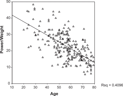
Table 1 Anthropometric data and movement parameters of Greek women in our population
Discussion
In this cross-sectional study, we measured musculoskeletal parameters to establish the changes with age in these variables. We also studied to what extent menopause had any influence on kinetic and kinematic parameters.
Changes in musculoskeletal system with age are becoming an important issue because of the increasingly elderly population. Reference values for kinetic and kinematic parameters might be useful in the clinical assessment of pathologies and the evaluation of therapeutic interventions.Citation10–Citation17
Fricke and Schoenau reviewed the literature and explained why anthropometric characteristics, mainly body height and body mass, are important factors which influence the recording of muscle function.Citation18 Besides anthropometry, hormones also influence muscle function. Low postmenopausal estrogen levels were associated with lower strength of the abductor pollicis muscle.Citation19
In our study population, body weight increased while height decreased with aging. In addition, younger generations tend to be taller meaning that the differences found between young and old women could be partially associated with differences in height and weight. In women, weight is often gained because of less activity, higher calorie intake, or a loss in lean muscle. In most cases it is probably a combination of all three factors that makes women gain weight with age. In this study, women tended to start gaining weight during the perimenopausal period before the menopause. This is thought to be caused when estrogen levels are starting to decline. In our population, the BMI, a very simple measurement of fat, was slowly rising during aging until the end of the sixth decade of life. This was a consequence of the increase in body weight and decrease in height. However, and in line with this study, other authors noticed that BMI decreases in later years of life and the reason of this decrease above 80 years of age is sarcopenia, an age-related muscle mass loss.Citation20–Citation22 Aging is associated with anatomical changes leading to physical impairment because of a gradual loss of bone and a progressive decline in muscle mass and power. Weight stability in elderly years, also found in this study, is often a mark of sarcopenia,Citation23 which is due in part to other age-related changes in body composition such as increased fat mass and BMI values (p < 0.001 versus groups 1–4).Citation24,Citation25
Velocity declined during aging because the critical factor may be a greater percentage of slow twitch muscle fibers in older people which reduces the maximum contraction speed.Citation26 In addition, the force we need for a movement against gravity is a summation of quickly released energy which has been previously stored in elastic elements during (eg, eccentric counter-movements), and currently generated muscle force by the actin-myosin-system.Citation8 During old age, the elastic modulus of the Achilles tendon declines and whole tendon stiffness is decreased.Citation27
There was a high statistical difference in force unrelated to age in every age decade of our sample. Sportive people have in this specific movement (two leg jumps at maximum height) a maximum force of 2.5 ± 0.3 times their body weight (Rawer Rainer, personal communication), which was not the case in our population of community dwelling women. Furthermore, we didn’t find any significant difference in force among age decades, but found an accelerated post-menopausal fall-off in power (p = 0.001) and strength (p = 0.08, NS). According to Skelton and colleagues, muscle power is lost more rapidly than force between the ages of 65 and 90 years (−3.5% per year for the former compared with about 1.8% for the latter).Citation28 Cross-sectional studies have shown that elderly individuals are weaker than young adultsCitation29–Citation31 and these reports are supported by longitudinal studies demonstrating a continual strength decline with aging,Citation32–Citation34 which is suggested to accelerate after the sixth decade of life.Citation35 Nevertheless, force parameter losses appear to be partially reversible.Citation36 For example, Morse and colleagues investigated the reversibility of the decline in specific force in old age in response to long-term (12 months) resistive loading of males aged over 70 years and found an increase (p = 0.05) in specific force.Citation37
Anthropometric characteristics, mainly body height and body mass, are important factors which influence the recording of muscle function. Therefore we need to evaluate power in relation to body size parameters.Citation18 Dependent on personal power and weight, the comparison between persons according to weight results in the power/weight parameter. Runge and colleagues showed a very good correlation between maximum power output per body weight and age for both sexes separately in a healthy sportive reference collective.Citation38 In Runge and colleagues’ cross-sectional study of more than 200 subjects aged between 18 and 88 years, the decline in power/weight parameter was more than 50% from the ages of 20 years to 80 years.Citation38 In the women of our study, there was a 56% fall from the 20–29 decade versus the 70–79 decade and an accelerated postmenopausal fall-off in power. The decline in personal power was continuous across the entire age range from the young to the very elderly women. Possible reasons are changes in body composition, reduction of skeletal mass, and tendon properties. Several factors have to be considered for the age-related decline in power output and are well summarized in the paper by Runge and colleagues.Citation38 According to these authors, fat mass and extracellular space increases with age and makes up a passive mass which does not contribute to strength or power, while muscle mass may be lost during aging to a different degree in different muscles. In sedentary and mildly active subjects, muscle thickness appears to decline 40% more with age in the vastus lateralis muscle than in the medial gastrocnemius muscle and this suggest that we need to consider locally specific patterns of muscle atrophy. A third group of explanatory mechanisms focuses on skeletal muscle. It has been recognized recently that, all things being equal, changes in muscle cross-sectional area (CSA) should affect output power more than proportionally. This effect could potentially explain why comparatively small reductions in muscle CSA with age in past studies were paralleled by a decline in power output. Moreover, and according to the same authors, aging appears to affect the muscle-fiber pennation angle.
Menopause has been linked to a reduction in lean mass (LM) and bone mineral density (BMD).Citation39–Citation41 It is easy to connect the loss of estrogens with the decline in muscle mass during aging. There is debate about the positive association of muscle mass and estrogens, but the strength of evidence in support of an anabolic effect of estrogens on skeletal muscle via meta-analysis outweighs the evidence of no effect.Citation42–Citation44
In conclusion, the study presents reference values measured by jumping mechanography in women. The data might be useful to assess pathologies and to study the effects of any therapeutic interventions according to muscles and power.
Acknowledgements
We would like to thank Johannes Willnecker and Rainer Rawer from Stratec, Pforzheim, Germany for technical advice and especially all the Greek women who took part in this study. The authors report no conflicts of interest.
Disclosures
Part of this paper was presented in the following congresses: 1) XXI Paulo Symposium on Preventing Bone Fragility and Fractures, Tampere, Finland, May 2006. Oral presentation: “Kinetic parameters in pre and postmenopausal women”. 2) 6th Mediterranean Congress of Physical and Rehabilitation Medicine. Vilamoura, Portugal, October 18–21, 2006. Oral presentation. “Study of locomotive parameters in physically competent women”. 3) 34th European Symposium on Calcified Tissues, May 2007, Copenhagen, Denmark. Poster presentation: “Physically competent women decline in kinetic parameters during aging”. 4) 6th International Workshop for Musculoskeletal and Neuronal Interactions, Cologne 2008. Oral presentation: “Evaluation of muscle function in pre and postmenopausal women”. 5) 16th European Congress of Physical and Rehabilitation Medicine, Bruges, June 2008, TESC competition. “Assessment of kinetic parameters in premenopausal and postmenopausal women with jumping mechanography”.
References
- RungeMMeasurement of human movements by mechanography. Abstracts bookBad Liebenzell, GermanyInternational Society of Musculoskeletal and Neuronal Interactions, Black Forest Forum52006
- LindleRSMetterEJLynchNAAge and gender comparisons of muscle strength in 654 women and men aged 20–93 yrsJ Appl Physiol199783158115879375323
- KallmanDAPlatoCCTobinJDThe role of muscle loss in the age-related decline of grip strength: cross-sectional and longitudinal perspectivesJ Gerontol A Biol Sci Med199045M82M88
- LarssonLGrimbyGKarlssonJMuscle strength and speed of movement in relation to age and muscle morphologyJ Appl Physiol197946451456438011
- CampbellAJBorrieMJSpearsGFRisk factors for falls in a community-based prospective study of people 70 years and olderJ Gerontol198944M112M1172738307
- AnianssonAZetterbergCHedbergMImpaired muscle function with aging: a background factor in the incidence of fractures of the proximal end of the femurClin Orthop1984191192210
- RungeMSchießlHRittwegerJKlinische Diagnostik des Regelkreises Muskel-Knochen am UnterschenkelOsteologie200212537
- RungeMSchachtEMultifactorial pathogenesis of falls as a basis for multifactorial interventionsJ Musculoskelet Neuronal Interact2005512713415951628
- RittwegerJSchiesslHFelsenbergDRungeMReproducibility of the jumping mechanography as a test of mechanical power output in physically competent adult and elderly subjectsJ Am Geriatr Soc20045212813114687327
- SamsonMMMeeuwsenIBCroweADessensJADuursmaSAVerhaarHJRelationships between physical performance measures, age, height and body weight in healthy adultsAge Ageing20002923524210855906
- TruebloodPRRubensteinLZAssessment of instability and gait in elderly personsCompr Ther19911720291742974
- ImmsFJEdholmOGStudies of gait and mobility in the elderlyAge Ageing1981101471567270322
- AndrewsAWThomasMBohannonRWNormative values for muscle strength obtained by hand-held dynamometry from individuals 50–79 years of agePhys Ther1996762482598602410
- RothsteinJMEchternachJLPrimer on Measurement: an introductory guide to measurement issuesAlexandria, VAAmerican Physical Therapy Association1993
- RutherfoldOWelshLEffects of isometric strength training on quadriceps muscle properties in over 55 year oldsEur J Appl Physiol199672219223
- JetteAMA home based exercise program for nondisabled older adultsJ Am Geriatr Soc1996446446498642153
- GreigAJYoungASkeltonDAExercise studies with healthy volunteersAge Ageing1994231851898085501
- FrickeOSchoenauEExamining the developing skeletal muscle: Why, what and how?J Musculoskelet Neuronal Interact2005522523116172513
- PhilipsSKBruceSAWoledgeRCForce and cross sectional area of adductor pollicis muscle in post menopausal women with and without hormone replacement therapyJ Phys1992446364367
- WalshMCHunterGRLivingstoneMBSarcopenia in premenopausal and postmenopausal women with osteopenia, osteoporosis and normal bone mineral densityOsteoporos Int200617616715995793
- RosenbergIRRoubenoffRStalking sarcopeniaAnn Intern Med19951237277287574231
- Iannuzzi-SucichMPrestwoodKMKennyAMPrevalence of sarcopenia and predictors of skeletal muscle mass in healthy, older men and womenJ Gerontol A Biol Sci Med Sci200257M772M77712456735
- GallagherDRutsEVisserMWeight stability masks sarcopenia in elderly men and womenAm J Physiol Endocrinol Metab2000279E366E37510913037
- MartiniGValentiRGiovaniSNutiRAge-related changes in body composition of healthy and osteoporotic womenMaturitas19972725339158074
- HughesVAFronteraWRRoubenoffREvansWJSinghMALongitudinal changes in body composition in older men and women: role of body weight change and physical activityAm J Clin Nutr20027647348112145025
- LexellJHuman aging, muscle mass, and fiber type compositionJ Gerontol A Biol Sci Med Sci19955011167493202
- NariciMVMaganarisCNReevesNMuscle and tendon adaptations to ageing and spaceflightJ Gravit Physiol20029137138
- SkeltonDAGreigCADaviesJMYoungAStrength, power and related functional ability of healthy people aged 65–89 yearsAge Ageing1994233713777825481
- HortobagyiTZhengDWeidnerMLambertNJWestbrookSHoumardJAThe influence of aging on muscle strength and muscle fiber characteristics with special reference to eccentric strengthJ Gerontol A Biol Sci Med Sci199550B399B4067583797
- RoosMRRiceCLConnellyDMVandervoortAAQuadriceps muscle strength, contractile properties, and motor unit firing rates in young and old menMuscle Nerve1999221094110310417793
- MacalusoANimmoMAFosterJECockburnMMcMillanNCDe VitoGContractile muscle volume and agonist-antagonist coactivation account for differences in torque between young and older womenMuscle Nerve20022585886312115975
- WinegardKJHicksALSaleDGVandervoortAAA 12-year follow-up study of ankle muscle function in older adultsJ Gerontol A Biol Sci Med Sci199651B202B2078630696
- LynchNAMetterEJLindleRSMuscle quality. I. Age-associated differences between arm and leg muscle groupsJ Appl Physiol1999861881949887130
- FronteraWRHughesVAFieldingRAFiataroneMAEvansWJRoubenoffRAging of skeletal muscle: a 12-yr longitudinal studyJ Appl Physiol2000881321132610749826
- NariciMVBordiniMCerretelliPEffect of aging on human adductor pollicis muscle functionJ Appl Physiol199171127712811757349
- ReevesNDNariciMVMaganarisCNEffect of resistance training on skeletal muscle-specific force in elderly humansJ Appl Physiol20049688589214578365
- MorseCIThomJMMianOSBirchKMNariciMVGastrocnemius specific force is increased in elderly males following a 12-month physical training programmeEur J Appl Physiol200710056357016858613
- RungeMRittwegerJRussoCRSchiesslHFelsenbergDIs muscle power output a key factor in the age-related decline in physical performance? A comparison of muscle cross section, chair-rising test and jumping powerClin Physiol Funct Imaging20042433534015522042
- AloiaJFMcGowanGMVaswaniANRossPCohnGDRelationship of menopause to skeletal and muscle massAm J Clin Nutr199153137813832035465
- LeyCJLeesBStevensonJCSex- and menopause associated changes in body fat distributionAm J Clin Nutr1992559509541570802
- DouchiTYamamotoSNackamuraSThe effect of menopause on regional and body lean massMaturitas1998292472529699196
- OttenbacherKJOttenbacherMEOttenbacherAJAchaAAOstirGVAndrogen treatment and muscle strength in elderly men: a meta-analysisJ Am Geriatr Soc2006541666167317087692
- HansenRDRajaCBaberRJLiebermanDAllenBJEffects of 20-mg oestradiol implant therapy on bone mineral density, fat distribution and muscle mass in postmenopausal womenActa Diabetol200340S191S19514618470
- BrownMSkeletal muscle and bone: effect of sex steroids and agingAdv Physiol Educ20083212012618539850