Abstract
Epiploic appendagitis is a rare and uncommon diagnosis that is frequently unknown to clinicians. Inflammation is usually acute and causes abrupt symptoms, but once the diagnosis is accurately made, most patients respond to pain control and conservative management. We report the case of a young woman presenting with acute primary epiploic appendagitis of the right colon. The inflammatory mass was unusually large and occurred a few months after surgery for gastric bypass. This case will give us the opportunity to discuss the clinical presentation of this disease, as well as the potential associations and risk factors and the means for adequate diagnosis and treatment.
Introduction
Epiploic appendagitis is a rare, self limited condition that involves inflammation of the epiploic appendages of the colon. Appendix epiploica are fat-containing peritoneal outpouchings arising from the serosal surface of the colon. They are 50–100 in number, 0.5–5.0 cm long and extend onto the anti-mesenteric border from the cecum to recto-sigmoid colon.Citation1,Citation2 Epiploicae are attached to the colonic wall by a vascular stalk that can be subject to torsion with secondary ischemia or detachment. The function of these appendages is currently unknown. A multitude of theories have been proposed, such as potential bacteriostatic properties, a role in colonic absorption, or a flexible cushion to protect the blood supply when the colon is collapsed.Citation3 Appendagitis is due to torsion along the longitudinal axis, this leads to vascular compromise and the appendages soon develop thrombosis, ischemia and finally necrosis. This results in inflammation of the surrounding area, the reason for which is unknown, but their shapes make appendages prone to torsion and hemorrhagic infarction.Citation4–Citation6 There are two recognized forms of this condition: spontaneous inflammation of the appendage causes primary epiploic appendages, while inflammation of the adjacent organs, including the colon, gall bladder and appendix, could lead to the secondary form. We report a case of acute primary epiploic appendagitis unusual in both its size and occurrence after a gastric bypass surgery.
Case report
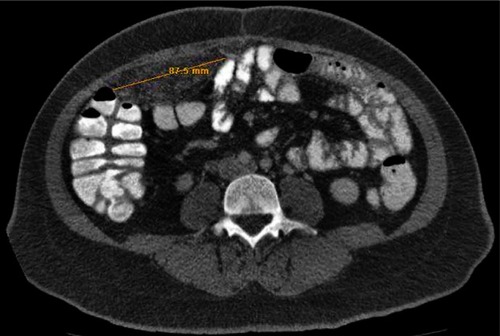
A 35-year-old woman presented complaining of progressive abdominal pain for the previous three days. The pain was severe and localized over the right quadrant with no specific radiation. She had feverishness but no chills, nausea but no vomiting. Her past medical history included thalassemia minor and obesity (previous BMI of 41) for which she had undergone a laparoscopic gastric bypass five months previously and had already lost 31 kg. Medications included multivitamins only. On physical examination she was found to be afebrile with normal vital signs. Her abdomen showed diffuse tenderness especially over the right mid quadrant and the feeling of a large ill defined mass. Her complete blood count and urine analysis were within normal range. An immediate CT scan of the abdomen was ordered and revealed a large (9 × 5 × 3.5 cm) oblong mass in the right mesentery inseparable from the abdominal wall. There was also stranding of fat within and surrounding it and some haziness in the abdominal rectus muscle (). The appearance was compatible with epiploic appendagitis of the right colon.
Figure 1 High resolution computed tomography showing a large mass in the right mesentery with stranding of fat within and surrounding it.
She was started on Ibuprofen 500 mg po twice daily and followed up closely. Seventy-two hours later her symptoms improved markedly and there was significant decrease in the size of the mass on palpation. She continued her treatment for a total duration of 14 days and she was completely symptom free at her two month follow up. The CT scan was not repeated as the symptoms had disappeared and the physical examination became normal.
Discussion
Epiploic appendagitis is a relatively rare inflammation of the small lobular masses of fat emanating from the serosal surface of the colon. Normally they are undetectable by imaging techniques except if ascites or inflammation are present.Citation7,Citation8 The reported age range for primary appendagitis is 12–82 years, with a peak of incidence in the fifth decade. A slight predilection for the male gender has been reported.Citation9 Presentation usually involves abrupt onset of focal abdominal pain mostly in the lower quadrants, which worsens with cough or stretching of the abdominal wall muscles.Citation10,Citation11 Associated symptoms are rare, bowel function and appetite are usually unchanged, and nausea and vomiting are rare.Citation4,Citation12 The patient usually does not appear to be very ill and the white blood cell count is either normal or mildly elevated.Citation13 Localized peritoneal signs may or may not be found on clinical examination. Only 10%–30% of patients do present with an abdominal mass like in our patient.Citation14 Localization, if present, is usually to the left lower quadrant, and this is based on the sigmoid colon harboring the largest number of epiploic appendages.Citation15 Due to similarities in presentation, this entity is often confused with diverticulitis and appendicitis. In fact a retrospective analysis reported epiploic appendagitis in 2.3%–7.1% of patients with clinically suspected colonic diverticulitis and 1% of patients with suspected appendicitis.Citation16 In addition, the differential diagnosis might include ovarian torsion, ovarian cyst rupture, ectopic pregnancy, Crohn’s disease, acute cholecystitis, intra-abdominal abscess and enteric infections.Citation17
Obesity has been reported to be a predisposing factor as well as strenuous physical exertion.Citation13 However; it is surprising to notice that no publications have attempted to isolate risk factors for the development of appendagitis. This is probably due to the fact that this is a rare diagnosis and therefore most case series are small in number. This will not allow for the detection of trends or predisposing factors. For instance, a recent study detailed five cases of primary appendagitis, but no trends or predisposing factors could be identified or isolated.Citation17 Our patient had a BMI of 41; however she lost more than 30 kg after her gastric bypass a few months earlier. So can the development of appendagitis be linked to the weight loss or the surgery per se? A connection will be very difficult to establish for the reasons mentioned above and for the fact that although bariatric surgery is becoming common, no center has ever reported an association similar to this one.
The diagnosis of epiploic appendagitis has become easier with the development of high resolution computed tomography (CT), and this has been recently supported in the literature.Citation18 On CT studies, round or ovoid lesions of fatty density measuring 1.5–3.5 cm in diameter with a hyper attenuating rim and surrounding ill-defined fat stranding are characteristically identified in the mesenteric fat adjacent to the colon. The mesenteric fat in these lesions is typically higher than normal-appearing mesenteric fat elsewhere in the abdomen.Citation19 The hyper attenuating ring is considered to be a characteristic sign.Citation2 Fat stranding is more pronounced than wall thickening because the para-colonic inflammatory changes are disproportionately more severe than the mild local reactive thickening of the adjacent colonic wall.Citation5,Citation20 In patients with acute abdominal pain this suggests a relatively narrow differential diagnosis: diverticulitis, omental infarction, appendicitis and less commonly mesenteric panniculitis.Citation21 Most of the inflammatory masses reported in the literature are 5 cm in size or less, however as previously mentioned our patient had a 9 cm sized mass which is unusual to say the least. In fact a previous report studied 52 patients with a diagnosis of acute appendagitis and found only 3 patients with a size >3.5 cm, the largest being 5.8 cm.Citation10 Does this carry any indication? There is no clear answer to this from the available published data.
Nonetheless even when the mass is evident and large in diameter, the presence of an experienced radiologist is essential for a prompt diagnosis. This is crucial to avoid unnecessary surgery. Previously, diagnosis was usually made post-operatively. However the experience gained over the past 20 years has shown that these patients respond well to conservative management.Citation22 Pain control, nonsteroidal anti-inflammatory drugs and close follow up are suggested, and surgery is kept only for those rare cases unresponsive to medical therapy.Citation23 In fact the effect of misdiagnosing this condition results in a total hospital cost up to six times higher than in cases where the correct diagnosis is made.Citation24 As clinicians become more aware of this entity, hospitalization, and improper use of antibiotics and possible surgical evaluation will be avoided, as was the case in our patient.
Conclusion
This case helps to illustrate a rare and uncommon diagnosis. The temporal occurrence after gastric bypass and weight loss is suspicious. However at this point causality cannot be established and therefore no conclusion can be drawn. Accurate diagnosis by an experienced radiologist through a CT of the abdomen is essential to avoid unnecessary admission and surgery.
Disclosure
The authors report no conflicts of interest in this work.
References
- GhahremaniGGWhiteEMHoffFLAppendices epilpoicae of the colon: radiologic and pathologic featuresRadiographics19921259771734482
- VriesmanALVBThe hyperattenuating ring signRadiology200322655655712563154
- MarinisTPCheckJHPrimary inflammation of the appendices epiploicaeAnn Surg194912953353717859336
- RiouxMLangisPPrimary epiploic appendagitis: clinical, US and CT findings in 14 casesRadiology19941915235268153333
- BlinderELedbetterSRybickiFPrimary epiploic appendagitisEmerg Radiol2002923123315290568
- SandrasegaranKMaglinteDDRajeshAAkisikFMPrimary epiploic appendagitis: CT diagnosisEmerg Radiol20041191415278705
- SandMGelosMBecharaFEpiploic appendagitis: clinical characteristics of an uncommon surgical diagnosisBMC Surg20071711
- SinghAJGervaisDAHahnPFSagarPMuellerPRNovellineRAAcute epiploic appendagitis and its mimicksRadiographics2005251521153416284132
- CarmichaelDHOrganCHJrEpiploic disorders: conditions of the epiploic appendagesArch Surg1985120116711714038060
- SinghAKGervaisDAHahnPFRheaJMuellerPRCT appearance of acute appendagitisAJR Am J Roentgenol20041831303130715505294
- BaherMEImaging and interventional techniques in acute left sided diverticulitisJ Gastrointest Surg2008121314131718270783
- Al JaberiTMGharaibehKIYaghanRJTorsion of abdominal appendages presenting with acute abdominal painAnn Saudi Med20002021121317322659
- ChowbeyPKSinghGSharmaATorsion of appendices epiploicae presenting as acute abdomen: laparoscopic diagnosis and therapyIndian J Gastroenterol200322686912696833
- ThomasJHRosotoFEPattersonLTEpiploic appendagitisSurg Gynecol Obstet197513823254808999
- MacariMLaksSHajduCCeacal epiploic appendagitis: an unlikely occurrenceClin Radiol20086389590018625354
- MollaERipollesTMartinezMJPrimary epiploic appendagitis: US and CT findingsEur Radiol199884354389510579
- SorserSAMaasLCYousifEMaasLEpiploic appendagitis: the great mimickerSouth Med J20091021214121720016426
- OzkurtHKaratagOKaraarslanERosanesIBasakMBavbeckCCT findings in epiploic appendagitisSurgery200714153053217383530
- PereiraJMSirlinCBPintoPSDisproportionate fat stranding: a helpful CT sign in patients with acute abdomenRadiographics20042470371515143223
- JainTShahTJinejaSTambiRPriamry epiploic appendagitis: radiologic diagnosis can avoid surgerySemin Roentgenol2008434618053822
- IssaIBaydounHMesenteric panniculitis: various presentations and treatment regimensWorld J Gastroenterol2009303827383019673029
- Van Breda VriesmanSCde Mol van OtterlooJCPuylaertJBEpiploic appendagitis: an underestimated self-limiting acute abdominal conditionNed Tijdschdr Geneeskd200714711131118
- PatelVGRaoAWilliamsRCecal epiploic appendagitis: a diagnostic and therapeutic dilemmaAm Surg20077382883017879696
- RaoPMRheaJTWittenbergJWarshawALMisdiagnosis of primary epiploic appendagitisAm J Surg199817681859683140