Abstract
We report on two unrelated Brazilian patients both presenting a very unusual association of ano/microphthalmia, cystic orbital anomaly, atypical clefting, and facial appendages in one patient. Clinical manifestations presented by these patients represent a MCA/MR syndrome of unknown etiology. Considerations about syndromic delineation, genetic aspects, and differential diagnosis are discussed.
Introduction
Anophthalmia/microphthalmia is part of a dozen of well-recognized autosomal recessive, dominant, X-linked, chromosomal and environmental syndromes (CitationGorlin et al 2001; CitationOMIM™ 2007). Its association with other developmental defects has been seen in several conditions, however, the occurrence of clinical signs such as atypical cleft of the face, facial appendages, cystic eye anomalies, and abnormal nares, isolated or in different combination associated to ano/microphthalmia are rarely seem (CitationMasuno et al 1997; CitationGuion-Almeida et al 2000; CitationGupta et al 2003; CitationSemerci et al 2003; CitationPascual-Castroviejo et al 2005; CitationRichieri-Costa and Ribeiro 2005). Diverse central nervous system (CNS) anomalies ranging from cystic lesions (CitationNaafs et al 1999; CitationPascual-Castroviejo et al 2005), pericallosal lipomas (CitationSemerci et al 2003), to holoprosencephaly (HPE) (CitationRichieri-Costa and Ribeiro 2005) have been reported in most of the papers on the issue. The patients here reported illustrate the main challenges concerning diagnosis, genetic counseling, and management involving these complex conditions.
Case reports
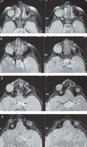
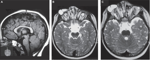
Case 1 (), male, born in 2000, was the first child of a 21-year-old G1P1 white woman and her 28-year-old nonconsanguineous husband. Pregnancy was uneventful, no toxic, infectious, or traumatic incidents or X-ray exposure were reported. Vaginal delivery was at term. Birth weight was 3.800 kg (75th centile), length 53 cm (75th centile), and head circumference was not recorded. On clinical examination at age 23 days, weight was 4.000 kg (50th centile), height was 52 cm (25th centile), and OFC was 38 cm (<98th centile). He presented with bilateral ano/microphthalmia, mild hypertelorism, bluish cystic formation in the right orbit; large ears, preauricular pit at right; broad nasal bridge, asymmetric and abnormally modeled nostrils with cleft nasal ala at left, and cleft lip and palate. Last examination at age 6 years showed, besides the surgical aspects of the corrected facial anomalies, marked neuropsychological delay (). Results of routine laboratory blood tests were normal. G-banded chromosomes were normal. Magnetic resonance imaging of the brain showed agenesis of the anterior part of the body of the corpus callosum, ectopic migration of gray matter from midline toward left lateral ventricle, and increased supracellar and pre-pontine cisterns (). Magnetic resonance imaging of the orbits showed: at right, two major cystic structures: a hyperintense, well demarcated, and homogeneous cystic mass laterally placed and a less intense, medially placed, both posteriorly displacing a severe microphthalmic globe; at left, the orbit is partially empty with fat and an elliptic microphthalmic globe. Optic nerves and external eye muscles are hypoplastic ().
Figure 1 A–D Clinical aspects of Case 1.
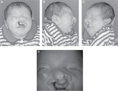
Figure 2 Case 1 at age 6 years. Note typical facial phenotype.
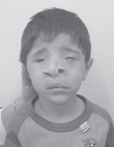
Figure 3 A–B Magnetic resonance imaging of the brain of case 1.
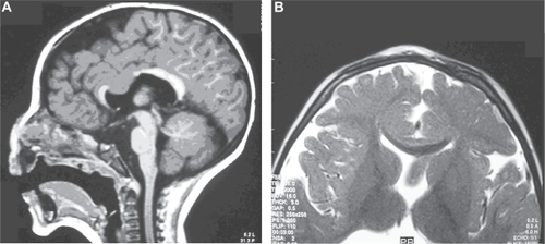
Figure 4 A–D Magnetic resonance imaging of the orbits of case 1.
Case 2 (), female, born in 2000, was the second child of a 29-year-old G2P2 white woman and her 34-year-old nonconsanguineous husband. Pregnancy was uneventful, no toxic, infectious, or traumatic incidents or X-ray exposure were reported. Vaginal delivery was at 37 weeks of gestation. Birth weight was 3.030 g (50th centile), length was 47 cm (10th centile), and head circumference was 32 cm (2nd centile). On clinical examination at the age of 7 days, weight was 2.960 g (10th centile), height was 48 cm (10th centile), and OFC was 33 cm (>2nd centile). She presented with bilateral ano/microphthalmia, cystic formation in the right orbit, a small appendage in the midline nasal tip, and two over the right nasal ala, broad nasal bridge, asymmetric and abnormally modeled nostrils with cleft nasal ala at right, large and low set ears, and cleft lip and palate. Last examination at age 6 years showed, besides the surgical aspects of the corrected facial anomalies, marked neuropsychological delay (). Results of routine laboratory blood tests were normal. G-banded chromosomes were normal. Magnetic resonance imaging of the brain showed increased supracellar and pre-pontine cisterns (). Magnetic resonance imaging of the orbits showed: at right, two major and well demarcated, hyperintense cystic structures, posteriorly displacing a severe microphthalmic globe; at left, a small globe is surrounded by hypointense material fatty characteristics. Optic nerves and external eye muscles are within normal range ().
Figure 5 A–D Clinical aspects of case 2. Note in 5D facial aspects before surgical correction.
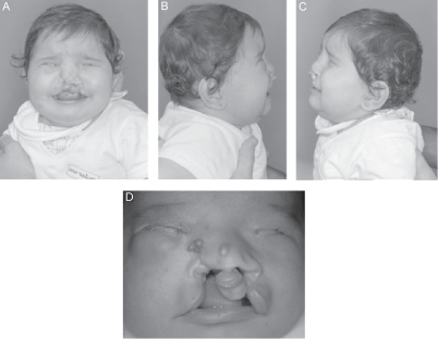
Figure 6 Case 2 at age 6 years. Note typical facial phenotype.
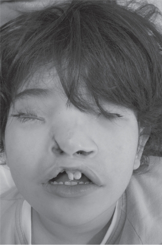
Figure 7 A–C Magnetic resonance imaging of the brain (7A) and of the orbits (7B–C) of case 2.
Discussion
Anophthalmia/microphthalmia, ocular cysts, CNS anomalies, and neuropsychological delay are part of some remarkable craniofacial syndromes such as: the cerebro-oculo-nasal syndrome (CONS) and the oculo-cerebro-cutaneous syndrome (OCCS). The CONS, a recurrent pattern syndrome of unknown etiology, is mainly characterized by ano/microphthalmia; abnormal nares, namely proboscis-like; facial appendages, and structural anomalies of the CNS in some cases (CitationRichieri-Costa and Guion-Almeida 1993; CitationErcal and Say 1998; CitationGuion-Almeida et al 2000; CitationRichieri-Costa and Ribeiro 2005). Ano/microphthalmia, orbital cysts, skin appendages, focal areas of skin hypoplasia, and variable types of CNS malformations, mainly intracerebral cysts, are the typical clinical features of OCCS (CitationMoog et al 2005). This condition shares several and nonspecific clinical signs with the encephalocraneocutaneous lipomatosis (ECCL), however, clinical observation suggests that these two syndromes have a different pathogenesis (CitationHunter 2006).
Recently, two patients presenting clinical signs close related with the ones here described were reported (CitationGupta et al 2003; CitationSemerci et al 2003). One of them, consisted of unilateral anophthalmia, Tessier number 3 cleft, skin appendages, and midline pericallosal lipoma (CitationSemerci et al 2003). Taking in to account the whole clinical picture, the authors suggested that this patient could represent either a clinical variability in CONS or a new entity. The other paper reports on a patient with bilateral microphthalmia with a defect of the left upper lid, and a bulging and bluish discoloration of the left lower lid corresponding to the orbital cyst; cleft of the right nostril, cleft lip/palate, and mild limb anomalies which led the authors to suggest the possibility that he has a previously undescribed syndrome related to the CONS (CitationGupta et al 2003).
The cases here described are close similar to that report by CitationGupta and colleagues (2003), and most likely they represent a new recognizable syndrome of unknown etiology. CNS anomalies, observed only in Case 1 of the present report, could be part of the clinical picture, however, the few number of cases do not permit a final definition of the syndrome spectrum. The cystic/multicystic anomalies, observed in the eye of the patients with this new syndrome, close resembles those observed in the embryo with absence of early lens structures when several folds of the retina are formed separated by patches of retinal pigmented epithelium (CitationAshery-Padan and Gruss 2001).
References
- Ashery-PadanRGrussP2001Pax6 lights-up the way for eye developmentCurr Opin Cell Biol137061411698186
- ErcalDSayB1998Cerebro-oculo-nasal syndrome: another case and review of the literatureClin Dysmorphol7139419571287
- GorlinRJCohenMMJrHennekamRCM2001Syndromes of the Head and NeckNew YorkOxford Univ Pr
- Guion-AlmeidaMLKokitsu-NakataNMRichieri-CostaA2000Clinical variability in cerebro-oculo-nasal syndrome: report on two additional casesClin Dytsmorphol92537
- GuptaPCPeraltaDParkerM2003Bilateral microphthalmia with cyst, facial clefts, and limb anomalies: A new syndrome with features of Waardenburg syndrome, cerebro-oculo-nasal syndrome, and craniotelencephalic dysplasiaAm J Med Genet117A725
- HunterAG2006Oculocerebrocutaneous and encephalocraniocutaneous lipomatosis syndromes: blind men and an elephant or separate syndromes?Am J Med Genet14070926
- MasunoMImaizumiKFukushimaY1997Median cleft of the upper lip and pedunculated skin masses associated with de novo reciprocal translocation 46,X,t(X; 16)q28; q11.2)J Med Genet3495249391896
- MoogUJonesMCBirdLM2005Oculocerebrocutaneous syndrome: the brain malformation defines a core phenotypeJ Med Genet429132115879499
- NaafsGGvan der VlietAMHewJM1999The oculocerebrocutaneous (Delleman-Oorthuys) syndromeNeuroradiology415599987771
- [OMIM™] National Center for Biotechnology Information2007Online Mendelian Inheritance in Man™ McKusick-Nathans Institute for Genetic Medicine, Johns Hopkins University (Baltimore, MD) and National Center for Biotechnology Information, National Library of Medicine (Bethesda, MD) [online] Accessed January 3, 2007. URL: http://www.ncbi.nlm.nih.gov/sites/entrez?db=OMIM
- Pascual-CastroviejoIPascual-PascualSIVelásquez-FraguaR2005Cerebrocutaneous (Delleman) syndrome: Report of two casesNeuropediatrics3650415776323
- Richieri-CostaAGuion-AlmeidaML1993Mental retardation, structural anomalies of the central nervous system, anophthalmia and abnormal nares: a new MCA/MR syndrome of unknown causeAm J Med Genet4770268266999
- Richieri-CostaARibeiroLA2005Cerebro-oculo-nasal syndrome, a disorder with some manifestations suggestive of the holoprosencephalic spectrum: new case and imaging review of previous casesAm J Med Genet1363523
- SemerciCNZorluPTopalY2003Is it a new syndrome or a clinical variability in cerebro-oculo-nasal syndrome?Am J Med Genet120A2535