Abstract
Purpose:
To describe the first case of anterior segment dysgenesis associated with factor VII congenital deficit (hypoproconvertinemia).
Method:
A 2-month-old child with factor VII deficiency was referred to our clinic because of corneal opacities. The child was born to consanguineous parents and severe factor VII deficiency was diagnosed on the third day of life because of gastrointestinal bleeding.
Result:
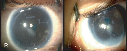
Ocular examination under anesthesia showed bilateral corneal opacities with adherent iris strands and peripheral anterior synechiae. The intra-ocular pressure was normal in both eyes and there were neither signs of cataracts nor glaucomatous optic nerve damage.
Conclusion:
This observation provide new information on the possible ocular findings in patients with hypoproconvertinemia. Based on this report, we suggest that careful coagulation screening should be considered in children who possess idiopathic anterior segment dysgenesis aiming at identifying the possible coagulation disorder.
Introduction
The purposes of this case report are double: (1) to provide new information on the possible ocular findings in patients with hypoproconvertinemia (factor VII deficit), (2) to suggest a careful coagulation screening in children who possess idiopathic anterior segment dysgenesis.
Case report
A 2-month-old female baby was admitted with seizures leading to a diagnosis of subdural hematoma due to factor VII deficiency. The child was born to consanguineous parents after a normal pregnancy and delivery. There was no history of maternal intrauterine infection, toxemia or exposure to alcohol or drugs. Severe hypoproconvertinemia was diagnosed on the third day of life (factor VII activity below 2% of normal) because of gastrointestinal bleeding. She had no associated systemic abnormalities, no mental retardation and there was no family history of bleeding disorder or ocular abnormality. After urgent decompressive craniectomy and evacuation of hematoma, the child was referred to our clinic because of corneal opacities. The child showed no symptoms of conjunctivitis or keratitis in the neonatal period. Both pupils constricted to light stimulus, and no afferent pupillary defect could be seen. Careful ocular examination under anesthesia showed bilateral corneal opacities with adherent iris strands and peripheral anterior synechiae (). On gonioscopy of the right eye, there were two localized iris adhesions to Schwalbe’s line, in the 2-O’clock and 6-O’clock meridians. Gonioscopy of the left eye showed a white iris membrane with peripheral iris adhesion to clear cornea visible between 4- and 9-O’clock positions. There was no sign of neovascularization or staining with fluorescein. The intra-ocular pressure was normal in both eyes and there were neither signs of cataracts, ocular inflammation nor glaucomatous optic nerve damage. Since the visual axis was not obstructed by corneal opacities, penetrating keratoplasty was not required. The patient was followed at two-month interval periods for a total of 12 months. Corneal opacities and intra-ocular pressure remained stable in both eyes during the follow-up period.
Figure 1 Operating room photographs of right eye (R) and left eye (L) showing paracentral corneal opacities with peripheral anterior synechiae and adhesions between the iris and the cornea.
Discussion
Hypoproconvertinemia is a rare autosomal recessive hemorrhagic disorder with a wide heterogeneous clinical pattern. The most severe form may be associated with severe hemorrhagic symptoms such as massive intracranial bleeding with a high mortality rate (CitationIngerslev and Kristensen 1998). To date, there are very little published data about the possible ocular findings associated with the bleeding disorder (CitationBrooks et al 2006). To the best of our knowledge, this is the first report of anterior segment dysgenesis observed in a child with hypoproconvertinemia. In our case, possible diagnoses that must be considered include Peters anomaly, Axenfeld-Rieger anomaly and posterior embryotoxon. Classically, Peters anomaly presents as a central defect consisting of central corneal leucoma and adherence of iris and lens capsule to the cornea (CitationPeters 1906; CitationMaillette de Buy Wenniger-Prick and Hennekam 2002), Axenfeld-Rieger anomaly is a dominantly inherited ocular malformation characterized by corectopia or polycoria with iris hypoplasia (CitationLines et al 2002), and posterior embryotoxon represents a defect of the angle with a prominent and anteriorly displaced Schwalbe line but without corneal opacification (CitationSim et al 2004). Another congenital abnormality called fibrous congenital iris membranes, can lead to white iris membrane with peripheral iris adhesions, but the abnormality is always unilateral and cause pupillary distortion with progressive occlusion or seclusion of the pupil (CitationRobb 2001). Finally, none of these diseases appears to be a convincing diagnosis in our case. Therefore, we suggest the possibility that the ocular malformation may be part of the general disorder. Another possibility is that the patient could have a variety of other autosomal recessive mutations in other ocular genes. According to this view, further genetic investigations are needed in order to definitively establish the relationship between the two disorders. In our observation, the dysplasia of the anterior chamber may potentially lead to progressive corneal opacification and/or glaucoma, therefore requiring keratoplasty or filtering surgery, respectively. This fact is particularly important because most factor VII-deficient patients do have a surgical risk due to excessive blood loss during surgical procedures. Consequently, we suggest that careful coagulation screening should be considered in children who possess idiopathic anterior segment dysgenesis aiming at identifying the possible coagulation disorder and prevent perioperative bleeding in affected patients.
Disclosure
Research grants: none.
Financial support: none.
Competing interests: the authors declare that they have no competing interests.
References
- BrooksBPMeckJMHaddadBR2006Factor VII deficiency and developmental abnormalities in a patient with partial monosomy of 13q and trisomy of 16p: case report and review of the literatureBMC Med Genet7216412230
- IngerslevJKristensenHL1998Clinical picture and treatment strategies in factor VII deficiencyHaemophilia4689969873815
- LinesMAKozlowskiKWalterMA2002Molecular genetics of Axenfeld-Rieger malformationsHum Mol Genet1111778412015277
- Maillette de Buy Wenniger-PrickLJHennekamRC2002The Peters’ plus syndrome: a reviewAnn Genet459710312119218
- PetersA1906Ueber angeborene Defektbildung der Descemetschen MembranKlin Mbl Augenheilkd442740
- RobbRM2001Fibrous congenital iris membranes with pupillary distorsionTrans Am Ophthalmol Soc99455011797319
- SimKTKarriBKayeSB2004Posterior embryotoxon may not be a forme fruste of Axenfeld-Rieger’s SyndromeJ AAPOS8504615492748