Abstract
Objective
To describe the corneal endothelial density and morphology in patients of Phramongkutklao Hospital and the relationship between endothelial cell parameters and other factors.
Methods
Four hundred and four eyes of 202 volunteers were included. Noncontact specular microscopy was performed after taking a history and testing the visual acuity, intraocular pressure measurement, Schirmer’s test and routine eye examination by slit lamp microscope. The studied parameters included mean endothelial cell density (MCD), coefficient of variation (CV), and percentage of hexagonality.
Results
The mean age of volunteers was 45.73 years; the range being 20 to 80 years old. Their MCD (SD), mean percentage of CV (SD) and mean (SD) percentage of hexagonality were 2623.49(325) cell/mm2, 39.43(8.23)% and 51.50(10.99)%, respectively. Statistically, MCD decreased significantly with age (p < 0.01). There was a significant difference in the percentage of CV between genders. There was no statistical significance between parameters and other factors.
Conclusion
The normative data of the corneal endothelium of Thai eyes indicated that, statistically, MCD decreased significantly with age. Previous studies have reported no difference in MCD, percentage of CV, and percentage of hexagonality between gender. Nevertheless, significantly different percentages of CV between genders were presented in this study.
Background
The cornea contains 5 layers. In the deepest layer, monolayer of corneal endothelial cell covers the posterior surface of descemet’s membrane. Corneal endothelium is metabolically active (Na+ – K+ pump) and responsible for keeping the corneal stroma in its usual hydrated state of 70% water to prevent stromal edema (CitationKrik 2005).
Nowadays specular microscope has made the in vivo evaluation of endothelium possible. It measures mean cell density (MCD), cell size variations (percentage of coefficient of variation [CV], polymegathism), and cell shape (polymorphism). The specular microscope has been used to establish and compare normative data for endothelium parameters among ethnic groups, as well as gender and age.
Due to the difference in endothelial parameters among various populations (CitationMatsuda et al 1985; CitationRao et al 2000; CitationSnellingen et al 2001; CitationKitagawa et al 2002; CitationPadilla et al 2004; CitationHashemian et al 2006; CitationZoega et al 2006) the study of normative data of each population is important. This study described the endothelial cell density and morphology of normal Thai eyes in relation to age, gender, and some risk factors such as smoking, underlying disease, intraocular pressure (IOP), and Schirmer’s test.
Materials and methods
The study population comprised 216 volunteers randomly selected from the visitors, outpatients, and staff of Phramongkutklao Hospital. Sample size was calculated using a minimum exceptional error (10%) of SD from normal Indian cell density data (CitationRao et al 2000).
Subjects were submitted to medical history examination and excluded from the study if they presented history of intraocular surgery or ocular trauma, corneal opacity, glaucoma, dry eye, inflammatory eye disease like uveitis, diabetes mellitus, use of contact lens, and family history of corneal decompensation. Additionally, to investigate the unknown effect of underlying diseases (such as hypertension, allergy, etc) and eye drugs (such as antihistamine) on corneal endothelium, such underlying diseases and eye drugs were included in the present study. According to the exclusion criteria, 14 volunteers were excluded. A total of 404 eyes from 202 volunteers (aged 20 to 80 years) were examined. All the subjects enrolled in this study originated from Thailand. Their national ID card and hospital record presented their Thai race and Thai nationality. Subjects enrolled in the study signed an informed consent form, and this study was approved by the Ethical Committee for Human Research, Phramongkutklao Hospital.
After taking a patient history, which included age, gender, smoking, underlying disease, and use of eye drop drugs, visual acuity measurement by Snellen chart, noncontact ocular tension measurement, modified Schirmer’s test 1 in 5 minutes, and slit-lamp biomicroscopy examination were examined. All volunteers underwent specular microscopy using a noncontact specular microscopy (SP2000: Topcon corporation, Japan). A single examiner performed all of the measurements. Corneal endothelial cell analysis was performed as follows: images from central cornea were taken of at least 50 contiguous cells and were manually marked with a mouse by the examiner for analysis by a built-in software program.
Statistical analysis
For statistical analysis, the SPSS program (version 11.5) was used. Baseline characteristics were calculated by descriptive statistics (eg, mean and standard deviation) and compared by using unpaired t-test for parametric data. Correlation between factors (age, gender, smoking, underlying disease, eye drop drug use, intraocular pressure, and Schirmer’s test) and MCD, percentage of CV and percentage of hexagonality were calculated by Pearson correlation.
In studies of the corneal endothelial cell morphology, which could be affected by multiple factors, the multiple regression analysis was used. p-value less than 0.05 were considered as the significance.
Results
Characteristics of the subjects were shown in . 202 patients with a mean (SD) age of 45.73 (16.12) years old participated. Ninety patients (44.6%) were male and one hundred and twelve patients (55.4%) were female. The most visual acuity was 20/20 (36.6%) and visual acuity range was 20/20 to light perception. Mean (SD) intraocular pressure was 13.76 (3.17) mmHg. Mean (SD) Schirmer’s test was 8.09 (6.13) mm.
Table 1 Characteristics of the subjects
describes the total data of corneal endothelial cell density and morphology and the characteristics of the studied population in gender difference. Their MCD (SD), mean percentage of CV (SD) and mean (SD) percentage of hexagonality were 2623.49(325) cell/mm2, 39.43(8.23)% and 51.50(10.99)%, respectively.
Table 2 Endothelial cell characteristics of the study population in different gender
Results of mean MCD, mean percentage of CV and mean percentage of hexagonality in different age groups are listed in . MCD was statistically significantly decreased in relation to increasing age (p < 0.01) ().
Figure 1 Correlation between MCD and age.
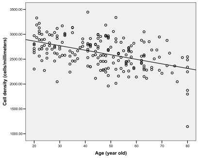
Table 3 Endothelial cell characteristics of the study population in different age groups
The correlation between parameters and others factors () are shown: the p-value of each factor related with MCD, percentage of CV, and percentage of hexagonality. There were significant correlations between age and MCD, and between age and percentage of hexagonality. Gender showed a significant correlation in MCD, percentage of CV and percentage of hexagonality. Moreover, there is a significant correlation between Schirmer’s test and percentage of hexagonality. However, no significant correlations between parameters and other factors such as intraocular pressure, smoking, underlying disease, and eye drop drug use were shown in this study.
Table 4 Correlation between parameters and other factors
The multiple regression analysis between parameters and univariate significant factors are showed in . However, there is statistical significance only between age and MCD, age and percentage of hexagonality, and gender and percentage of CV.
Table 5 Multiple regression analysis between parameters and univariated significant factors
Discussion
Endothelial cell analysis is important for corneal function and viability assessment. The clinical uses include the assessment of donor corneal endothelium, the effects of intraocular surgery and the essential in evaluating the safety of the first time intraocular or corneal surgical procedures and intraocular lens.
Many studies have been published on endothelial cell density and morphology in relation to age, gender, and ethnicity. Although it is clear that significant differences in corneal endothelial properties do exist among races and ethnic groups, normative data that is derived from the underlying population of each country is still necessary. This study reports endothelial cell data in Thai population.
Noncontact specular microscopy is used in many studies. Image capturing of the endothelium cells and calculation of cornea endothelial cells by a unique method does not require touching the cornea. This patented procedure eliminates the risk of transmitted infectious diseases and reduces potential physical injury to the eye. The disadvantage of this method is less control over patient eye movement.
The endothelium has been reported with confocal microscopy. Comparison of endothelial cell count density using confocal and contact specular microscopy was studied by CitationKlais and colleagues (2003), no statistically significant difference of endothelial cell density between confocal and specular microscopy.
In our study, the endothelial cell parameters were within normal range. The results have shown that with increasing age, MCD and % CV tended to decrease. There were conflicting reports about the relationship between gender and endothelial cell characteristics. Many studies have not found any statistical differences between them (CitationLaing et al 1976; CitationHirst et al 1980; CitationMatsuda et al 1985; CitationLandesz et al 1995; CitationAAO 1997; CitationHashemian et al 2006) as our data showed no differences between MCD, hexagonality, and gender. However we have found that females have a higher percentage of CV than males. As it was only one significant parameter, we could not assume that the female quality of corneal endothelium was less than that of males.
We did not find that smoking, underlying disease, eye drop drug usage, IOP, and Schirmer’s test affected the corneal endothelium. CitationZoega and colleagues (2006) had studied the risk factors for cornea guttata and found that smoking more than 20 pack-years increased the risk of developing corneal guttata more than 2-fold but our study did not find correlation between them. This could be for two reasons. Firstly, there was no real significance. Secondly, our number of pack-years was less than the previous study so we did not find the difference.
While we collected the data, we found abnormal endothelial parameters like Fuchs’ dystrophy. A 50 year old female had been examined. Her right eye had MCD 1564.8 cell/mm2, CV 75.3%, and hexagonality 26%. Her left eye had MCD 1836.6 cell/mm2, CV 56.9%, and hexagonality 40%. The incidence of abnormal endothelial cell was calculated about 1:202 (0.5%).
We compared MCD with the previous studies and found the difference (). In our study, MCD was similar to the Indian population (CitationRao et al 2000) but less than the Japanese and American populations (CitationMatsuda et al 1985).
Table 6 Comparison of endothelial cell density
Conclusions
The normative data of the corneal endothelium of Thai eyes indicated and confirmed that MCD was decreased with increasing age and there are statistically significant differences in polygonality between genders. Relationship between endothelial cell parameters and some factors like smoking, underlying disease, eye drop drug usage, IOP, and Schirmer’s test were not found.
MCD in Thai population was similar to the Indian population compared with the previous studies. However, it was less than in the Japanese and American populations.
Acknowledgments
The authors would like to thank Dr. Praweena Sopapornamorn and Miss Worarachanee Imjaijitt for their valuable comments on this paper.
References
- [AAO] American Academy of Ophthalmology1997Corneal endothelial photography. Three-year revisionOphthalmology104136059261327
- HashemianMNMoghimiSFardMA2006Corneal endothelial cell density and morphology in normal Iranian eyesBMC Ophthalmol6916519812
- HirstLWFerrisFLIIIStarkWJ1980Clinical specular microscopyInvest Ophthalmol Vis Sci19247350130
- KitagawaKKojimaMSasakiH2002Prevalence of primary cornea guttata and morphology of corneal endothelium in aging Japanese and SingaporeanOphthalmic Res34135812097795
- KlaisCMBührenJKohnenT2003Comparison of endothelial cell count using confocal and contact specular microscopyOphthalmologica2179910312592045
- KrikRW2005Basic and clinical science course section 8 2004–2005; External disease and corneaSan FranciscoAmerican Academy of Ophthalmology323
- LaingRASandstromMMBerropsiAR1976Changes in corneal endothelium as a function of ageExp Eye Res2258794776638
- LandeszMSiertsemaJVVan RijG1995Comparative study of three semiautomated specular microscopesJ Cataract Refract Surg21409168523285
- MatsudaMYeeRWEdelhauserHF1985Comparison of the corneal endothelium in an American and a Japanese populationArch Ophthalmol10368703977679
- PadillaMDSibayanSAGonzalesCS2004Corneal endothelial cell density and morphology in normal Filipino eyesCornea231293515075881
- RaoSKRanjanSPFoglaR2000Cornea endothelial cell density and morphology in normal Indian eyesCornea19820311095057
- SnellingenTRaoGNShresthaJK2001Quantitative and morphological characteristics of the human corneal endothelium in relation to age, gender, andethnicity in cataract populations of south AsiaCornea2055811189005
- ZoegaGMFujisawaASasakiH2006Prevalence and risk factors for cornea guttata in the Reykjavik Eye studyOphthalmology113565916581419