Abstract
Classical anatomical teaching reports the presence of the lateral palpebral raphe formed at the union in the preseptal and orbital parts of the orbicularis oculi muscle, or by the tendon adhering these to the underlying zygomatic bone. The lateral palpebral raphe has been shown to be absent in Asian cadavers. The current study uses both evidence from the anatomical dissection of five eyelids from three Caucasian cadavers, and histological assessment of the lateral canthus of 13 eyelids from seven Caucasian cadavers to illustrate the absence of the lateral palpebral raphe in Caucasian population.
The presence or absence of the lateral palpebral raphe has long been a source of contention. A famous medical dictionaryCitation1 generally defines the “raphe” as “the line of union of two contiguous, bilaterally symmetrical structures”. In addition, in the following part of the same dictionary,Citation1 the “lateral palpebral raphe” is defined as “a narrow fibrous band in the lateral part of the orbicularis oculi muscle formed by the interlacing of fibers passing through the upper and lower eyelids”. Traditionally, the lateral palpebral raphe has been thought to be the common tendon of the septal and orbital parts of the orbicularis oculi muscle,Citation2 or the linear tendon of these parts attaching to the zygomatic bone.Citation3 In contrast, there are also reports refuting the presence of the lateral palpebral raphe, suggesting that the orbicularis oculi muscle in the lateral part is continuous without tendinous intercalation.Citation4 That the lateral palpebral raphe is formed purely by the interlacing of the muscle fibers is another opinion.Citation5 Thus, there is no consensus regarding the lateral palpebral raphe.
The purpose of the present study was, therefore, to elucidate whether the lateral palpebral raphe existed in the Caucasian lateral canthus.
Materials and methods
We took both gross dissection and microscopic examination to elucidate whether lateral palpebral raphe existed or not, in which the decision was made by the single examiner (HK).
In gross dissection, five eyelids of three Caucasian cadavers (2 males: 1 right, 2 left; 1 female: 1 right, 1 left) aged from 81 to 89 years at death (average, 85.0 years), fixed in 10% buffered formalin, were examined. Westcott scissors were used to remove the lateral half of the upper and lower eyelid skin, and the orbicularis oculi muscle exposed. The connective tissue on the orbicularis muscle fibers was removed as completely as possible. The lateral part of the orbicularis oculi muscle was then examined to ascertain the presence or absence of the lateral palpebral raphe.
The method for removing the eyelid samples for the microscopic examination are stated elsewhere.Citation6 Full thickness sections of eyelids fixed in 10% buffered formalin, from 13 postmortem eyelids of seven Caucasians (2 males: 2 right and 2 left; aged 78 and 88 years at death; 5 females: 5 right and 4 left; aged 80, 81, 90, and 101 years at death) were examined. Since some samples were broken during the microscopic specimen production, they were excluded. We made axial sections with 1 mm inferior from the lower eyelid margin,Citation6 and included the part corresponding to the interlacing of the lateral orbicularis oculi muscle. Sliced specimens were dehydrated and embedded in paraffin, and cut into 7-μm-thickness sections, after which the sliced sections were stained with Masson trichrome.Citation6 Microscopic photographs were taken with a digital camera system attached to the microscope (Moticam 2000; Shimadzu Rika Kikai, Tokyo, Japan).
All cadavers were registered with the University of Adelaide, and proper consents and approvals were obtained prior to use. Methods for securing human tissues were humane and complied with the tenets of the Declaration of Helsinki.
Results
In gross dissection, the lateral part of the orbicularis oculi muscle was continuous without tendinous intercalation in all specimens (). A linear tendinous structure, namely the lateral palpebral raphe,Citation1 was not identified in all five samples. Although an oblique union was seen in the pretarsal and preseptal portions of the orbicularis oculi muscle, the lack of any true linear union between the muscle fibers of the superior and inferior portions of the orbicularis oculi muscle belies its classificationCitation1 as the lateral palpebral raphe.
Figure 1 Macroscopical examination (left: cephalad, bottom: lateral). The lateral palpebral raphe is not identified in the lateral part of the orbicularis oculi muscle. The superior and inferior orbicularis oculi muscles were continuous without any tendinous intercalation between them. Central white tissue on the preseptal orbicularis oculi muscle is fascia.
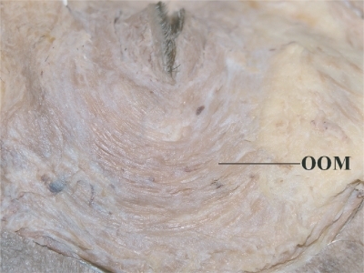
Microscopically, in all specimens, the orbicularis oculi muscle was also seen to be continuous without any tendinous interruption ().
Figure 2 Microscopical sanple (left: lateral, up: anterior). The orbicularis oculi muscle continuously situated and the lateral palpebral raphe is not identified.

Discussion
The lateral palpebral raphe was not identified in the lateral part of the orbicularis oculi muscle in any of the samples examined. Whilst this has been previously documented in Asian samples,Citation4 our findings appear to confirm its absence in Caucasian eyelids, although they were very old, and show concordance between these two racial groups.
The absence of the lateral palpebral raphe may have physiological significance. The interlacing of the superior and inferior orbicularis oculi muscle fibers may allow a moderate degree of medial movement of the lateral canthus with blinking, allowing the tear film to effectively spread medially for efficient corneal wetting. Some lateral fixation remains, however, to overcome the medial pull of the lateral canthus during blinking, maintaining the efficiency of the blinking movement.
Further studies may elucidate the true nature of the lateral portion of the orbicularis oculi muscle. Whilst this report documents the absence of the lateral palpebral raphe in the specimens examined, as drawbacks of the current study are the small sample numbers and the very advanced age specimens, assessment of a larger number of specimens from a more widely variable age range may add to our understanding of this contested feature of eyelid anatomy.
Disclosure
The authors report no conflicts of interest or financial interest related to this manuscript.
References
- Stedman’s Medical Dictionary27th edBaltimore, MDLippincott Williams & Wilkins20001518
- BronAJTripathiRCTripathiBJWolff’s Anatomy of the Eye and Orbit8th edLondon, UKChapman and Hall Medical19973084
- KikkawaDOLemkeBNOrbital and eyelid anatomyDortzbachRKOphthalmic Plastic Surgery – Prevention and Management of ComplicationsNew York, NYRaven Press1994129
- KakizakiHZakoMNakanoTNo raphe identified in the orbicularis oculi muscleOkajimas Folia Anat Jpn200481939615747875
- WhitnallSEAnatomy of the Human Orbit and Accessory Organs of Vision (reprint). New York, NYRobert E. Krieger Publishing Company Inc1979132
- KakizakiHZakoMNakanoTMicroscopic findings of lateral tarsal fixation in AsiansOphthal Plast Reconstr Surg200824131135