Dear Sir,
Evaluation of the pupils for a relative afferent pupillary defect can sometimes be challenging in poor lighting or in patients with dark irides. Near-ultraviolet light causes fluorescence of the lens and highlights the pupil, thus facilitating the pupil examination. Near-ultraviolet light is safe to use in a clinical setting and performs well when compared to examination with a conventional light source.
The pupillary examination, including the relative afferent pupillary defect (RAPD) and evaluation of anisocoria, is a standard component of medical examinations, and is an essential part of the ophthalmologic and neurologic exam. Ideally, this clinical test should be performed in dim lighting with a transilluminator as the light source; however, pocket flashlights or pen lights are also commonly used. At times, it can be challenging to view the pupil well, especially in suboptimal lighting and in patients with dark irides. One technique that can be used in this setting is to observe the red reflex with a direct ophthalmoscope and note changes in the size of this reflex as the light is “swung” from eye to eye. However, this method has its own limitations, especially given the relatively dim light stimulus. We propose a new simple technique that may help in sub-optimal viewing conditions.
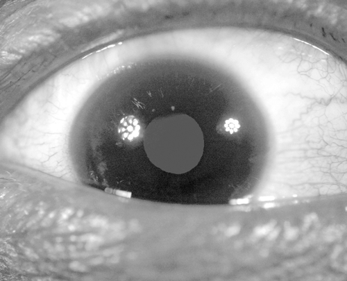
We have found that using a near-ultraviolet light source can assist in performing the pupillary exam. It has long been known that the human lens fluoresces at certain wavelengths of incident light. Lens fluorescence has been found to occur with frequencies of light between 350 and 450 nm.Citation1 (Wavelengths smaller than 380 nm are considered ultraviolet, and wavelengths slightly longer than this may be termed as near-ultraviolet. Radiation in this range is commonly known as “black-light.”) Fluorescence of the lens has been shown to increase with aging,Citation2 and also with certain disease states, notably diabetes.Citation3 Patients may perceive the emitted fluorescent light as glare. This light can also be appreciated by an observer as a green-white glow to the lens. The fluorescent glow of the lens effectively highlights the margins of the pupil and affords a clear view of pupillary response to light ). The pupil reacts to light in this setting by two mechanisms. There is some transmission of incident light to the retina, and the fluorescent light emitted from the lens stimulates the retina as well.
FIGURE 1 Pupil examination facilitated by near-ultraviolet fluorescence of the lens. Note that two near-ultraviolet light sources were used for the purpose of this photograph.
With recent advances in light emitting diode (LED) technology, devices that provide a near-ultraviolet light source are now ubiquitous. There are many such flashlights and penlights readily available at a low cost. These devices are generally marketed as “black-light” flashlights and are commonly used for verifying IDs or currency and forensic applications.
We evaluated one particular flashlight (9 LED 400 nm, distributed by LED Wholesalers). The total irradiance of this device was found to be equivalent to that of a Finnoff transilluminator. (6.6 mW/cm2) Also the emission spectrum was measured and found to have a peak at 405 nm with a relatively narrow band width from approximately 380–430 nm. The International Commission on Non-Ionizing Radiation Protection (ICNIRP) identified photochemical injury to the cornea, lens or retina as possible risks of exposure to ultraviolet or blue light.Citation4 Based on guidelines from the ICNIRP a safe exposure limit was calculated to be longer than 12 min per day. This is well beyond any likely clinical exposure and the safe exposure limit set by the ICNIRP is a conservative limit.
In our experience, the near-ultraviolet light is reliable for detecting a RAPD when compared to a conventional light source. Even with the use of neutral density filters the near-ultraviolet light performs similar to conventional light. However, as progressively darker filters are used the fluorescent effect becomes less and less pronounced. We have found that near-ultraviolet light works best as a “yes or no” screening test to evaluate for the presence of RAPD. It is also a useful as an initial evaluation for the presence of anisocoria.
This “black-light” method does have some limitations. The phenomenon of lens fluorescence is age dependent; therefore this would not be as effective in young patients. Also, the safety of this exposure to the developing eye is less well known, and this technique may not be advisable for very young patients. In addition, aphakic or pseudophakic patients will not have any fluorescence without their natural lens, so they cannot be evaluated with this technique. It is conceivable that a patient may have an asymmetry in the amount of fluorescence produced by each eye. If that were the case, then the variation in the amount of fluorescent light could result in the appearance of a pseudo-RAPD. (A similar phenomenon sometimes occurs with a media opacity with conventional light.) Although this is a theoretical possibility, further evaluation would be needed to determine if this is a true clinical concern. Finally, using a “blue” light source, instead of full-spectrum white light, may preferentially stimulate a different population of retinal ganglion cells known as melanopsin-expressing retinal ganglion cells, and the RAPD test done with different wavelengths may not be entirely equivalent, for example photoreceptor disease may result in a different pupillary response than ganglion cell disease when red and blue light are used.Citation5 However, these latter considerations are unlikely to affect the basic RAPD test to determine if a RAPD is present or not.
In conclusion, using a near-ultraviolet or “black” light source is a simple method to supplement a standard pupil examination. This technique is most beneficial in situations where it is difficult to view the pupil clearly, such as emergency departments and intensive care units, where the lighting is suboptimal and especially with patients with dark irides. The exposure from a pocket flashlight is within established safety limits, and the examination appears to be reliable compared to the standard method.
ACKNOWLEDGMENTS
We appreciate the assistance of Richard Williams, Ph.D., Keith Bonin Ph.D. and Chad McKell from Wake Forest University, Department of physics with measurements of irradiance, emission spectrum and safety calculations.
Declaration of interest: The authors report no conflicts of interest. The authors alone are responsible for the content and writing of the paper.
REFERENCES
- Zuclich J, Previc F, Novar B, Edsall P. Near-UV/blue light-induced fluorescence in the human lens: potential interference with visual function. J Biomed Opt 2005;10(4):44021.
- Occhipinti J, Mosier M, Burstein N. Autofluorescence and light transmission in the aging crystalline lens. Ophthalmologica 1986;192(4):203–209.
- Mosier M, Occhipinti J, Burstein N. Autofluorescence of the crystalline lens in diabetes. Arch Ophthalmol 1986;104:1340–1343.
- Sliney D, Aron-Rosa D, DeLori F, Fankhauser F, Landry R, Mainster M, Marshall J, Rassow B, Stuck B, Trokel S, West TM, Wolffe M; International Commission on Non-Ionizing Radiation Protection. Adjustment of guidelines for exposure of the eye to optical radiation from ocular instruments: statement from a task group of the International Commission on Non-Ionizing Radiation Protection (ICNIRP). Appl Optics 2005;44(11):2162–2176.
- Kawasaki A, Kardon RH. Intrinsically photosensitive retinal ganglion cells. J Neuroophthalmol 2007;27(3):195–204.