Abstract
Purpose: To compare the effectiveness of ultrasound (US)-guided percutaneous 915 MHz microwave (MW) ablation with the 2450 MHz MW ablation for large hepatocellular carcinoma (HCC) (>4 cm in diameter).
Materials and methods: Patients with HCC >4 cm in diameter who underwent US-guided percutaneous MW ablation with curative intention between March 2007 and December 2008 (39) were randomly divided into two groups, 915 MHz MW group and 2450 MHz MW group. We compared the results of ablation between the two groups.
Results: Fewer antenna insertions for each tumour were required in the 915 MHz MW group (3.69 ± 0.6) than in the 2450 MHz MW group (4.71 ± 1.61) (p = 0.01). According to the follow-up contrast-enhanced imagings, technique effectiveness rate was 85.7% (18/21) and 73.7% (14/19) in the 915 MHz MW group and 2450 MHz MW group, respectively (p = 0.44). The rate of local tumour progression (LTP) was 14.3% (3/21) and 26.3% (5/19) in the 915 MHz MW group and 2450 MHz MW group, respectively (p = 0.44). There were no deaths and no thrombosis of major vessels in any patient.
Conclusions: Compared with 2450 MHz MW ablation, our initial experience showed that percutaneous 915 MHz MW ablation with cooled-shaft antennae was safe and could achieve a high technique effectiveness rate with fewer insertion numbers in the treatment of large HCC. Therefore, percutaneous 915 MHz MW ablation may provide a new method for the treatment of large HCC.
Introduction
Hepatocellular carcinoma (HCC) is one of the most common malignancies in the world and is estimated to cause more than 500,000 deaths worldwide annually Citation[1]. Its incidence has been increasing worldwide due to the widespread occurrence of hepatitis B virus (HBV) and hepatitis C virus (HCV) infection Citation[2], Citation[3]. Although HCC can be treated successfully by surgical resection, the majority of patients are not candidates for resection because of advanced tumour stage at the time of diagnosis and underlying liver cirrhosis Citation[4], Citation[5]. Although orthotropic liver transplantation offers the chance for therapeutic success Citation[6], its performance is limited by a shortage of donor organs. Furthermore, HBV or HCV infection recurs after transplantation, leading to severe liver damage Citation[7]. One promising alternative for these patients is thermal ablation, such as radiofrequency (RF) ablation and MW ablation. RF ablation has been widely used in the management of HCC and has achieved encouraging results for small HCC less than 3 cm in diameter Citation[8–10]. But it is relatively difficult to ablate large hepatic tumours in respect that the treatment of large tumours is cumbersome and time consuming, requiring multiple sequential overlapping ablations to ensure adequate coverage Citation[11], Citation[12]. And high recurrence rates remain the problem Citation[13], Citation[14]. The reasons for RF failures are likely multifactorial, but an important factor is the inability of the RF electrode to heat substantially above 100°C because of increased tissue impedance from charring and tissue desiccation around it Citation[15].
MW ablation is one of the effective localised thermal ablation methods which has been used widely in east Asia. Compared with RF ablation, microwave energy does not appear to be limited by charring and tissue desiccation, and is less affected by the heat-sink effect of local blood vessels Citation[16]; thus, MW ablation could generate a larger ablation zone than RF ablation Citation[16–18]. So it provides a potential method for ablating large HCC. The MW ablation techniques and equipment have undergone remarkable progress in recent years since non-cooled 2450 MHz MW equipment was applied in ablation for hepatic tumours in 1994 Citation[19]. The cooled-shaft antenna of 2450 MHz MW could achieve a larger ablation zone and avoid skin injury compared with non-cooled 2450 MHz MW, which has led to exciting clinical results in the treatment of HCC Citation[20], Citation[21]. In 2003, a 915 MHz MW ablation system (Vivant Medical, Mountain View, CA) was reported Citation[17]. After further improvement of this 915 MHz MW ablation system, Simon et al. reported that a mean maximal ablation diameter of 5.5 cm (range 5.0–6.5 cm) was obtained by intraoperative application of three antennae spaced 1.5–2.5 cm apart Citation[22]. In 2007, a new triaxial antenna of 915 MHz MW ablation designed for percutaneous use was reported, and a large ablation zone was obtained Citation[18], Citation[23]. The 915 MHz MW may penetrate deeper than the 2450 MHz MW which may yield larger ablation zones Citation[24], Citation[25]. Our previous in vivo experiments in the porcine liver have shown that 915 MHz MW with cooled-shaft antenna could yield a significantly larger ablation zone than 2450 MHz MW Citation[26]. However, to our knowledge, there have been no clinical studies to compare the effectiveness of 915 MHz and 2450 MHz MW ablation for local control of large HCC. Therefore, the purpose of our study was to compare the effectiveness of ultrasound (US)-guided percutaneous 915 MHz MW ablation and 2450 MHz MW ablation with cooled-shaft antennae for large HCC (>4 cm in diameter).
Materials and methods
Patients
Between March 2007 and December 2008, 39 consecutive patients with 40 HCC nodules larger than 4 cm in diameter were referred to our department for MW ablation therapy. Participating patients included 28 men and 11 women aged 43–80 years (mean age, 58.69 ± 10.75 years). The diagnosis of HCC was confirmed in all patients with US-guided needle biopsy. Inclusion criteria for our study were unresectable HCC or patients’ refusal to undergo surgery; three or fewer nodular hepatic lesions with a maximum nodule diameter of 4–8 cm; absence of portal vein thrombosis; prothrombin time of less than 25 s, prothrombin activity higher than 40%, and platelet count higher than 40 cells × 109/L. Our study was approved by our institutional human research review committee. Written informed consent was obtained from all patients.
The patients were randomly assigned (using sealed envelopes) to the 915 MHz MW group (n = 20) or the 2450 MHz MW group (n = 19). Of the 20 patients treated with 915 MHz MW ablation, 19 had a solitary nodule and one had two nodules; thus, a total of 21 nodules were treated in these patients. Of the 19 patients treated with 2450 MHz MW ablation, all patients had a solitary nodule; thus, a total of 19 nodules were treated in these patients. Of all the nodules in the two groups, some were adjacent to gastrointestinal tract (3 in the 915 MHz group, 2 in the 2450 MHz group), gallbladder (1 in 915 MHz group, 1 in 2450 MHz group), diaphragm (3 in 915 MHz group, 4 in 2450 MHz group), and major intrahepatic branches of vessels (2 in 915 MHz group, 2 in 2450 MHz group), because of larger diameter of the tumours. There was no significant difference in clinical backgrounds between the two groups ().
Table I. Clinical characteristics of 39 patients treated with 915 MHz and 2450 MHz MW ablation.
Power calculations had indicated that 18 cases would be required per group to detect a difference of 30% in the number of antenna insertions with a power of 90% (α = 0.05, β = 0.1).
Preablation imaging work-up
All patients had undergone US, contrast-enhanced US, and contrast-enhanced computed tomography (CT) or gadolinium-enhanced magnetic resonance imaging (MRI) to delineate the target tumour before MW ablation. The maximum diameters of the index tumours were measured on contrast-enhanced US image, because the borders of lesions were more accurate in contrast-enhanced US than in conventional US. US and contrast-enhanced US were performed using a Sequoia 512 system (Acuson, Mountain View, CA) with 3.5–5.0 MHz multifrequency transducers. US contrast agent was Sonovue (Bracco, Milano, Italy). All CT studies were carried out with the same multi-detector row CT (Lightspeed 16; GE Medical Systems, Milwaukee, WI) and contrast medium (iopromide, Ultravist 300; Schering, Berlin). All MRI studies were carried out with the same 1.5-T unit (Signa Echo-Speed, GE Medical Systems), contrast medium (Magnevist, Schering; 0.1 mmol/kg of body weight) and sequences.
Microwave ablation device
The 915 MHz MW unit (KY-2100, Kangyou Medical, Nanjing China) consists of two independent MW generators, two flexible coaxial cables and two water-pumping machines which can drive two 15-gauge cooled-shaft antennae (2.2 cm antenna tip) simultaneously. The 2450 MHz MW unit (KY-2000, Kangyou Medical) consists of three independent MW generators, three flexible coaxial cables and three water pumping machines, which can drive three 15-gauge cooled-shaft antennae (1.1 cm antenna tip) simultaneously. The generators in each of the two MW units are capable of producing 1–100 W of power. The cooled-shaft antennae are coated with Teflon which is used to prevent adhesion. Inside the antenna shaft there are dual channels through which distilled water is circulated by a peristaltic pump, continuously cooling the shaft to prevent overheating. The MW system is also equipped with thermocouple needles which can monitor the temperature of its placement point in real-time during ablation.
Microwave ablation procedures
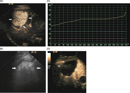
All treatments were performed in our institution and were carried out under US guidance with the patients under unconscious intravenous anaesthesia (propofol, 6–12 mg/kg/h; ketamine, 1–2 mg/kg) in the operating room. All procedures were performed by two experienced doctors (L.P., Y.X.L.); both had more than 10 years’ experience in MW ablation of HCC. A detailed protocol including the placement of the antennae, power output setting, emission time and appropriate approach was worked out for each patient on an individual basis before treatment. In general, MW ablation was performed at 60 W using two to three cooled-shaft antennae simultaneously, the distance between which was no more than 3 cm (mean 2.37 ± 0.4 cm; range 1.7–3.0 cm) for 915 MHz MW and no more than 2.5 cm (mean 2.03 ± 0.3 cm; range 1.6–2.4 cm) for 2450 MHz MW. A thermal monitoring system attached to the MW unit was used during ablation. One or two 21 gauge MW thermal monitoring needles were inserted in the margin of targeted tumour (proximity to the tumour periphery about 5 mm) under US guidance. During MW ablation we monitored the hyperechoic area of ablation using grey-scale sonography and thermal monitoring to decide the endpoint of treatment. The treatment session would be ended if the transient hyperechoic zone between antennae on grey-scale US merged and covered the target region; meanwhile the temperature of the thermal needles achieved target temperature (over 60°C for lesions in safe location and keeping 54°C∼60°C for more than 3 min for lesions adjacent to the gastrointestinal tract, diaphragm, gallbladder, etc.) according to our previous clinical experiences Citation[27], Citation[28]. When withdrawing the antennae, the needle tracks were routinely cauterised to avoid tumour seeding and bleeding. Every patient received contrast-enhanced US 10 min after ablation with two to three antennae, to evaluate treatment response. All contrast-enhanced US were performed by one doctor (Y.X.L. who has 4 years experience of more than 2000 cases on contrast-enhanced US). Possible residual tumour was doubted if any abnormal nodular hypervascular region existed at the peripheral region of ablation. Additional treatment was performed with one to three cooled-shaft antennae depending on the size of the residual tumour. Treatment was not considered complete until the entire tumour showed no enhancement on contrast-enhanced US (). Emission time and power of each generator were recorded. Emission energy was the sum of each generator's emission energy during the treatment of one patient. Emission time was the working time of generators.
Figure 1. Microwave ablation images in an 80-year-old man who had HCC. (A) The contrast-enhanced US arterial phase pre-ablation image shows a 5.8 × 5.0 cm HCC (arrow). (B) The US image shows the hyperechoic region syncretized and covered the target region (arrow). (C) The curve of temperature monitoring during microwave ablation shows that the temperature measured of the tumour periphery is over 60° (yellow curve). (D) The contrast-enhanced US arterial phase image post-ablation 2 days shows a 6.9 × 6.1 cm non-enhancing zone enveloping the tumour (arrows).

Therapeutic efficacy assessment and follow-up
Therapeutic efficacy was assessed by contrast-enhanced imaging and serum tumour marker levels after the treatment. The technique effectiveness was defined as the ‘complete ablation’ of the macroscopic tumour proved by imaging follow-up after ablation. Local tumour progression (LTP) was defined as incompletely treated viable tumour continuing to grow or a new tumour (or in the case of hepatocellular carcinoma, ‘daughter’ or ‘satellite’ tumours) growing at the original site during follow-up. The number of antenna insertions was defined as the total number of antenna placements for each tumour until this tumour was ablated completely. The follow-up period was calculated starting from the beginning of MW ablation in all patients. Contrast-enhanced US and contrast-enhanced CT or MRI were repeated at 1 month and at 3 months intervals within 1 year and then at 6-month intervals after MW ablation. If abnormal peripheral nodular enhancement of the ablation area was found during follow-up and it was likely to be LTP, further MW ablation was performed and biopsy was performed simultaneously to confirm it if patients agreed.
Statistical analysis
Data analysis was performed using SPSS 11.0 for windows (Chicago, IL) and the continuous data were expressed as means ± standard deviations (SD). Independent samples t-test was used to compare the means between two groups and Fisher's exact test was undertaken to compare the technique effectiveness rate and the rate of local tumour progression and other ratio between two groups. The level of statistical significance was set at P-value less than 0.05.
Results
Outcome of microwave ablation
All patients were successfully treated. The follow-up time was 6–24 months (median, 9 months) in both 915 MHz MW group and 2450 MHz MW group. In the 915 MHz MW group 18 of 21 tumours (85.7%) and in the 2450 MHz MW group 14 of 19 tumours (73.7%) achieved complete ablation confirmed by follow-up imaging of 6–24 months (). There were no significant differences in technique effectiveness rate between the 915 MHz MW group and the 2450 MHz MW group (p = 0.44). The number of antenna insertions for each tumour was 3.69 ± 0.6 in the 915 MHz MW group, and was 4.71 ± 1.61 in the 2450 MHz MW group. Although the size of tumours in the two groups were not significantly different, fewer antenna insertions were required in the 915 MHz MW group than in the 2450 MHz MW group (p = 0.01). In the two groups there were no significant differences in mean emission energy and mean emission time ().
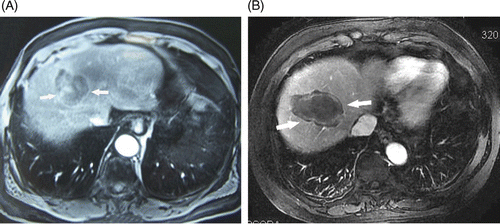
Figure 2. Transverse contrast-enhanced T1-weighted MR images in a 74-year-old man who underwent microwave ablation of HCC. (A) Hepatic arterial phase pre-ablation image shows a 5.9 × 4.2 cm HCC (arrow). (B) Hepatic arterial phase image post-ablation 6 months shows a 7.1 × 5.1 cm non-enhancing zone of hypointensity enveloping the tumour (arrow).

Table II. Comparison of ablation results in the 915 MHz and 2450 MHz MW groups.
Complications
There were no deaths and no thrombosis of major vessels in either group. Two cases (one in the 915 MHz MW group, one in the 2450 MHz MW group) with a large sub-diaphragmatic tumour had a large amount of pleural fluid requiring aspiration. Two patients (one in the 915 MHz MW group, one in the 2450 MHz MW group) who had an ablated lesion adjacent to the surface of the liver had moderate right upper quadrant pain (grade 2) requiring oral analgesics (oxycodone) and the symptoms gradually disappeared one week later. Thirteen cases (seven in the 915 MHz MW group, six in the 2450 MHz MW group) had non-infective high fever, while the temperature reduced to normal within 2–7 days without special treatments. There was no obvious differentiation in minor complications between the 915 MHz MW group and the 2450 MHz group (p > 0.05). One case of subcutaneous tumour seeding of needle track occurred 6 months after MW ablation in the 2450 MHz MW group.
Local tumour progression
Local tumour progression was found in 3 of 21 tumours (14.3%) in the 915 MHz MW group and 5 of 19 tumours (26.3%) in the 2450 MHz MW group on follow-up contrast-enhanced imaging. All cases of LTP were confirmed by biopsy which was performed simultaneously with re-treatment. There were no significant differences in the rate of local tumour progression between the 915 MHz MW group and the 2450 MHz MW group (p = 0.44). Of the three cases of local tumour progression in the 915 MHz MW group, two cases were located in the vicinity of diaphragm and one case contacted with the hepatic capsule. They were completely ablated by further MW ablation. Of the five cases in the 2450 MHz MW group, three cases were in the vicinity of the diaphragm, one case was adjacent to the para-umbilical veins and one case contacted with the hepatic capsule. They were also completely ablated by further MW ablation. In five cases of α-fetoprotein levels over 200 ng/L in the 915 MHz MW group, four cases decreased to normal level in 1 month after ablation, and one case decreased to below 200 ng/L, but was still abnormal and had LTP in 1 month after ablation. In six cases of α-fetoprotein levels over 200 ng/L in the 2450 MHz MW group, four cases decreased to normal level 1 month after ablation, and two cases were still abnormal and had LTP 3 months after ablation.
Discussion
RF ablation or MW ablation has been used as a potentially curative method in the treatment of small liver cancer Citation[29]. For lesions larger than 4 cm in diameter, transarterial chemoembolisation (TACE) is a commonly used method at present for patients who are non-surgical candidates. However, it is difficult for TACE to achieve complete tumour necrosis in which the complete necrosis rate was below 50% Citation[30], Citation[31]. At present, RF and MW (2450 MHz) ablation have also been used in larger hepatic lesions by multiple sequential overlapping ablations to ensure adequate coverage Citation[12], Citation[32], Citation[33]. Several MW techniques have recently been developed to increase the coagulation volume, such as 915 MHz MW generator and cooled-shaft MW antennae. It is possible to increase the power output to enlarge the volume of coagulation for the cooled-shaft MW antennae, at the same time avoiding skin injury Citation[20]. In vivo experiments in the porcine liver by Sun et al. showed that 915 MHz cooled-shaft MW antenna could yield a significantly larger ablation zone than that of 2450 MHz Citation[26]. Some studies showed that 915 MHz MW ablation is a reliable, efficient, and safe technique to perform hepatic tumour ablation and could obtain large ablation zones Citation[18], Citation[22], Citation[23], Citation[34]. The application of new 915 MHz cooled-shaft antennae may bring us a promising method for the percutaneous ablation of large HCC.
In our 2450 MHz MW group, the number of antenna insertions was 4.71 ± 1.61 (range 4–9), which was coincident with Lu's report Citation[32] that five to seven antenna insertions were needed in US-guided percutaneous 2450 MHz MW ablation for tumours of 4.1 cm to 6 cm. Fewer antenna insertions were required in the 915 MHz MW group than in the 2450 MHz MW group, although the size of tumours in the two groups had no significant difference. The 915 MHz MW may penetrate deeper than the 2450 MHz MW; therefore, theoretically 915 MHz MW may produce a larger ablation zone Citation[24], Citation[25]. Sun et al. Citation[26] reported that a 915 MHz MW antenna could yield a larger ablation zone than 2450 MHz MW antenna at the same emission energy. When multiple antennae were simultaneously inserted into the tumour, the distance between antennae could be further in the 915 MHz MW group than in the 2450 MHz MW. So compared with 2450 MHz MW, when ablating the same size tumour, fewer antenna insertions of 915 MHz MW were required. There were some advantages for 915 MHz MW due to its fewer antenna insertions: first, fewer antenna insertions means less invasions to patients; second, fewer antenna insertions needs less expenditure for patients; third, 915 MHz MW may decrease the difficulty of technical procedure; furthermore, Kuang Citation[20] reported that an increased frequency of antenna insertions to treat multiple or large nodules resulted in significantly higher complication rates, therefore, fewer antenna insertions may decrease the incidence rate of complications.
Lu et al. reported the technique effectiveness rate of 2450 MHz MW ablation for HCC larger than 3 cm in diameter was 83.3% Citation[35]. Martin et al. reported the technique effectiveness rate of 915 MHz MW ablation for hepatic tumours with median size of 3 cm was 100% Citation[34]. In our study, according to the follow-up contrast-enhanced imaging, technique effectiveness rate was 85.7 % (18/21) and 73.7% (14/19) in 915 MHz MW group and in 2450 MHz MW group, respectively, and there was no significant difference between the two groups (p = 0.44). The rate of LTP was 26.3% in our 2450 MHz MW group, which was coincident with previous reports Citation[35]. There were no significant differences in the rate of LTP between the 915 MHz MW group and the 2450 MHz MW group. However, not enough cases were included and LTP is affected by many risk factors and the clinical experiences of physicians, so, the comparison of LTP between 2450 MHz and 915 MHz MW ablation needs further study according to more cases and longer follow-up.
Besides, although we didn’t have a comparison study between 915 MHz MW ablation and RF ablation, the technique effectiveness rate in our study was higher than or similar to previous reports Citation[12], Citation[13] of percutaneous RF ablation, whereas fewer numbers of insertions were needed compared with previous report of RF ablation for large HCC.
In our study, there was no obvious difference in immediate complications between the 915 MHz MW group and the 2450 MHz MW group. The incidence rate of complications was low in our study. One reason was the application of the cooled-shaft antennae which could easily avoid skin burn. Another reason of low incidence rate of complications was that we performed the percutaneous MW ablation assisted with temperature monitoring through which damage to adjacent organs could be avoided. Our result showed that both 915 MHz and 2450 MHz MW with cooled-shaft antennae were safe in the percutaneous treatment of large HCC.
This study had some limitations. First, we were unable to accurately measure the energy delivered to the tissue; the reported emission energy was the sum of each generator's emission energy. It is known that energy attenuation in coaxial cables at 915 MHz is less than at 2450 MHz, so it is possible that more energy was delivered to the tissue at 915 MHz. Second, only 39 patients included in our study and follow-up period is relatively short. Further study of a larger sample with long-term follow-up is needed. Third, this was only a single centre study; a multi-centre study would be more convincing.
In conclusion, compared with 2450 MHz MW ablation, our study showed percutaneous 915 MHz MW ablation with cooled-shaft antennae was safe and could achieve a high technique effectiveness rate with fewer insertion numbers in the treatment of large HCC. Percutaneous 915 MHz MW ablation may provide a new way for the treatment of large HCC.
Declaration of interest: The authors report no conflicts of interest. The authors alone are responsible for the content and writing of the paper.
References
- Parkin DM, Bray F, Ferlay J, Pisani P. Global cancer statistics, 2002. CA Cancer J Clin 2005; 55: 74–108
- El-serag HB, Mason AC. Rising incidence of hepatocellular carcinoma in the United States. N Engl J Med 1999; 340: 745–750
- Bosch FX, Ribes J, Borras J. Epidemiology of primary liver cancer. Semin Liver Dis 1999; 19: 271–285
- Lai EC, Fan ST, Lo CM, Chu KM, Liu CL, Wong J. Hepatic resection for hepatocellular carcinoma: An audit of 343 patients. Ann Surg 1995; 221: 291–298
- Bismuth H, Chiche L, Adam R, Castaing D, Diamond T, Dennison A. Liver resection versus transplantation for hepatocellular carcinoma in cirrhotic patients. Ann Surg 1993; 218: 145–151
- Bruix J, Fuster J, Llovet JM. Liver transplantation for hepatocellular carcinoma: Foucault pendulum versus evidence-based decision. Liver Transpl 2003; 9: 700–702
- Forman LM, Lewis JD, Berlin JA, Feldman HI, Lucey MR. The association between hepatitis C infection and survival after orthotopic liver transplantation. Gastroenterology 2002; 122: 889–896
- Lu DS, Yu NC, Raman SS, Limanond P, Lassman C, Murray K, Tong MJ, Amado RG, Busuttil RW. Radiofrequency ablation of hepatocellular carcinoma: Treatment success as defined by histologic examination of the explanted liver. Radiology 2005; 234: 954–960
- Goldberg SN, Gazelle GS, Mueller PR. Thermal ablation therapy for focal malignancy: A unified approach to underlying principles, techniques, and diagnostic imaging guidance. Am J Roentgenol 2000; 174: 323–331
- Xu HX, Xie XY, Lu MD, Chen JW, Yin XY, Xu ZF, Liu GJ. Ultrasoundguided percutaneous thermal ablation of hepatocellular carcinoma using microwave and radiofrequency ablation. Clin Radiol 2004; 59: 53–61
- Dodd GD, Frank MS, Aribandi M, Chopra S, Chintapalli KN. Radiofrequency thermal ablation: Computer analysis of the size of the thermal injury created by overlapping ablations. Am J Roentgenol 2001; 177: 777–782
- Chen MH, Yang W, Yan K, Zou MW, Solbiati L, Liu JB, Dai Y. Large liver tumors: Protocol for radiofrequency ablation and its clinical application in 110 patients–Mathematic model, overlapping mode, and electrode placement process. Radiology 2004; 232: 260–271
- Livraghi T, Goldberg SN, Lazzaroni S, Meloni F, Ierace T, Solbiati L, Gazelle GS. Hepatocellular carcinoma: Radiofrequency ablation of medium and large lesions. Radiology 2000; 214: 761–768
- Kuvshinoff BW, Ota DM. Radiofrequency ablation of liver tumors: Influence of technique and tumor size. Surgery 2002; 132: 605–611
- Goldberg SN, Gazelle GS, Solbiati L, Rittman WJ, Mueller PR. Radiofrequency tissue ablation: Increased lesion diameter with a perfusion electrode. Acad Radiol 1996; 3: 636–644
- Wright AS, Sampson LA, Warner TF, Mahvi DM, Lee FT, Jr. Radiofrequency versus microwave ablation in a hepatic porcine model. Radiology 2005; 236: 132–139
- Wright AS, Lee FT, Jr, Mahvi DM. Hepatic microwave ablation with multiple antennae results in synergistically larger zones of coagulation necrosis. Ann Surg Oncol 2003; 10: 275–283
- Brace CL, Laeseke PF, Sampson LA, Frey TM, van der Weide DW, Lee FT, Jr. Microwave ablation with multiple simultaneously powered small-gauge triaxial antennas: Results from an in vivo swine liver model. Radiology 2007; 244: 151–156
- Seki T, Wakabayashi M, Nakagawa T, Itho T, Shiro T, Kunieda K, Sato M, Uchiyama S, Inoue K. Ultrasonically guided percutaneous microwave coagulation therapy for small hepatocellular carcinoma. Cancer 1994; 74: 817–825
- Kuang M, Lu MD, Xie XY, Xu HX, Mo LQ, Liu GJ, Xu ZF, Zheng YL, Liang JY. Liver cancer: Increased microwave delivery to ablation zone with cooled-shaft antenna–Experimental and clinical studies. Radiology 2007; 242: 914–924
- Liang P, Wang Y, Yu XL, Dong BW. Malignant liver tumors: Treatment with percutaneous microwave ablation–Complications among cohort of 1136 patients. Radiology 2009; 251: 933–940
- Simon CJ, Dupuy DE, Iannitti DA, Lu DSK, Yu NC, Aswad BI, Busuttil RW, Lassman C. Intraoperative triple antenna hepatic microwave ablation. Am J Roentgenol 2006; 187: W333–340
- Brace CL, Laeseke PF, Sampson LA, Frey TM, van der Weide DW, Lee FT, Jr. Microwave ablation with a single small-gauge triaxial antenna: In vivo porcine liver model. Radiology 2007; 242: 435–440
- Metaxas AC, Meredith RJ. Industrial Microwave Heating. Peter Peregrinus, London 1983; 281–282
- Lee Chin, Michael Sherar. Changes in dielectric properties of ex vivo bovine liver at 915 MHz during heating. Phys Med Biol 2001; 46: 197–211
- Sun Y, Wang Y, Ni X, Gao Y, Shao Q, Liu L, Liang P. Comparison of ablation zone between 915- and 2450-MHz cooled-Shaft microwave antenna: Results in in vivo porcine livers. Am J Roentgenol 2009; 192: 511–514
- Dong BW, Liang P, Yu XL, Su L, Yu D, Cheng Z, Zhang J. Percutaneous sonographically guided microwave coagulation therapy for hepatocellular carcinoma: Results in 234 patients. Am J Roentgenol 2003; 180: 1547–1555
- Zhou P, Liang P, Yu X, Wang Y, Dong B. Percutaneous microwave ablation of liver cancer adjacent to the gastrointestinal tract. J Grastrointest Surg 2009; 13: 318–324
- Bruix J, Hessheimer AJ, Forner A, Boix L, Vilana R, Llovet JM. New aspects of diagnosis and therapy of hepatocellular carcinoma. Oncogene 2006; 25: 3848–3856
- Sergio, A, Cristofori, C, Cardin, R, Pivetta G, Ragazzi R, Baldan A, Girardi L, Cillo U, Burra P, Giacomin A, et al. Transcatheter arterial chemoembolization (TACE) in hepatocellular carcinoma (HCC): The role of angiogenesis and invasiveness. Am J Gastroenterol 2008; 103: 914–921
- Miraglia R, Pietrosi G, Maruzzelli L, Petridis I, Caruso S, Marrone G, Mamone G, Vizzini G, Luca A, Gridelli B. Efficacy of transcatheter embolization/chemoembolization (TAE/TACE) for the treatment of single hepatocellular carcinoma. World J Gastroenterol 2007; 13: 2952–2955
- Lu MD, Chen JW, Xie XY, Liu L, Huang XQ, Liang LJ, Huang JF. Hepatocellular carcinoma: US-guided percutaneous microwave coagulation therapy. Radiology 2001; 221: 167–172
- Yu Z, Liu W, Fan L, Shao J, Huang Y, Si X. The efficacy and safety of percutaneous microwave coagulation by a new microwave delivery system in large hepatocellular carcinomas: Tour case studies. Int J Hyperthermia 2009; 25: 392–398
- Martin RC, Scoggins CR, McMasters KM. Microwave hepatic ablation: Initial experience of safety and efficacy. J Surg Oncol 2007; 96: 481–486
- Lu MD, Xu HX, Xie XY, Yin XY, Chen JW, Kuang M, Xu ZF, Liu GJ, Zheng YL. Percutaneous microwave and radiofrequency ablation for hepatocellular carcinoma: A retrospective comparative study. J Gastroenterol 2005; 40: 1054–1060