Abstract
The goal of this review is to provide an overview on laser treatment of benign skin tumours and recent developments in this field. Ablational laser systems are established treatment devices for benign skin tumours. They obtain good cosmetic results with mostly minimal side-effects. Recently, fractional laser devices or combination of laser therapy with photodynamic therapy has gained attention in this field. However, there is a lack of randomised, controlled trials for laser treatment of benign skin tumours.
Introduction
Treatment with different laser systems has become an integral part of dermatological therapy. The group of indications regarding benign tumours and organoid nevi is very heterogeneous. For these indications, patients often search help for cosmetic reasons. Therefore, high standards for treatment are required. Ablative and semi-ablative laser devices are mainly used for superficial ablation of tissue without damage of the surrounding structures. The ablation depth of the laser depends on wavelength, fluence rate and spot size. The most common or classic treatment devices include the erbium:yttrium-aluminium-garnet (Er:YAG) and the carbon dioxide (CO2) laser. Although the argon laser is only of historical importance it is still used for the ablation of benign skin tumours but can frequently be replaced by the potassium titanium oxide phosphate (KTP) laser. Argon lasers are gas ion lasers and emit a continuous-wave blue-green visible light at peak wavelengths of 488 and 514 nm. Pigmented lesions can be damaged selectively with the argon laser with rapid wound healing Citation[1]. The Er:YAG laser with a 2940 nm wavelength is well established for ablation of soft and hard biological tissue because the wavelength is highly absorbed in water. It is capable of ablating thin layers of skin precisely, especially in delicate locations, with minimal collateral damage Citation[2]. The CO2 laser is widely used for medical surgery. At a wavelength of 10,600 nm the energy is mainly absorbed by the extracellular fluid of biologic structures which leads to a non-specific vaporisation and coagulation of tissue. Ultra-pulsed CO2 lasers emit short light pulses (600–900 µs) with high peak energies so that tissue can be ablated very precisely layer by layer Citation[3].
Further treatment devices for benign skin tumours include the alexandrite, ruby, neodymium:YAG (Nd:YAG), and the diode laser. Alexandrite lasers emit light at wavelengths ranging from 700 to 830 nm. As these wavelengths are absorbed by melanin and dyes, but not significantly by blood, it can be used for the destruction of melanin-containing structures. The ruby laser has an emission wavelength of 694 nm and the laser light is absorbed by melanin and dark pigments.
The Nd:YAG laser is the most important solid-state laser with a wavelength of 1064 nm. Doubling the frequency of 1064 nm by a potassium titanyl phosphate crystal results in green light-emitting KTP lasers (532 nm) Citation[4]. Quality (Q)-switched lasers have extremely short pulse times of about 10 ns which is much shorter than the relaxation time of the target chromophore. Q-switched Nd:YAG lasers are especially suited for treatment of tattoos, but also other pigmented lesions Citation[4].
In diode lasers electrical energy is transformed directly into laser light. Laser diodes are available from the ultraviolet (UV) spectrum into the visible spectrum until the near-infra red (IR) spectrum for surgical applications. Diode lasers are non-ablative devices but they are capable of inducing thermal damage in the middle to upper dermis without causing scarring or epidermal damage Citation[5].
The 585 nm pulsed dye laser (PDL) is usually no appropriate device for ablational therapy of benign skin tumours. Its main indication is for the treatment of vascular lesions Citation[6]. However, it produces a non-specific thermal effect so that the PDL has proved to be effective for the treatment of xanthelasmas Citation[7]. It has the further advantage of not producing an open wound in contrast to ablative laser systems.
There is a wide range of different laser devices for the treatment of benign skin tumours. Their application depends on the indication as well as the experience of the practioner.
Indications
The group of benign skin tumours that might be treated by laser therapy is rather heterogeneous. These tumours can be roughly classified as epidermal tumours/nevi, adnexal, fibrous, and neural tumours, as well as xanthelasmas and cysts.
Epidermal, dermal and organoid nevi/tumours
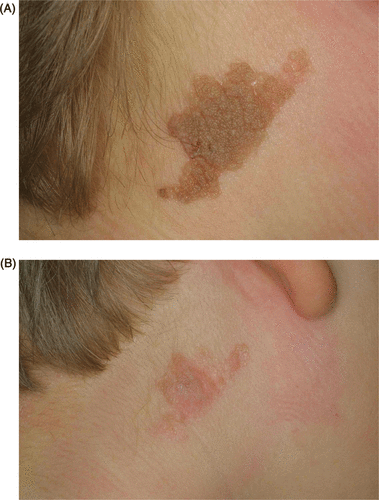
Epidermal nevi are the most common manifestation of cutaneous mosaicism (). They are present at birth and affect only the epidermis, whereas organoid nevi involve adnexal structures. A distinction must be made between papillomatous soft and hard verrucous lesions. Inflammatory linear verrucous epidermal nevus (ILVEN) is an unusual form of epidermal nevus marked by an additional inflammatory component. Complete removal of epidermal nevi remains difficult. Whereas aggressive approaches like surgical excision result in scar formation, superficial means of removal often result in recurrence Citation[8].
Figure 1. (A) Epidermal nevus in the cervical region of an 11-year-old boy. (B) Result after three sessions of CO2 laser (cw mode), 10 W of power, 2 mm spot size.

Different types of lasers have been reported to be effective in the treatment of epidermal nevi. The first report on the successful laser treatment of verrucous, epidermal nevi was published in 1984 Citation[9]. In two patients, localised verrucous nevi were removed completely and in another patient with a systemic verrucous nevus, a marked improvement was obtained after several argon laser sessions. Paradela et al. Citation[10] treated a total of 25 patients with epidermal nevi with the CO2 laser in the super-pulsed mode (2 W/cm2). The mean number of treatment sessions was four (range: 1 to 28). Good results were achieved in 92% of soft, flattened nevi and in only 33% of patients with verrucous lesions. These authors believe that the thickness of the nevus is the most determining factor for the cosmetic result. Hohenleutner et al. Citation[8] found the argon laser and the continuous-wave (cw) carbon dioxide laser able to remove epidermal nevi of various types and textures in 43 patients. Papillomatous, soft variants could be treated successfully with the argon laser without scarring whereas hard, keratotic lesions responded to CO2 laser therapy but with a tendency to scar formation (). The CO2 laser offers the advantage of intraoperative haemostasis whereas bleeding might complicate treatment with the Er:YAG laser Citation[11]. Various case reports have proved the efficacy of the CO2 laser Citation[11], Citation[12]. Park et al. Citation[13] treated 20 patients with verrucous epidermal nevi either with the variable pulsed or the dual-mode Er:YAG laser. In 15 out of 20 patients, successful removal of the verrucous epidermal nevus could be obtained after one treatment session. All patients showed post-operative erythema and 15% had pigmentary changes. Five patients showed a relapse within one year.
Further treatment devices for epidermal nevi include the long-pulsed ruby laser Citation[14] and the pulsed dye laser for treating itch in ILVEN Citation[15]. In conclusion, the CO2 laser (cw/pulsed) has proved to be a good therapy option, especially for treating large areas with limited bleeding. The Er:YAG laser offers a good alternative for smaller lesions with comparable results although bleeding might hamper the visibility and thus exact ablation. Alternative treatment strategies for ILVEN include trichloroacetic acid peeling Citation[16], topical calcipotriol Citation[17] and cryosurgery. Photodynamic therapy (PDT) has been reported to be successful in the treatment of a verrucous epidermal nevus Citation[18].
Nevus sebaceous usually presents at birth as a yellow patch and tends to thicken during puberty, so that patients then often request their removal for cosmetic reasons. As in epidermal nevi, the CO2 laser offers a good therapy modality to ablate such lesions Citation[19]. Recently, the combination of CO2 laser treatment followed by photodynamic therapy has proved to be effective for ablation of nevus sebaceous on the face in 12 patients Citation[20]. However, several treatment sessions (2–9) were necessary and two patients showed partial recurrences shortly after completion of treatment.
Seborrhoeic keratoses are very frequent benign epithelial tumours that are especially annoying when situated on the head and neck area or in case of extensive occurrence. Recurrence after curettage, shave excision, cryoablation, or chemical peelings can be observed. As these lesions might be confused with malignant tumours such as melanoma or basal cell carcinoma, laser therapy should be performed only by experienced dermatologists or after histological examination of the lesion. A recent study Citation[21] investigated the efficacy of the 532-nm diode laser to remove 1567 seborrhoeic keratoses in 326 patients. The lesions were coloured with a red marker or ferric subsulphate to enhance the ablative effects of laser treatment. Complete resolution occurred in 93% of lesions after one single treatment, whereas 7% of lesions required a second laser treatment. Other suitable laser devices include the CO2, erbium, argon, and alexandrite laser Citation[1], Citation[22–24]. Dermatosis papulosa nigra is very common among people with skin type V and VI and is considered a variant of seborrhoeic keratoses. In 2008, Schweiger and colleagues reported the use of a long-pulsed Nd:YAG laser for dermatosis papulosa nigra in two patients who achieved 70 to 90% clearance of the treated lesions Citation[25]. Kundu et al. Citation[26] reported the efficacy of a KTP laser for dermatosis papulosa nigra in 14 patients, with 96% of patients achieving greater than 50% improvement. A case report describes the successful treatment of dermatosis papulosa nigra with a 1.550 nm fractionated erbium-doped fibre laser resulting in greater than 75% improvement Citation[27].
Er:YAG and CO2 lasers can be used for ablation of benign, papillomatous dermal nevi Citation[28]. However, a number of case reports have been published wherein melanoma has been diagnosed after laser ablation of presumed benign skin lesions Citation[29], Citation[30]. In patients with a history of melanoma or evolution of a pigmented lesion, laser therapy should be avoided or a biopsy should be performed before laser treatment. Alternative treatment options for benign epidermal/dermal tumours include surgical excision, cryotherapy and chemical peelings with a risk of scar formation and hypopigmentation. Surgical excision is especially difficult in extensive lesions.
Adnexal tumours
Therapy options for adnexal tumours are limited and include chemical peelings, surgical excision, electrocauterisation and laser therapy. Hair follicle tumours occur most often on the face and scalp. Trichoblastomas are the most common secondary tumours in nevus sebaceous. They are often difficult to distinguish from basal cell carcinoma, both clinically and histologically. Therefore, surgical excision and histological examination is mandatory in these cases. Multiple trichoblastomas might occur in a context of Brooke-Spiegler syndrome along with trichoepitheliomas, spiradenomas and cylindromas Citation[31]. In the case of multiple tumours, non-surgical treatments are favoured. Lopiccolo et al. Citation[32] treated a patient with Brooke-Spiegler syndrome after histological examination of facial trichoepitheliomas either with Er:YAG fractionated ablative laser resurfacing and photodynamic therapy (PDT), resurfacing combined with imiquimod, resurfacing alone, or PDT alone. Ablative laser resurfacing in combination with PDT was rated best by the patient. Treatment with CO2 laser alone, argon laser or the combination of Er:YAG and CO2 laser has also been reported for this indication Citation[33–36]. Multiple trichodiscomas and fibrofolliculomas, or so-called mantleomas, are typical for Birt-Hogg-Dubé syndrome. They present as flat white papules, most commonly on the face and chest, and might be treated successfully with the CO2 or Er:YAG laser Citation[37], Citation[38].
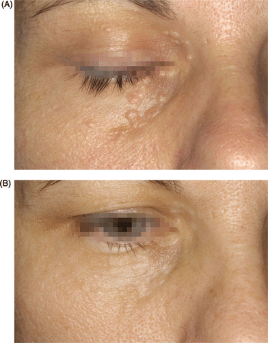
Syringomas are probably the most common adnexal tumours arising from sweat glands. They typically appear in the periorbital or perinasal region of young women and are almost always multiple (). Recently it has been shown that familial syringoma is an autosomal dominant disorder and the syringoma gene has been mapped to chromosome 16q22 Citation[39]. Several studies and case reports have proved the effectiveness of the CO2 laser in the treatment of syringomas Citation[40–42]. Park et al. Citation[41] treated 11 patients with syringomas with the CO2 laser using a multiple-drilling method. In small lesions, a single hole was made in the centre of the lesion, whereas for conglomerate lesions, multiple holes were made within the lesions at distinct distances. Patients received 1 to 4 treatment sessions at 4- to 6-week intervals. Clinical response was excellent in seven patients and good in four patients without occurrence of any serious side effect. Fractional laser devices are widely used for skin rejuvenation but they might also be beneficial for the treatment of syringomas. Cho et al. Citation[42] treated 35 patients with periorbital syringomas with an ablative 10,600 nm fractional CO2 laser. After two treatment sessions at 1-month interval, 8.6% showed ≥75% improvement, 42.9% had marked improvement and 34.3% had moderate improvement. Five patients (14.3%) had transitory hyperpigmentation after the treatment. Fractional CO2 laser treatment seems to be a promising new method for the treatment of syringomas with minimal side-effects.
Figure 2. (A) Syringomas in the periorbital region of a 41-year-old woman. (B) Result after four treatment sessions with the argon laser, 1.3 W of power, 2 mm spot size, 300 ms pulse duration.

Sebaceous gland hyperplasia is an extremely common finding in seborrhoeic sun-damaged skin. Besides cryotherapy and chemical peelings, ablational laser devices (argon (), CO2 laser, Er:YAG laser), the PDL and 1450 nm diode laser lead to good cosmetic results Citation[43–46]. A rather new therapy option is the combination of PDT with CO2 laser treatment that was tested in four patients. CO2 laser therapy was performed before PDT to reduce lesion size and facilitate penetration of the photosensitiser. Three of four patients showed marked improvement without any side-effects Citation[47].
In conclusion, there is a wide range of possible laser devices for ablation of adnexal tumours with good cosmetic results and a low incidence of side-effects.
Fibrous tumours
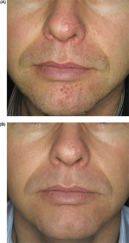
Angiofibromas can occur as solitary lesions or aggregated in a context of tuberous sclerosis, then often misnamed as adenoma sebaceum (). Further typical lesions of tuberous sclerosis include fibromas appearing in the sub- or periungual region, so-called Koenen tumours. Fibrous papules of the nose occur mostly isolated as small, red papules on the tip of the nose.
Figure 3. (A) Angiofibromas in the facial region of a 41-year-old man with tuberous sclerosis. (B) Results after five treatment sessions with the argon laser, 1.3 W of power, 2 mm spot size, 300 ms pulse duration.

Boixeda et al. Citation[48] treated 10 patients with facial angiofibromas with CO2, argon () or PDL. Excellent results were obtained in seven patients and good in three patients, with CO2 laser treatment being superior to argon laser therapy. Other authors described successful treatment of angiofibromas with argon laser surgery Citation[49–51]. Song et al. Citation[52] tried CO2 laser resurfacing in two Asian patients with angiofibromas with a flash scanner because it produces less thermal damage than the CO2 laser defocused mode. Furthermore, it enables controlled depth vaporisation for more precise ablation of lesions. Both patients showed remarkable cosmetic improvement, but post-treatment erythema persisted about 2 months.
The treatment of Koenen tumours is difficult due to their location next to the nail plate. There is a great risk of injury of the nail plate and consecutive disturbed nail growth. Therapeutic options include surgical excision, electrodessication, shave excision and phenolisation Citation[53], as well as CO2 laser treatment Citation[54]. CO2 laser therapy of Koenen tumours proved to be similar to conventional surgical resection in terms of cosmetic outcome. However, bleeding was negligible and operating time was considerably shorter for laser treatment compared to surgical excision Citation[54].
Fibrous papules of the nose are common benign lesions that might be treated by tangential excision or laser ablation. There are no studies comparing different laser devices for this indication. In our experience, the argon laser is a very suitable device for ablation of those small papules. Ablation with the pulsed CO2 laser or the Er:YAG laser might be a treatment alternative.
Ablative laser devices like the CO2 or the argon laser are the most suitable devices for ablation of benign fibrous tumours. Surgical excision is only an alternative treatment in cases of solitary lesions.
Neural tumours
Neurofibromas may occur as solitary molluscoid nodules that can be pushed back into the skin like a bell button. When multiple, they are an important marker for neurofibromatosis and a condition of cosmetic concern. In contrast to surgical treatment, the CO2 laser can rapidly remove large numbers of lesions with high patient satisfaction Citation[55], Citation[56]. After opening the epidermis, the lesion can be squeezed out and ablated. A complete removal down to the base should be done to prevent recurrences. However, the risk of scar formation after CO2 laser therapy has been reported in the literature Citation[57], Citation[58]. Elwakil et al. Citation[59] treated 12 patients with multiple neurofibromas either with a long-pulsed Nd:YAG laser (74% of lesions, thickness ≤ 5 mm) or bulkier lesions (26%, thickness ≥ 5 mm) with a cw Nd:YAG laser. Patients received 1 to 2 treatment sessions. Regarding flat lesions, a regression of 76% to 100% could be obtained in 43% of lesions, and 34% of lesions showed a regression of 51% to 75%. Hyperpigmentation occurred in 4.8% of lesions. Regarding bulkier lesions, 68% of lesions had a regression between 51% and 100%. In this group, wound infection and ulceration occurred in 1.5% and 3%, respectively, whereas hyperpigmentation occurred in 4.5% of lesions. There were no recurrences during a follow-up of 14 months.
Both Nd:YAG and CO2 lasers are useful devices for ablation of neurofibromas. However, there is a risk of scar formation so that patients have to be informed about these possible side-effects. Test spots are mandatory before treatment of larger areas.
Xanthelasmas
Xanthelasmas represent lipid deposits in the skin and are sometimes the first clinical marker of hypercholesterolemia. They present as flat, yellow plaques and are most often found in the periorbital region. When performing laser therapy, protection of the eyes is mandatory, for example by insertion of steel eye shields or sterile rubberised contact lenses. The classic laser devices for treatment of xanthelasmas include CO2, argon, and Er:YAG lasers Citation[2], Citation[3], Citation[60]. Furthermore, KTP, Nd:YAG, PDL, and diode lasers have been applied successfully Citation[7], Citation[61–63]. Fractional photothermolysis has recently been reported as a new therapy strategy in a case report Citation[64].
Raulin et al. Citation[3] treated 23 patients with 52 periorbital xanthelasmas with an ultra-pulsed CO2 laser and could remove all lesions by a single treatment (4 to 7 passes). Three patients developed a recurrence during a follow-up of 10 months. No side-effects occurred besides transient pigmentary changes. The cw CO2 laser has a higher risk of scar formation and should be used only cautiously Citation[65]. Basar et al. Citation[60] treated 24 patients in one to four sessions with an argon laser and obtained good cosmetic results in 85% of patients. The Er:YAG laser also represents an effective and safe therapy option for this indication Citation[2]. The Q-switched Nd:YAG laser as well as the KTP laser have obtained good results after one to three treatment sessions with minimal side-effects in two studies Citation[61], Citation[62]. Another study could not reproduce the good effects of the Q-switched Nd:YAG laser in 37 patients and these authors do not recommend it for the treatment of xanthelasmas Citation[66].
In contrast to ablative laser systems, PDL treatment does not produce an open wound. In the treatment of xanthelasmas it causes an unspecific thermal effect. A recently published study Citation[7] has investigated the efficacy of PDL treatment for xanthelasma palpebrarum. Twenty patients received five treatment sessions (two passes per session) with a short-pulsed PDL. About 90% of patients had good or excellent clearance of their lesions and patient satisfaction was high. Hyperpigmentation occurred in 7.9% of lesions and was still present at the end of the final 4-week follow-up. Park et al. Citation[63] investigated the safety and efficacy of a 1450 nm diode laser in 16 patients with xanthelasmas. A moderate or marked improvement was seen in 75% of patients, whereas mild focal transient hyperpigmentation occurred in five patients.
Besides laser ablation, xanthelasmas might be treated by surgical excision, with a risk of scar formation or ectropion, or by the use of trichloroacetic acid Citation[67]. Recurrence of xanthelasmas is common, regardless of the mode of treatment. Most often several treatment sessions are necessary. New treatment options like diode laser therapy or fractional photothermolysis have to be investigated in larger controlled, randomised studies.
Cysts
Cysts are well-circumscribed tumours that can be divided into true cysts (epithelial, glandular and developmental cysts) and pseudocysts. Eruptive vellus hair cysts are epithelial cysts that are closely related to steatocystoma multiplex. CO2 and Er:YAG lasers have been used with variable success to treat this entity Citation[68–71]. Coras et al. Citation[70] treated a patient with dense facial eruptive vellus hair cysts with the pulsed Er:YAG laser. The lesions recurred shortly after therapy and the authors concluded that the therapy is not successful in regions where the depth of ablation is limited, owing to the risk of atrophy and scarring. Kageyama et al. Citation[72] treated two patients with truncal eruptive vellus hair cysts successfully with the Er:YAG laser. After penetration of the cyst wall and digital expression of the cyst content, three additional passes were performed to ablate the posterior cyst wall. There was no recurrence during a 13-month follow-up. Individual cases report good results with the Er:YAG and CO2 laser for the treatment of steatocystoma multiplex Citation[68], Citation[69].
Eccrine hidrocystomas are benign, solitary glandular cystic tumours that can be treated by surgical measures or laser therapy. Case reports have described the successful treatment of hidrocystomas with diode lasers, CO2 and PDL Citation[5], Citation[73], Citation[74]. The PDL targets vascular structures and is mainly used for the treatment of port wine stains and haemangiomas Citation[6]. Lee et al. Citation[73] postulated that PDL therapy for eccrine hidrocystomas leads to destruction of dermal cystic structures by non-selective heating energy diffused out from the selectively photothermolysed adjacent microvasculatures. However, the pulse duration seems to play an important role. Tanzi and Alster Citation[75] used a PDL with a pulse duration of 1.5 ms and needed four treatment sessions at 6- to 8-week intervals to obtain good cosmetic results. Lee et al. Citation[73] used a PDL with a pulse duration of 6 ms and obtained clearing after a single treatment session during a follow-up period of 11 months. Other authors could not reproduce the good results with the PDL using a pulse duration of 3 ms in two Asian women Citation[76]. Therefore, further controlled studies are needed to investigate the efficacy of different laser systems in the treatment of hidrocystomas.
Mucoid dorsal cysts are pseudocysts because they lack an epithelial lining. They may be painful and cause a longitudinal furrow in the nail when arising on the nail fold. In the literature there are only two reports on the successful treatment of mucoid dorsal cysts with the CO2 laser Citation[77], Citation[78]. Karrer et al. Citation[77] treated four out of six patients successfully with the CO2 laser without any complications or side-effects. The cyst was penetrated, the content squeezed out and the cyst completely vaporised under protection of the underlying nail matrix. As a consequence of surgical excision, scar formation and deformation of the nail might occur. Thus, CO2 laser therapy might be used as first line therapy. Oral mucous cysts might be treated by the CO2 laser, as well Citation[79].
In conclusion, the CO2 laser is the most common laser for the treatment of cysts. However, non-ablative lasers like PDL or diode lasers might be an alternative for this indication.
Contraindications for laser therapy of benign skin tumours and alternative treatment options
Accurate diagnosis of benign skin tumours is necessary before initiating laser therapy, as this treatment cannot be monitored by histology. In case of the slightest doubt on the benign nature of the lesion, a biopsy must be performed for histopathological examination. Otherwise serious medical complications and legal impacts for the practitioner may arise. Pigmented lesions in patients with a history of melanoma or dysplastic nevi should not be treated by laser therapy. The use of dermatoscopy contributes to the exact clinical classification of pigmented lesions, but even experienced dermatologists are at risk of clinical misdiagnosis. Therefore, laser treatment of pigmented skin lesions is discussed controversially. Furthermore, it is currently not known whether removal or treatment of nevi with laser devices will affect the risk of malignant transformation of these lesions. However, laser treatment to remove or lighten nevi has been used successfully. In the literature, there are several reports of pseudomelanoma following laser treatment of melanocytic nevi Citation[30], Citation[80–82].
Surgical excision, shaving, electrocautery, cryotherapy or dermabrasion are sometimes good alternatives for removal of benign skin tumours. Especially surgical treatments have the advantage of a histopathological confirmation of the suspected diagnosis. However, scar formation and the risk of keloid formation in special localisations limit patients’ acceptance.
Conclusions
Many different laser systems have shown to be effective in the treatment of benign skin tumours. Often, multiple treatment sessions are necessary and recurrences are possible. Patients must be informed about possible side-effects as they are mainly treated for cosmetic reasons.
There must be no doubt about the benign nature of a tumour before performing a laser treatment. If there is any doubt, histological evaluation to clarify the benign or malignant nature of the lesion must be done first.
An optimal treatment of benign skin tumours combines complete destruction of lesions with minimal side-effects. Ablative laser systems are usually used for this indication. Recently, non-ablative laser systems such as the PDL and diode lasers have been used for the treatment of benign skin tumours as well Citation[7], Citation[73]. Fractional laser devices represent a promising addition to the dermatologist's armamentarium. Furthermore, combinational approaches such as laser treatment and PDT are new therapeutic options.
However, there is a lack of randomised, controlled trials in the treatment of benign skin tumours comparing laser therapy with conventional therapy options.
Declaration of interest: The authors report no conflicts of interest. The authors alone are responsible for the content and writing of the paper.
References
- Apfelberg DB, Maser MR, Lash H, Rivers JL. Progress report on extended clinical use of the argon laser for cutaneous lesions. Lasers Surg Med 1980; 1: 71–83
- Borelli C, Kaudewitz P. Xanthelasma palpebrarum: Treatment with the erbium:YAG laser. Lasers Surg Med 2001; 29: 260–264
- Raulin C, Schoenermark MP, Werner S, Greve B. Xanthelasma palpebrarum: Treatment with the ultrapulsed CO2 laser. Lasers Surg Med 1999; 24: 122–127
- Anderson RR, Margolis RJ, Watenabe S, Flotte T, Hruza GJ, Dover JS. Selective photothermolysis of cutaneous pigmentation by Q-switched Nd:YAG laser pulses at 1064, 532, and 355 nm. J Invest Dermatol 1989; 93: 28–32
- Echague AV, Astner S, Chen AA, Anderson RR. Multiple apocrine hidrocystoma of the face treated with a 1450-nm diode laser. Arch Dermatol 2005; 141: 1365–1367
- Landthaler M, Hohenleutner U. Laser therapy of vascular lesions. Photodermatol Photoimmunol Photomed 2006; 22: 324–332
- Karsai S, Czarnecka A, Raulin C. Treatment of xanthelasma palpebrarum using a pulsed dye laser: A prospective clinical trial in 38 cases. Dermatol Surg 2010; 36: 610–617
- Hohenleutner U, Landthaler M. Laser therapy of verrucous epidermal naevi. Clin Exp Dermatol 1993; 18: 124–127
- Landthaler M, Haina D, Waidelich W, Braun-Falco O. Argon laser therapy of verrucous nevi. Plast Reconstr Surg 1984; 74: 108–113
- Paradela S, Del Pozo J, Fernandez-Jorge B, Lozano J, Martinez-Gonzalez C, Fonseca E. Epidermal nevi treated by carbon dioxide laser vaporization: A series of 25 patients. J Dermatolog Treat 2007; 18: 169–174
- Alam M, Arndt KA. A method for pulsed carbon dioxide laser treatment of epidermal nevi. J Am Acad Dermatol 2002; 46: 554–556
- Boyce S, Alster TS. Co2 laser treatment of epidermal nevi: Long-term success. Dermatol Surg 2002; 28: 611–614
- Park JH, Hwang ES, Kim SN, Kye YC. Er:YAG laser treatment of verrucous epidermal nevi. Dermatol Surg 2004; 30: 378–381
- Baba T, Narumi H, Hanada K, Hashimoto I. Successful treatment of dark-colored epidermal nevus with ruby laser. J Dermatol 1995; 22: 567–570
- Alster TS. Inflammatory linear verrucous epidermal nevus: Successful treatment with the 585 nm flashlamp-pumped pulsed dye laser. J Am Acad Dermatol 1994; 31: 513–514
- Toyozawa S, Yamamoto Y, Kaminaka C, Kishioka A, Yonei N, Furukawa F. Successful treatment with trichloroacetic acid peeling for inflammatory linear verrucous epidermal nevus. J Dermatol 2010; 37: 384–386
- Zvulunov A, Grunwald MH, Halvy S. Topical calcipotriol for treatment of inflammatory linear verrucous epidermal nevus. Arch Dermatol 1997; 133: 567–568
- Sim JH, Kang Y, Kim YC. Verrucous epidermal nevus successfully treated with photodynamic therapy. Eur J Dermatol 2010; 20: 814–815
- Ashinoff R. Linear nevus sebaceus of Jadassohn treated with the carbon dioxide laser. Pediatr Dermatol 1993; 10: 189–191
- In SI, Lee JY, Kim YC. Topical photodynamic therapy for nevus sebaceous on the face. Eur J Dermatol 2010; 20: 590–592
- Culbertson GR. 532-nm diode laser treatment of seborrheic keratoses with color enhancement. Dermatol Surg 2008; 34: 525–528; discussion: 528
- Fitzpatrick RE, Goldman MP, Ruiz-Esparza J. Clinical advantage of the CO2 laser superpulsed mode. Treatment of verruca vulgaris, seborrheic keratoses, lentigines, and actinic cheilitis. J Dermatol Surg Oncol 1994; 20: 449–456
- Hruza GJ. Laser treatment of epidermal and dermal lesions. Dermatol Clin 2002; 20: 147–164
- Mehrabi D, Brodell RT. Use of the alexandrite laser for treatment of seborrheic keratoses. Dermatol Surg 2002; 28: 437–439
- Schweiger ES, Kwasniak L, Aires DJ. Treatment of dermatosis papulosa nigra with a 1064 nm Nd:YAG laser: Report of two cases. J Cosmet Laser Ther 2008; 10: 120–122
- Kundu RV, Joshi SS, Suh KY, Boone SL, Huggins RH, Alam M, White L, Rademaker AW, West DP, Yoo S. Comparison of electrodesiccation and potassium-titanyl-phosphate laser for treatment of dermatosis papulosa nigra. Dermatol Surg 2009; 35: 1079–1083
- Katz TM, Goldberg LH, Friedman PM. Dermatosis papulosa nigra treatment with fractional photothermolysis. Dermatol Surg 2009; 35: 1840–1843
- Hammes S, Raulin C, Karsai S, Bernt R, Ockenfels HM. [treating papillomatous intradermal nevi: Lasers – yes or no? A prospective study]. Hautarzt 2008; 59: 101–107
- Giacomel J, Zalaudek I, Mordente I, Nicolino R, Argenziano G. Never perform laser treatment of skin tumors with clinical ‘EFG’ criteria. J Dtsch Dermatol Ges 2008; 6: 386–388
- Gottschaller C, Hohenleutner U, Landthaler M. Metastasis of a malignant melanoma 2 years after carbon dioxide laser treatment of a pigmented lesion: Case report and review of the literature. Acta Derm Venereol 2006; 86: 44–47
- Scholz IM, Numann A, Froster UG, Helmbold P, Enk AH, Naher H. New mutation in the cyld gene within a family with Brooke-Spiegler syndrome. J Dtsch Dermatol Ges 2010; 8: 99–101
- Lopiccolo MC, Sage RJ, Kouba DJ. Comparing ablative fractionated resurfacing, photodynamic therapy, and topical imiquimod in the treatment of trichoblastomas of brooke-spiegler syndrome: A case study. Dermatol Surg 2011; 37: 1047–1050
- Sajben FP, Ross EV. The use of the 1.0 mm handpiece in high energy, pulsed CO2 laser destruction of facial adnexal tumors. Dermatol Surg 1999; 25: 41–44
- Flores JT, Apfelberg DB, Maser MR, Lash H. Trichoepithelioma: Successful treatment with the argon laser. Plast Reconstr Surg 1984; 74: 694–698
- Martins C, Bartolo E. Brooke-Spiegler syndrome: Treatment of cylindromas with CO2 laser. Dermatol Surg 2000; 26: 877–880; discussion: 881
- Rallan D, Harland CC. Brooke-spiegler syndrome: Treatment with laser ablation. Clin Exp Dermatol 2005; 30: 355–357
- Kahle B, Hellwig S, Schulz T. [multiple mantleomas in Birt-Hogg-Dube syndrome: Successful therapy with co2 laser]. Hautarzt 2001; 52: 43–46
- Gambichler T, Wolter M, Altmeyer P, Hoffman K. Treatment of Birt-Hogg-Dube syndrome with erbium:YAG laser. J Am Acad Dermatol 2000; 43: 856–858
- Wu WM, Lee YS. Autosomal dominant multiple syringomas linked to chromosome 16q22. Br J Dermatol 2010; 162: 1083–1087
- Frazier CC, Camacho AP, Cockerell CJ. The treatment of eruptive syringomas in an African American patient with a combination of trichloroacetic acid and CO2 laser destruction. Dermatol Surg 2001; 27: 489–492
- Park HJ, Lee DY, Lee JH, Yang JM, Lee ES, Kim WS. The treatment of syringomas by CO2 laser using a multiple-drilling method. Dermatol Surg 2007; 33: 310–313
- Cho SB, Kim HJ, Noh S, Lee SJ, Kim YK, Lee JH. Treatment of syringoma using an ablative 10,600-nm carbon dioxide fractional laser: A prospective analysis of 35 patients. Dermatol Surg 2011; 37: 433–438
- Landthaler M, Haina D, Waidelich W, Braun-Falco O. A three-year experience with the argon laser in dermatotherapy. J Dermatol Surg Oncol 1984; 10: 456–461
- Gonzalez S, White WM, Rajadhyaksha M, Anderson RR, Gonzalez E. Confocal imaging of sebaceous gland hyperplasia in vivo to assess efficacy and mechanism of pulsed dye laser treatment. Lasers Surg Med 1999; 25: 8–12
- Schonermark MP, Schmidt C, Raulin C. Treatment of sebaceous gland hyperplasia with the pulsed dye laser. Lasers Surg Med 1997; 21: 313–316
- No D, McClaren M, Chotzen V, Kilmer SL. Sebaceous hyperplasia treated with a 1450-nm diode laser. Dermatol Surg 2004; 30: 382–384
- Kim SK, Do JE, Kang HY, Lee ES, Kim YC. Combination of topical 5-aminolevulinic acid-photodynamic therapy with carbon dioxide laser for sebaceous hyperplasia. J Am Acad Dermatol 2007; 56: 523–524
- Boixeda P, Sanchez-Miralles E, Azana JM, Arrazola JM, Moreno R, Ledo A. CO2, argon, and pulsed dye laser treatment of angiofibromas. J Dermatol Surg Oncol 1994; 20: 808–812
- Arndt KA. Adenoma sebaceum: Successful treatment with the argon laser. Plast Reconstr Surg 1982; 70: 91–93
- Pasyk KA, Argenta LC. Argon laser surgery of skin lesions in tuberous sclerosis. Ann Plast Surg 1988; 20: 426–433
- Landthaler M, Haina D, Waidelich W. [Argon laser therapy of sebaceous adenoma]. Hautarzt 1982; 33: 340–342
- Song MG, Park KB, Lee ES. Resurfacing of facial angiofibromas in tuberous sclerosis patients using CO2 laser with flashscanner. Dermatol Surg 1999; 25: 970–973
- Mazaira M, del Pozo Losada J, Fernandez-Jorge B, Fernandez-Torres R, Martinez W, Fonseca E. Shave and phenolization of periungual fibromas, Koenen's tumors, in a patient with tuberous sclerosis. Dermatol Surg 2008; 34: 111–113
- Berlin AL, Billick RC. Use of CO2 laser in the treatment of periungual fibromas associated with tuberous sclerosis. Dermatol Surg 2002; 28: 434–436
- Roenigk RK, Ratz JL. CO2 laser treatment of cutaneous neurofibromas. J Dermatol Surg Oncol 1987; 13: 187–190
- Becker DW, Jr. Use of the carbon dioxide laser in treating multiple cutaneous neurofibromas. Ann Plast Surg 1991; 26: 582–586
- Ostertag JU, Theunissen CC, Neumann HA. Hypertrophic scars after therapy with CO2 laser for treatment of multiple cutaneous neurofibromas. Dermatol Surg 2002; 28: 296–298
- Algermissen B, Müller U, Katalinc D, Berlien HP. CO2 laser treatment of neurofibromas of patients with neurofibromatosis type 1: Five years experience. Med Laser Appl 2001; 16: 265–274
- Elwakil TF, Samy NA, Elbasiouny MS. Non-excision treatment of multiple cutaneous neurofibromas by laser photocoagulation. Lasers Med Sci 2008; 23: 301–306
- Basar E, Oguz H, Ozdemir H, Ozkan S, Uslu H. Treatment of xanthelasma palpebrarum with argon laser photocoagulation. Argon laser and xanthelasma palpebrarum. Int Ophthalmol 2004; 25: 9–11
- Berger C, Kopera D. [ktp laser coagulation for xanthelasma palpebrarum]. J Dtsch Dermatol Ges 2005; 3: 775–779
- Fusade T. Treatment of xanthelasma palpebrarum by 1064-nm q-switched Nd:YAG laser: A study of 11 cases. Br J Dermatol 2008; 158: 84–87
- Park EJ, Youn SH, Cho EB, Lee GS, Hann SK, Kim KH, Kim KJ. Xanthelasma palpebrarum treatment with a 1,450-nm-diode laser. Dermatol Surg 2011
- Katz TM, Goldberg LH, Friedman PM. Fractional photothermolysis: A new therapeutic modality for xanthelasma. Arch Dermatol 2009; 145: 1091–1094
- Apfelberg DB, Maser MR, Lash H, White DN. Treatment of xanthelasma palpebrarum with the carbon dioxide laser. J Dermatol Surg Oncol 1987; 13: 149–151
- Karsai S, Schmitt L, Raulin C. Is Q-switched neodymium-doped yttrium aluminium garnet laser an effective approach to treat xanthelasma palpebrarum? Results from a clinical study of 76 cases. Dermatol Surg 2009; 35: 1962–1969
- Rohrich RJ, Janis JE, Pownell PH. Xanthelasma palpebrarum: A review and current management principles. Plast Reconstr Surg 2002; 110: 1310–1314
- Mumcuoglu CT, Gurel MS, Kiremitci U, Erdemir AV, Karakoca Y, Huten O. Er:YAG laser therapy for steatocystoma multiplex. Indian J Dermatol 2010; 55: 300–301
- Rossi R, Cappugi P, Battini M, Mavilia L, Campolmi P. CO2 laser therapy in a case of steatocystoma multiplex with prominent nodules on the face and neck. Int J Dermatol 2003; 42: 302–304
- Coras B, Hohenleutner U, Landthaler M, Hohenleutner S. Early recurrence of eruptive vellus hair cysts after Er:YAG laser therapy: Case report and review of the literature. Dermatol Surg 2005; 31: 1741–1744
- Krahenbuhl A, Eichmann A, Pfaltz M. CO2 laser therapy for steatocystoma multiplex. Dermatologica 1991; 183: 294–296
- Kageyama N, Tope WD. Treatment of multiple eruptive hair cysts with erbium:YAG laser. Dermatol Surg 1999; 25: 819–822
- Lee HW, Lee DK, Lee HJ, Chang SE, Lee MW, Choi JH, Moon KC, Koh JK. Multiple eccrine hidrocystomas: Successful treatment with the 595 nm long-pulsed dye laser. Dermatol Surg 2006; 32: 296–297
- Madan V, August PJ, Ferguson J. Multiple eccrine hidrocystomas – Response to treatment with carbon dioxide and pulsed dye lasers. Dermatol Surg 2009; 35: 1015–1017
- Tanzi E, Alster TS. Pulsed dye laser treatment of multiple eccrine hidrocystomas: A novel approach. Dermatol Surg 2001; 27: 898–900
- Choi JE, Ko NY, Son SW. Lack of effect of the pulsed-dye laser in the treatment of multiple eccrine hidrocystomas: A report of two cases. Dermatol Surg 2007; 33: 1513–1515
- Karrer S, Hohenleutner U, Szeimies RM, Landthaler M. Treatment of digital mucous cysts with a carbon dioxide laser. Acta Derm Venereol 1999; 79: 224–225
- Huerter CJ, Wheeland RG, Bailin PL, Ratz JL. Treatment of digital myxoid cysts with carbon dioxide laser vaporization. J Dermatol Surg Oncol 1987; 13: 723–727
- Frame JW. Carbon dioxide laser surgery for benign oral lesions. Br Dent J 1985; 158: 125–128
- Boer A, Wolter M, Kaufmann R. [Pseudomelanoma following laser treatment or laser-treated melanoma?]. J Dtsch Dermatol Ges 2003; 1: 47–50
- Dummer R, Kempf W, Burg G. Pseudo-melanoma after laser therapy. Dermatology 1998; 197: 71–73
- Hwang K, Lee WJ, Lee SI. Pseudomelanoma after laser therapy. Ann Plast Surg 2002; 48: 562–564