To the Editor,
Previously we reported the first clinical kilovoltage (kV) cone-beam CT (CBCT) imaging during volumetric modulated arc therapy (VMAT) using a linac with an on-board CBCT unit (Elekta Synergy, Crawley, UK) Citation[1], The effect on CBCT image quality during rotational treatments was first presented in ESTRO conference in 2004 Citation[2] and the first clinical in-treatment CBCT images were acquired for rotational lung treatment Citation[3]. The purpose of in-treatment CBCT imaging is direct verification of time-averaged tumor position during treatment. Reported standard deviation of intrafraction prostate movements for 20 patients during 10 fractions was 1.4 mm in cranio-caudal direction Citation[4], which may support the validity of the time-averaged CBCT images. In this letter, prostate displacements immediately before and after treatment relative to the position during VMAT delivery have been reported for the first time. As was described in our previous articles Citation[1], Citation[3], the current Synergy system does not allow simultaneous delivery of kV CBCT beams and MV rotational beams. A method for disabling this interlock was therefore investigated and it was deactivated with the first author's responsibility.
A treatment planning system, ERGO + + 1.7.1 (Elekta 3DLine, Milano) was employed to create a VMAT plan for a prostate cancer patient. A single arc consisting of 73 fixed beams was defined with 5 degree spacing. More detailed VMAT delivery and CBCT procedures were described in our previous article Citation[1].
Tumor registration was performed between a planning CT image and a CBCT image immediately after patient set-up. CBCT imaging was conducted four times a day once a week for six weeks; the timings were before couch adjustment, after couch adjustment, during VMAT delivery, and immediately after the treatment. The couch adjustment was done by automatic bone matching between planning CT and the CBCT images followed by manual operation based on calcification inside the prostate organ. Subsequently, off-line image registration was performed between two CBCT images of the same day to evaluate prostate displacements using the above mentioned matching procedures.
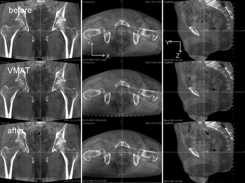
shows CBCT prostate images of a particular day for a patient with calcification inside the prostate organ. Images in the first row were acquired immediately before VMAT treatment after couch adjustment, the second row refers to images during VMAT delivery, and the third row images were taken immediately after the treatment. Crossing lines indicate the isocenter.
Figure 1. Cone-beam CT images of a prostate cancer patient with calcification inside the prostate organ. Images in the first row were acquired immediately before VMAT treatment after couch adjustment, the second row refers to images during VMAT delivery, and the third row images were taken immediately after the treatment. Crossing lines indicate the isocenter.
The mean and standard deviation of displacements at the time of pre-treatment against the in-treatment position of all the six days for the patient were 0.1±0.2 mm, −0.3±0.4 mm, and −0.4±0.6mm in lateral, vertical, and longitudinal directions. The mean and standard deviation of displacements at the time of post-treatment against the in-treatment position of all the six days for the same patient were 0.2±0.3 mm, −0.8±0.7 mm, and −0.3±0.6 mm in lateral, vertical, and longitudinal directions. No statistical conclusion can be drawn using the present limited data sets.
In conclusion, displacements of the prostate gland immediately before and after treatment relative to the position during VMAT delivery were reported for the first time using kV CBCT, which may facilitate decision making for subsequent treatment margin or dose adjustment.
Acknowledgements
Declaration of interest: Dr. Nakagawa receives research funding from Elekta K.K.A
References
- Nakagawa K, Haga A, Shiraishi K, Yamashita H, Igaki H, Terahara A, et al. First clinical cone-beam CT imaging during volumetric modulated arc therapy. Radiother Oncol 2009; 90: 422–3
- Williams, P, Sykes, J, Moore, CJ. The effects of radiation scatter from simultaneous MV irradiation on kV fluoroscopic and x-ray volume imaging with the Elekta Synergy system, ESTRO, Amsterdam 2004. Radiother Oncol 2004;73(Suppl 1)
- Nakagawa K, Yamashita H, Shiraishi K, Igaki H, Terahara A, Nakamura N, et al. Verification of in-treatment tumor position using kilovoltage cone-beam computed tomography: A preliminary study. Int J Radiat Oncol Biol Phys 2007; 69: 970–3
- Haskå TM, Honoré H, Muren LP, Høyer M, Pousen PR. Intrafraction changes of prostate position and geometrical errors studied by continuous electronic portal imaging. Acta Oncol 2008; 47: 1351–7