To the Editor,
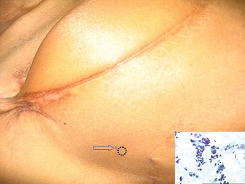
A 28-year-old female was diagnosed as right-sided locally-advanced breast cancer. She underwent neoadjuvant chemotherapy for two cycles with cyclophosphamide, adriamycin and 5 flourouracil (CAF). She subsequently underwent right radical mastectomy. Histopathology revealed a pathological tumour size of 9 cm, infiltrating duct carcinoma, (ER+, PR+). Sixteen of 17 axillary nodes were positive with perinodal extension. She received four further cycles of chemotherapy with CAF and then received adjuvant radiotherapy to the chest wall and supraclavicular fossa to a dose of 50 gray/25 fractions/5 weeks. She was then put on adjuvant tamoxifen. Four months after completion of radiotherapy, she presented with a 5 mm nodule (dotted circle) in the area around the lateral tattoo mark (arrow) put for delineating radiotherapy borders during radiotherapy simulation (). Since the nodule was firm and aroused suspicion, a FNAC was done. This revealed loosely cohesive and singly scattered pleomorphic ductal cells with high nuclear:cytoplasmic ratio, hyperchromatic nuceli and prominent nucleolisation, seen in a few; against an amorphous and a necrotic background suggestive of duct carcinoma ( inset). The systemic work up (chest x-ray, USG abdomen/pelvis) was normal. She was planned for wide excision of the nodule.
Figure 1. The lateral aspect of the right chest wall of the patient. Note the tattoo mark (arrow) and the area of the recurrent nodule (dotted circle). (Inset) Smear shows loosely cohesive and singly scattered pleomorphic ductal cells with high Nuclear:Cytoplasmic ratio, hyperchromatic nuceli and prominent nucleolisation, seen in a few; against an amorphous and a necrotic background. Papanicolaou stain × 200
It is well known that post mastectomy local recurrences in the chest wall occur predominantly in the scar site. But why does the recurrence involve most commonly the scar site and not other areas of the chest wall? A postulate is that most malignant cells can get attracted by chemotaxis to the area of the scar which is actively healing [Citation1,Citation2]. Hence this area becomes a concentration zone for malignant cells which can lead to a local recurrence subsequently. In our case too, the tattoo mark could have been a source of such attraction for malignant cells. Since this area was at the radiotherapy beam edge, it did not get a full dose of radiation and manifested as a recurrence.
Not only can such stimuli be a potential attraction for local malignant cells, they can also be a focus for deposition of circulatory tumour cells. Surgeons have long been concerned that cancer may be disseminated by shedding of tumour cells into the bloodstream during surgery [Citation3]. A study employed the technique of reverse transcription and polymerase chain reaction to detect circulating tumour cells in peripheral venous blood of patients with breast cancer preoperatively (nine malignant and three benign). In the group of patients with malignant disease, tumour cells were detected in one patient before operation and four patients during operation [Citation4]. Similar findings were reported on another study which used immunocytochemistry for cytokeratins to detect tumour cells in effluent blood from breast carcinomas in 18 patients undergoing surgery [Citation5]. These circulatory cells (along with the local malignant cells in skin adjacent to mastectomy incision, if any) could all potentially get attracted to the mastectomy scar area (or, in our case, the tattoo mark) as this site is liberating factors for chemotaxis [Citation6]. Future research in prevention of local recurrences could focus on this aspect.
Declaration of interest: The authors report no conflicts of interest. The authors alone are responsible for the content and writing of the paper.
Related Research Data
References
- Sawyer C, Sturge J, Bennett DC, O'Hare MJ, Allen WE, Bain J, . Regulation of breast cancer cell chemotaxis by the phosphoinositide 3-kinase p110delta. Cancer Res 2003; 63:1667–75.
- Müller A, Homey B, Soto H, Ge N, Catron D, Buchanan ME, . Involvement of chemokine receptors in breast cancer metastasis. Nature 2001;410:50–6.
- Wiksell H, Schässburger KU, Janicijevic M, Leifland K, Löfgren L, Rotstein S, . Prevention of tumour cell dissemination in diagnostic needle procedures. Br J Cancer 2010 Nov 2 (in press).
- Brown DC, Purushotham AD, Birnie GD, George WD. Detection of intraoperative tumor cell dissemination in patients with breast cancer by use of reverse transcription and polymerase chain reaction. Surgery 1995;117:95–101.
- Choy A, McCulloch P. Induction of tumour cell shedding into effluent venous blood breast cancer surgery. Br J Cancer 1996;73:79–82.
- Liu Y, Sun R, Wan W, Wang J, Oppenheim JJ, Chen L . The involvement of lipid rafts in epidermal growth factor-induced chemotaxis of breast cancer cells. Mol Membr Biol. 2007;24:91–101.