Abstract
Background. A recent study in our laboratory found significant differences in scapular kinematics between the affected and unaffected sides of women reporting shoulder pain following treatment for breast cancer. An earlier smaller study from our laboratory found reduced muscle activity from four key muscles and an association with greater shoulder pain and disability. The aims of this study were to: correlate altered muscle activity from a larger sample with observed movement deviations; compare within subject movement and muscle deviations in survivors with healthy variation; explore the impact of a mastectomy vs. a wide local excision (WLE) on the observed deviations. Method. Cross-sectional study. One hundred and fifty-five women treated for unilateral carcinoma of the breast and 21 age-matched healthy women were included in the study. All patients filled out the Shoulder Pain and Disability Index (SPADI). Three-dimensional (3D)-kinematic data and EMG muscle activity were recorded during scaption on the affected and unaffected side. The association between kinematic data, EMG data, SPADI and covariates was determined using a two stage, random effects mixed multiple regression technique. Results. All scapula kinematic and muscle EMG parameters in both arms were altered in breast cancer survivors when compared to healthy participants. Altered movement patterns were different for left vs. right side affected. Mastectomy patients demonstrated greater movement deviations and reported significantly higher levels of pain than WLE patients. Conclusion. Shoulder morbidity is bilateral, greater in patients having a mastectomy and is present for up to six years post-surgery. This study and others now provide ample evidence to support prospective surveillance programmes that can be integrated into Survivorship Programmes.
Women treated for breast cancer often complain of shoulder pain and decreased function from surgery to six years post-surgery, 10–55% of women show restricted shoulder range of movement (ROM), 22–38% complain of shoulder pain, and 42–56% report difficulties with lifting the upper limb or reaching above the head [Citation1–6]. These symptoms are often long lasting [Citation7–11] and significantly reduce quality of life and the ability to return to work [Citation7]. Recent studies comparing shoulder morbidity five to six years after surgery between women that had undergone sentinel lymph node biopsy (SLNB) or axillary lymph node dissection (ALND) respectively reported that from 9% to 25% complained of shoulder pain, 14% to 24% experienced shoulder stiffness, 3% to 7% were unable to raise their arm above the shoulder, and 11% to 23% had shoulder or arm weakness [Citation5,Citation6]. In spite of this evidence rehabilitation provision is either non-existent or very limited in most European countries.
Function of the upper limb requires adequate mobility of the shoulder, including the scapula. Measurement of three-dimensional (3D) shoulder motion in asymptomatic subjects has demonstrated that the scapula upwardly rotates, posteriorly tilts and externally rotates during arm elevation [Citation12]. However, when scapulothoracic motion is disproportionate to glenohumeral motion, the potential exists for microtrauma and long-term pain. For example, scapula retraction has been found to cause an increase in the subacromial space, which is thought to allow improved excursion of the humeral head and the rotator cuff under the coracoacromial arch, preventing impingement [Citation13]. It is suggested that subtle changes in this space may result in compression of the subacromial structures such as the bursa and rotator cuff during arm elevation. A recent study in our laboratory [Citation10] found significant differences in scapular kinematics between the affected and unaffected sides of women reporting shoulder pain following treatment for breast cancer. Interestingly, these differences were found to be side dependent, that is, when the left side was affected differences were found to be greater than when the right side was affected; moreover, women with left affected shoulders demonstrated a decrease in upward rotation (UR) and external rotation (ER), and an increase in posterior tilt (PT), whereas women with right affected shoulders presented an increase in UR, PT and ER on the affected side when compared to the unaffected side. An earlier study on this population found reduced muscle activity in rhomboid, trapezius, and serratus anterior muscles on the affected shoulder compared to the unaffected side, and an association between decreased muscle activity and greater shoulder pain and disability [Citation9]. These reports from our laboratory of movement deviations and altered muscle activity were based on a small sample and within subject comparisons and did not compare to a healthy population or carry out a group comparison for mastectomy and wide local excision (WLE).
The primary aim of this study was to evaluate whether or not the differences observed in shoulder kinematics and muscle activity between the affected and unaffected side in women following treatment for breast cancer can be explained in part by normal variation. Secondary aims were: 1) to evaluate the effect of surgery type (mastectomy vs. WLE) on the observed changes in movement and muscle activity; and 2) to look for associations between these data and the following covariates: degree of humeral elevation and direction of movement (up/down), age, time since surgery, medical treatment protocol, shoulder pain and disability index (SPADI), chemotherapy, handedness, and whether left or right side was affected.
Method
Following Ethical clearance by the Oxfordshire Local Research Ethics Committee (A02,064), a cross sectional study comparing patients with shoulder pain treated for unilateral carcinoma of the breast and a sample of healthy women was conducted.
Participants
One hundred and fifty-five women with shoulder pain after a breast cancer operation and 21 healthy women volunteered to take part in the study. The inclusion/exclusion criteria for the group of women with a history of breast cancer surgery are listed in . In the comparison group, women were included if they had no history of cancer, shoulder or neck pain on either side.
Table I. Inclusion and exclusion criteria.
Instrumentation
Described in detail in Shamley et al. [Citation9,Citation10].
Kinematic data. The 3 Space Fastrak® 3D motion analysis system was used to measure shoulder kinematics. This system is formed by a transmitter that emits an electromagnetic field and four receivers. Within a 76 cm source-to-sensor separation the RMS system accuracy is 0.3–0.8 mm for position and 0.15 degree for orientation [Citation14,Citation15].
Three sensors were attached to the skin and a fourth sensor was used to digitise the subject's thorax, scapula and humerus. Global and local coordinate systems were set up as described by the International Society of Biomechanics (ISB) protocol [Citation16].
Electromyography. Muscle activity of the pectoralis major, serratus anterior, rhomboids and upper trapezius muscles was measured using surface electromyography (SEMG). In standing, pre-gelled silver-silver chloride SEMG electrodes (Maersk Medical) were attached over the prepared skin sites, parallel to the muscle fibres as previously described [Citation17]. Reference electrodes were placed on electrically neutral tissues. SEMG signal quality was verified by having the participant perform a resisted contraction in the manual muscle testing position specific to each of the muscles being tested.
Arm elevation trials
Patients were instructed to elevate their arm in the plane of the scapula (40° from the coronal plane) at a pace dictated by a metronome, where a complete cycle of elevation and depression of the arm took 8 s. A flat surface oriented in this plane guided the subject's arm through the movement. Full kinematic data of three repeated elevation and depression movements of the arm were collected. Prior to data collection the movements were demonstrated by the researcher and the patient was given three practice movements. This process was repeated with each arm, which side was measured first was randomly selected.
Shoulder pain and disability index
All patients completed the SPADI pain and disability questionnaire immediately prior to the arm elevation trials. The SPADI comprises 13 visual analogue scales, five make reference to pain and eight to disability. The SPADI questionnaire has been found to be both sensitive and reliable to measure shoulder dysfunction [Citation18].
Reliability
Two observers blind to the SPADI questionnaire data carried out the kinematic and SEMG data collection. Reliability was assessed by carrying out a repeat of all measures on a different day for a randomly selected sample of five subjects.
Data reduction and analysis
The MotionMonitorTM software was used to simultaneously collect and synchronise shoulder SEMG and kinematic data. Furthermore, this software allowed the output from the 3 Space Fastrak® to be transformed into angular rotations of the scapula and the humerus relative to the trunk as determined by the ISB protocol [Citation16]. Scapular rotation was plotted as a dependent variable against thoracohumeral elevation as the independent variable. Analysis of the data only included thoracohumeral elevation of up to 110°, as the error of the scapular sensor increases beyond this point. In order to allow for international comparisons to be made terminology used to describe scapula motion in this report differs from our previous report and includes internal/external rotation (pro/retraction) and upward/downward rotation (lat/medial rotation). Anterior/posterior tilt remains the same.
For EMG data a normalisation reference was collected for 1 min at rest for each muscle. Following this, average root mean square (RMS) movement values minus the RMS resting value were determined. Data of all three arm elevation trials were averaged for the scapular position and muscle EMG reading at every 10° interval of thoracohumeral elevation.
Statistical analysis
Fastrak parameters for affected minus unaffected sides were the dependent variable and clinical and demographic data were the independent variables. In order to simultaneously model all three scapular motions of the same patient a two-stage, linear mixed model analogous to the model proposed by Weiner et al. (2002) was used [Citation19]. Stage one utilised a linear mixed model fitted to each scapular motion in order to determine residuals values representing the amount of variation in that particular scapular motion that cannot be explained by collective effect of all predictor variables. In stage two each dependent variable was modelled by another linear mixed model while the residuals which were obtained in stage one were included in the model as risk factors.
General linear regression models were used to assess any differences between scapulae movements and mastectomy vs. WLE. Bland-Altman methods were used to determine intra-rater reliability for Fastrak and EMG measures.
Results
Demographic and clinical details are shown in . The number of patients with left and right sides affected were closely represented. Intra-rater reliability for Fastrak and SEMG procedures was 0.98.
Table II. Demographic and clinical data for study sample (n = 176).
Kinematic data
Healthy vs. patients
Healthy scapulae showed greater ER and greater UR on the right vs. the left. These differences between left and right sides were also observed after treatment for breast cancer, however several differences were observed between patients and healthy participants. The increased IR seen on the left side in patients is accompanied by an increase in SA activity and a decrease in UT activity which contributes significantly to the difference seen between the affected and unaffected sides (). The activity of these muscles was not shown to contribute to the side differences observed in healthy participants (data not shown). Having received chemotherapy further contributes significantly to the difference seen between the affected and unaffected shoulders in patients ().
Table III. Two stage, random effects multiple linear regression for associations between scapula internal/external rotation and covariates.
Patients demonstrated greater upward rotation on the affected sides compared to healthy participants (p < 0.0001, CI 4.82–8.51 for left hand; CI 3.91–7.70 for right hand). This increase was greater if the left side was affected and is explained in part by a decrease in PM activity and an increase in SA activity ().
Table IV. Two stage, random effects multiple regression for associations between scapula upward/downward rotation and covariates.
Left affected shoulders showed a small increase in posterior tilt except over the critical phase of elevation/depression (80°–120°) where affected shoulders showed a shift into anterior tilt compared to healthy shoulders but this was not statistically significant (p > 0.1, CI −0.82–2.20). Differences between the affected shoulders and healthy shoulders were mirrored on the unaffected side. That is, unaffected shoulders of patients also showed greater upward rotation (left hand p < 0.001, CI 4.61–8.23; right hand p < 0.001, CI 3.05–6.92) and decreased posterior tilt (left hand p > 0.34, CI -0.40–1.15; right hand p < 0.001, CI 0.98–2.97) than the healthy shoulders. Thus both shoulders in patients demonstrated movement deviations over and above normal variation. Differences between tilt of affected and unaffected shoulders in patients were significantly associated with pain and disability and reduced SA activity ().
Table V. Two stage, random effects multiple regression for associations between scapula anterior/posterior tilt and covariates.
Mastectomy vs. WLE
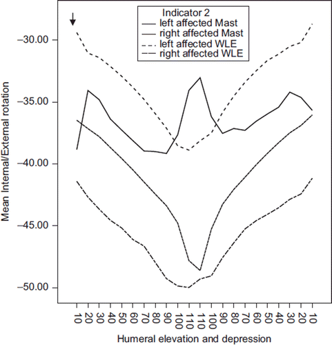
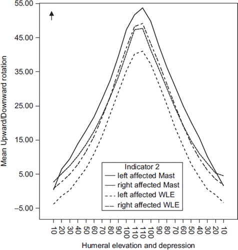
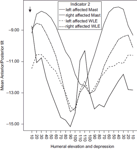
represent the patterns of scapulothoracic movement during elevation and depression of the arm. Although the Mast group did not appear to contribute to the differences seen in the linear regression mixed models () there are some clear differences that emerge when considering the patterns of movement and muscle activity as seen graphically at 10° intervals of arm elevation (–3). The magnitude of IR and UR on the left affected side in the Mast group is larger than left affected WLE (IR p < 0.001, CI 10.4–16.56; UR p < 0.001, CI 2.26–7.71) ( and ).
Figure 1. Mean scapula internal/external rotation plotted against humeral elevation and depression for mastectomy and wide local excision (WLE) patients. Arrow represents direction of external rotation.

Figure 2. Mean scapula upward/downward rotation plotted against humeral elevation and depression for mastectomy and wide local excision (WLE) patients. Arrow represents direction of upward rotation.

Figure 3. Mean scapula posterior/anterior tilt plotted against humeral elevation and depression for mastectomy and wide local excision (WLE) patients. Arrow represents direction of posterior tilt.

Mast patients had greater upward rotation in both left and right affected shoulders compared to the unaffected, whereas in WLE patients the increase was only observed in the right affected shoulder; the left affected shoulder of WLE patients demonstrated reduced upward rotation.
The left affected shoulders of both groups of patients demonstrated increased posterior tilt (). During the critical zone right WLE and left Mast patients show a larger shift in direction of tilt towards a more functional range.
Larger movement deviation is seen in the Mast group which also report significantly higher levels of pain (p < 0.03, CI 3.78–67.57).
Muscle activity
With the exception of UT in the mast group, all affected shoulders demonstrated significantly greater muscle activity on the left side when compared to the healthy left side (UT p < 0.05, CI 2.38–15.01 for WLE; PM p < 0.001, Rhom p < 0.001, SA p < 0.001, CI 6.09–12.9 for WLE and p < 0.05, CI 1.02–9.02 for mast). Compared to healthy shoulders, mastectomy patients demonstrated significantly increased activity in the right affected shoulders for UT (p < 0.001, CI 9.8–21.51), Rhom (p < 0.001, CI 11.1–15.71) and SA (p < 0.001, CI 7.16–16.26). Whereas in the case of WLE patients such increases were not observed in SA and PM activity on the right affected shoulders, where a decrease was noted. Both groups show a reversal of the healthy pattern seen in PM and SA for left and right sides.
Discussion
This paper reports altered muscle activity and shoulder kinematics in both shoulders of women treated for breast cancer compared to healthy women. Pain and movement deviations are greater in patients treated with mastectomy vs. WLE. This study has allowed us to place previous findings [Citation9,Citation10] in the context of normal variation and the more invasive management of mastectomy.
The results of this study would be strengthened by a larger, age matched control group.
Decreased posterior tilt and internal rotation of the scapula reported here have been noted in patients with impingement syndrome [Citation20,Citation21]. Evidence suggests that internal rotation of the scapula decreases the subacromial space thus resulting in a compression of the superior part of the rotator cuff and the subacromial bursa [Citation22]. The more internally rotated position of the scapula may also cause a secondary lateral rotation of the humerus, increasing the risk of an internal impingement of the posterior part of the rotator cuff [Citation23]. It is also postulated that increased anterior tilt further compromises the subacromial space, resulting in external impingement [Citation24]. In our study decreased posterior tilt was associated with high levels of reported pain.
The increased upward rotation of the scapula in our patient groups is in agreement with the findings of Crosbie et al. [Citation25] who reported an increase in upward rotation of the scapula following mastectomy for breast cancer. This is believed to represent a compensatory mechanism for a dysfunctional motion of the glenohumeral joint [Citation23]. This altered scapulohumeral rhythm has been observed in patients with shoulder capsulitis [Citation26] and full-thickness rotator cuff tears [Citation27]. Where stiffness of the glenohumeral joint or an inability of the rotator cuff to efficiently move the head of the humerus is present, patients appear to compensate by increasing the amount of scapula upward rotation during elevation of the arm.
Changes in scapula kinematics compared to healthy subjects were seen to a greater extent in patients following a mastectomy, and were even greater if the affected shoulder of mastectomy patients was the left. Whilst the statistical analysis of Crosbie et al. [Citation25] did not specifically differentiate between left affected and right affected shoulders, the authors acknowledge from the interpretation of their results that kinematic changes were greater on left affected sides. Mastectomy patients also demonstrated significantly greater pain than patients treated with a WLE.
Since left affected mastectomy shoulders demonstrated greatest pain, the different patterns of movement on the left may represent the greater need for compensatory scapular motion. In both patient groups internal rotation was increased on the left side whereas it was decreased on the right. Thus the increased internal rotation on the left might contribute to the greater presence of pain on the left side by causing either an internal or external impingement as stated above. In our previous paper [Citation10], when all patients were analysed together, we found decreased upward rotation in the left affected side, our subgroup analysis reported in this paper allows us to conclude that the difference was caused by the decrease in upward rotation observed in the left side of WLE patients. Interestingly, the changes in posterior tilt and upward rotation described for the affected shoulder were also seen in the unaffected shoulders of patients. Similar findings were also reported by Crosbie et al. [Citation25] who found a bilateral increase in upward rotation in patients following unilateral mastectomy.
UT and SA function as a force couple to rotate the scapula in an upward direction [Citation28], therefore an increase in the activity of these muscles in a patient population that show increased upward rotation of the scapula appears reasonable. The finding of increased UT activity in patients with shoulder dysfunction is quite consistent among the literature and has been observed in patients with impingement [Citation20] and frozen shoulder [Citation29]. In contrast, and unlike our study that found increased SA activity, studies assessing shoulder pathologies have reported a decrease in SA activity [Citation20]. This may be explained by the fact that these studies reporting decreased SA activity also reported a decrease in upward rotation of the scapula.
PM activity may be contributing to the increased level of pain seen in the Mast group. Increased PM activity in this group was accompanied by greater changes in scapular internal rotation and posterior tilt compared to healthy subjects, especially on the left side. It is well known that PM has an anterior tilting and internal rotating effect on the scapula [Citation30].
Large differences between the affected shoulder of both patient groups and healthy subjects were observed with regards to RH activity. RH is an external rotator of the scapula [Citation30], and despite the fact that RH activity was substantially increased in the affected shoulders, these still remained internally rotated when compared to healthy shoulders; this may be a reflection of decreased myofascial tissue extensibility of the anterior part of the pectoral girdle and the axilla in patients following treatment for breast cancer.
In line with the findings regarding scapular kinematics, EMG differences between patients and healthy subjects were mirrored on the asymptomatic shoulder of patients. There is evidence in the literature for the occurrence of this phenomenon [Citation31], which suggests an effect in the central nervous system that results in the reorganisation of motor control and movement patterns. However, these patients have received treatment with known systemic effects and as such the potential for cancer treatment to induce bilateral morbidity via changes in circulating levels of cytokines and growth factors (GF) cannot be ignored.
Indeed, the role of inflammatory cytokines and GF in the development of shoulder conditions such as rotator cuff disease, impingement and adhesive capsulitis (frozen shoulder) is a growing area of research which has not examined these factors in relation to co-morbidities such as breast cancer [Citation32,Citation33]. Given we have identified movement patterns in breast cancer patients that mimic these shoulder conditions it seems feasible to place this evidence in the context of current biomedical research.
A relationship between IL-1, IL-1β, COX-1, COX-2, substance-P, subacromial bursitis and rotator cuff disease has been shown by several authors [Citation34,Citation35]. Sakai et al. [Citation36] have shown marked hyperplasia of the blood vessels and fibroblasts of subacromial bursa in patients with rotator cuff tendonitis compared to patients with anterior instability. IL-1, TNF-α, bFGF and TGF-β were all over expressed in blood vessels and fibroblast cytoplasm. TGF-β was the only one that showed increased staining in the matrix and is a potent immunosuppressive cytokine whose levels undergo significant elevation after radiation therapy both in the short term (three weeks) and in the longer term (up to one year) [Citation37]. Irradiation is known to result in overproduction of proinflammatory mediators which leads to both vascular injury [Citation38] and activation of the inflammatory cascade [Citation39]. The associated cytokines released then participate in a number of physiological responses, which include the pain response and the development of fibrosis.
Less is known about the effects of chemotherapy on healthy tissues but there are general side effects associated with most forms of chemotherapy including fatigue, bilateral joint pain and myalgia. Our results show a significant association between chemotherapy and a number of altered patterns of movement.
In order to target at risk groups future research must attempt to elucidate the relevance of candidate markers of radiotherapy and chemotherapy damage in the development of shoulder pain and dysfunction.
Conclusion
Shoulder morbidity is bilateral, greater in patients having a mastectomy and is present for up to six years post-surgery. This study and others now provide ample evidence to support prospective surveillance programmes that can be integrated into Survivorship Programmes. In the absence of dedicated resources creative use of triage systems, DVDs and online self-assessment and self-referral pathways must be considered if we are to meet the needs of this patient group.
Acknowledgements
We would like to thank the Oxford Hospitals Research Charities for providing the funds for this research. Thank you to Professor Paula Ludewig for her critical review. The authors report no conflicts of interest. The authors alone are responsible for the content and writing of the paper.
Declaration of interest: The authors report no conflicts of interest. The authors alone are responsible for the content and writing of the paper.
References
- McNeely M, Cambell KL, Courneya KS, Dabbs K, Mackey J, Ospina M, . Exercise interventions for upper limb dysfunction due to breast cancer surgery. Cochrane Libr 2008;1–6.
- Karki A, Simonen R, Malkia E, Selfe J. Impairments, activity limitations and participation restrictions 6 and 12 months after breast cancer operation. J Rehabil Med 2005;37: 180–8.
- Lauridsen M, Overgaard M, Overgaard J, Hessov I, Cristiansen P. Shoulder disability and late symptoms following surgery for early breast cancer. Acta Oncol 2008;47: 569–75.
- Miedema B, Hamilton R, Tatemichi S, Thomas-McLean R, Towers A, Hack T, . Predicting recreational difficulties and decreased leisure activities in women's 6–12 months post breast cancer surgery. J Cancer Surviv 2008;2:262–8.
- Krane-Ocada R, Wascher R, Elashoff D, Giuliano A. Long-term morbidity of sentinel biopsy versus complete axillary dissection for unilateral breast cancer. Ann Surg Oncol 2008;15:1996–2005.
- Husted A, Haugaardc K, Soerensend J, Bokmande S, Friisf E, Holtvegg H, . Arm morbidity following sentinel lymph node biopsy or axillary lymph node dissection: A study from the Danish Breast Cancer Cooperative Group. Breast 2008;17:138–47.
- Rietman J, Dijkstra P, Hoekstra H, Eisman W, Szabo B, Groothoff J, . Late morbidity after treatment of breast cancer in relation to daily activities and quality of life: A systematic review. Eur J Surg Oncol 2003;29:229–38.
- Katz J, Poleshuck EL, Katz J, Andrus CH, Hogan LA, Jung BF, . Risk factors for acute postoperative pain and its persistence following breast cancer surgery: A prospective study. Pain 2005;119:16–25.
- Shamley DR, Srinaganathan R, Weatherall R, Oskrochi R, Watson M, Ostlere S, . Changes in muscle size and activity following treatment for breast cancer. Br Cancer Res Treatment 2007;106:19–27.
- Shamley D, Sriniganathan R, Oskrochi R, Lascurain-Aguirrebena I, Sugden E. Three-dimensional scapula-thoracic motion following treatment for breast cancer. Br Cancer Res Treat 2009;118:315–22.
- Hayes SC, Rye S, Battistutta D, DiSipio T, Newman B. Upper-body morbidity following breast cancer treatment is common, may persist longer-term and adversely influence quality of life. Health Qual Life Outcome 2010;8:92.
- Ludewig PM, Cook TM, Nawocszenski DA. Three-dimensional scapula orientation and muscle activity at selected positions of humeral elevation. J Orthop Sports Phys Ther 1996;24:57–65.
- Michener L, McClure P, Karduna A. Anatomical and biomechanical mechanisms of subacromial impingment syndrome. Clin Biomech 2003;18:369–79.
- Polhemus Inc. 3 Space Fastrak user's manual, revision F. Colchester: VT; 1993.
- Karduna A, McClure P, Michener L. Dynamic measurements of three dimensional scapular kinematics: A validation study. J Biomed Eng 2001;123:184–91.
- Wu G, van der Helm F, Veeger H, Makhsous M, Van Roy P, Anglin C, . ISB recommendation on definitions of joint coordinate systems of various joints for the reporting of human joint motion—Part II: Shoulder, elbow, wrist and hand. J Biomech 2005;38:981–92.
- Ludewig P, Cook T, Nawoczenski D. Three-dimensional scapular orientation and muscle activity at selected positions of humeral elevation. J Orthop Sports Phys Ther 1996;24:57–65.
- Williams JW Jr, Holleman DR Jr, Simel DL. Measuring shoulder function with the Shoulder Pain and Disability Index. J Rheumatol 1995;22:727–32.
- Weiner BJ, Alexander JA, Shortell SM. Management and governance processes in community health coalitions: A procedural justice perspective. Health Educ Behav 2002;29:737.
- Ludewig P, Cook T. Alterations in shoulder kinematics and associated muscle activity in people with symptoms of shoulder impingement. Phys Ther 2000;80:276–91.
- Hebert L, Moffet H, McFadyen B, Dionne C. Scapular behavior in shoulder impingement syndrome. Arch Phys Med Rehab 2002;83:60–9.
- Solem-Bertoft E, Thuomas K, Westerberg C. The influence of scapular retraction and protraction on the width of the subacromial space. Clin Orthop Related Res 1993:99–103.
- Ludewig P, Reynolds J. The association of scapular kinematics and glenohumeral joint pathologies. J Orthop Sports Phys Ther 2009;39:90–104.
- Seitz A, McClure P, Finucane S, Boardman N, Michener L. Mechanisms of rotator cuff tendinopathy: Intrinsic, extrinsic, or both? Clin Biomech 2011;26:1–12.
- Crosbie J, Kilbreath S, Dylke E, Refshauge K, Nicholson L, Beith J, . Effects of mastectomy on shoulder and spinal kinematics during bilateral upper-limb movement. Phys Ther 2011;90:679–92.
- Fayad F, Robi-Brami A, Yazbeck C, Hanneton S, Lefevre-Colau M, Gautheron V, . Three-dimensional scapular kinematics and scapulohumeral rhythm in patients with glenohumeral osteoarthritis or frozen shoulder. J Biomech 2008;41:326–32.
- Mell A, LaScalza S, Guffey P, Ray J, Maciejewski M, Carpenter J, . Effect of rotator cuff pathology on shoulder rhythm. J Shoulder Elbow Surg 2005;14:58S–64S.
- Magarey M, Jones M. Dynamic evaluation and early management of altered motor control around the shoulder complex. Manual Ther 2003;8:195–206.
- Lin J, Wu Y, Wang S, Chen S. Trapezius muscle imbalance in individuals suffering from frozen shoulder syndrome. Clin Rheum 2005;24:569–57.
- Ackland DC, Pandy MG. Moment arms of the shoulder muscles during axial rotation. J Ortho Res 2011;29:658–67.
- Falla D, Farina D, Graven-Nielsen T. Experimental muscle pain results in reorganization of coordination among trapezius muscle subdivisions during repetitive shoulder flexion. Exptl Brain Res 2007;178:385–93.
- Blaine TH, Kim Y, Voloshin I, Chen D, Murakami K, Chang S-S, . The molecular pathophysiology of subacromial bursitis in rotator cuff disease. J Shoulder Elbow Surg 2005;14:84S–89S.
- Ishii H, Brunet JA, Welsh RP, Uhthoff HK. ‘Bursal reactions’ in rotator cuff tearing, the impingement syndrome and calcifying tendinitis. J Shoulder Elbow Surg 1997;6:131–6.
- Gotoh M, Hamada K, Yamakawa H, Yanagisawa K, Nakamura M, Yamazaki H, . Interleukin-1 induced subacromial synovitis and shoulder pain in rotator cuff diseases. Rheum Oxford 2001;40:995–1001.
- Kobayashi M, Itoi E, Minagawa H, Miyakoshi N, Takahashi S, Tuoheti Y, . Expression of growth factors in the early phase of supraspinatus tendon healing in rabbits. J Shoulder Elbow Surg 2006;15:371–7.
- Sakai H, Fujita K, Sakai Y, Mizuno Z. Immunolocalization of cytokines and growth factors in subacromial bursa of rotator cuff tear patients. Kobe J Med Sci 2001;47:25–3.
- Chan BP, Fu S, Qin L, Lee K, Rolf CG, Chan K. Effects of basic fibroblast growth factor (bFGF) on early stages of tendon healing. A rat patella tendon model. Acta Orthop Scan 2000;71:513–8.
- Fajardo LF. The pathology of ionizing radiation as defined by morphologic patterns. Acta Oncol 2005;44:13–22.
- Rubin P, Johnston CJ, Jacqueline P, McDonald S, Finkelstein JN. A perpetual cascade of cytokines postirradiation leads to pulmonary fibrosis. Int J Radiat Oncol Biol Phys 1995; 33:99–109.