To the Editor,
Radiotherapy dose escalation improves patient outcome in non-small cell lung cancer (NSCLC) [Citation1–5]. To achieve this, motion compensation techniques are often used, e.g. active breathing control (ABC) which allows for relative tumour immobilisation during inspiration breath-hold [Citation6–8]. Although both reduction in dose-volume parameters determining lung toxicity and good reproducibility have been demonstrated with ABC, inter-patient variability exists with significant tumour motion being reported. As a result, image guidance and tumour-directed localisation have been recommended [Citation9,Citation10].
Image guidance with kilovoltage cone beam computerised tomography (CBCT) has been shown to provide accurate bony and soft tissue setup information [Citation11,Citation12]. Deep-inspiration breath-hold with CBCT is also feasible [Citation8] and has been integrated as a verification tool for correction protocols when treating patients with ABC [Citation13]. Current CB imaging software allows automatic matching within a user defined region of interest (ROI) but the selected ROI volume is important and can affect image registration accuracy [Citation14–16]. For patients with lung tumours undergoing free-breathing radiotherapy, studies have shown that ROIs centred on the carina and spine can produce accurate CBCT image registration with advanced tumours [Citation17] whilst soft tissue tumour matching is more accurate with early-staged tumours [Citation18]. With ABC-gated radiotherapy, the optimal automatic registration method for image-guided setup correction has not yet been established. The aim of this study was therefore to evaluate the effect of using four different ROIs (with the Synergy XVI R4.2 software, Elekta, Crawley, UK) with ABC-gated radiotherapy in order to determine the optimum volume for registration.
Methods
The study was approved by the local institution's Committee for Clinical Research.
Twenty-eight patients with NSCLC (Stage IA to IIIB; 10 early staged and 18 locally advanced, Supplementary Table I to be found online at http://informahealthcare.com/doi/abs/10.3109/0284186X.2013.861610) treated radically with radiotherapy using the ABC device, between February 2009 and September 2009, were evaluated. Patients were positioned in a supine position on a winged lung board (Civco, Oncology Systems Ltd, UK). Planning CT scans were acquired in one mid deep-inspiration breath (75% of maximum inspiratory volume) using 2 mm slice thickness and conformal planning was used to deliver a dose of 64 Gy in 32 fractions. The CBCT images were acquired through a rotation of 200° in around 70 seconds and was acquired continuously while the patient was using the ABC device with short breaths in between, usually 2–4 breath-holds of 15–20 seconds each [Citation13]. The CBCT acquired on the first fraction of treatment were used to assess the automatic registration to the reference planning CT scan using the grey-value registration algorithm [Citation19]. We defined the following ROIs using Synergy XVI R4.2 software, where all ROIs are cuboids, each observer made their own ROI for the automatic match:
1) The PTV + 2 cm, the current standard in our institution for radical lung cancer patients being treated with ABC.
2) PTV+ Bone. This included the PTV plus the closest bony anatomy adjacent to the lung tumour (vertebrae, ribs, scapula or clavicle depending on the tumour localisation) with 2 cm margin in the superior and inferior borders of the PTV.
3) The ipsilateral Hemithorax: lateral borders included the chest wall and the mediastinum; anterior border included the sternum and the posterior limit included the vertebrae; superior and inferior borders were limited by the maximum length of the collimator (13, 18 and 26 cm) centred on the isocentre.
4) The Carina. This included the carina with a superior-inferior/anterior-posterior/lateral margin of 3 cm.
These ROI definitions were set out in written guidelines to perform an automatic grey-scale match. The clinical oncologists were able to use a manual match where necessary and a consensus agreement confirmed visually between two clinical oncologists was used as the gold standard match. The amount of translational shifts and angular rotations after automatic matching were recorded by five independent radiation technologists without manual adjustments. The ROIs were scored as being a successful match for a given patient if the displacements in each of the three planes lie within 2 mm of the clinicians’ consensus measurements for each of the five radiation technologists. Thus, for each of the four ROIs, success was described in terms of a percentage match. For the ROI to be useful in automatic image registration, a successful match should occur in 95% of registrations; less than 75% was deemed unacceptable. The proposal was that an ROI would not be considered optimal for automatic matching if there were less than 25 successful matches (alpha 1-side = 5%, power = 90%). Reproducibility between independent users for each ROI was assessed by comparing the means between them [intra-class correlation (ICC), two-way random effects model] [Citation20]. The differences between the ROIs displacements after matching was assessed by the Mann-Whitney test to rank the ROIs and confidence of ROI definition was recorded by the users.
Results
Five independent observers (radiation technologists) performed registrations on 28 patients resulting in 560 observations.
Degree reliability of successful matching
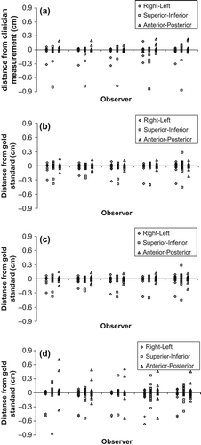
The regions of interest that resulted in > 24 (25/28) successful matches were PTV+ Bone and Hemithorax. PTV only and Carina resulted in 23/28 successful matches. shows the differences from the five observers and the clinician measurements using each ROI.
Figure 1. (a) Distance from clinician measurement for five observers and using ‘PTV only’ region of interest. (b) Distance from clinician measurement for five observers and using ‘PTV+ bone’ region of interest. (c) Distance from clinician measurement for five observers and using ‘Hemithorax’ region of interest. (d) Distance from clinician measurement for five observers and using ‘Carina’ region of interest.

Failed matches were documented in eight of 28 patients (). In three of these patients, there was only one observer failing to match for an individual ROI (PTV + 2 cm, Carina, Hemithorax). In four patients, all five observers using the Carina ROI failed to achieve a successful match.
Table I. The patients and number of observers where failed matches occurred in each region of interest.
Reproducibility
Reproducibility for all four ROIs was very good for translational (ICC > 0.94) and good for rotational (> 0.75) measurements. Hemithorax matching achieved the highest level of reproducibility for linear couch displacement (ICC = 0.99), followed by the PTV + 2 cm (ICC = 0.99–0.98). The Carina was the poorest with an ICC of 0.90–0.98.
Confidence in ROI definition
The confidence in defining the ROI was highest for PTV + 2 cm (94%), followed by Hemithorax (82%), Carina (68%) and PTV+ Bone (68%).
The mean time of automatic match for each ROI, measured in 10 patients, was 10.3, 9.6, 25 and 5.9 seconds for PTV + 2 cm, PTV+ Bone, Hemithorax and Carina, respectively.
Discussion
Reduction in setup errors and treatment verification is increasing important with dose escalation in lung cancer radiotherapy. In free-breathing radiotherapy, internal anatomical surrogates and different CBCT volumetric images ROI have been investigated for image registration accuracy [Citation17,Citation18,Citation21]. The situation with motion management using ABC gating is potentially different where there is relative tumour and chest wall immobilisation.
In our study, we have shown that for accurate registration for ABC-gated radiotherapy it is not always sufficient to rely on the automatic match and manual registration to the tumour is occasionally required for certain situations. Although Hemithorax and PTV+ bone ROIs produced successful matches, significant heterogeneity existed. Two groups of patients failed (): 1) small peripheral tumours; and 2) situations where there was collapse and consolidation or significant change in tumour size between planning and treatment. In the first group, there were four patients with T1N0M0 tumours (3 UL and 1LL); the failed matches in the UL tumours with the carina ROI is consistent with studies where the carina was not an optimal surrogate for tumour position in UL tumours [Citation21]. In the second group of four patients, three had central tumours with associated collapse and consolidation and one had a significant change in tumour size between planning and treatment. This is consistent with the literature where reported concerns of tumour deformation and migration affecting image registration accuracy [Citation17,Citation22]. There was no particular ROI which produced better registration between central and peripheral tumours in ABC-gated radiotherapy; this compares with non-gated radiotherapy where the Carina was deemed satisfactory for locally advanced tumours [Citation17] and soft tissue matching around the PTV for peripheral tumours [Citation18].
As only day one CBCT images were assessed in our study, future investigation throughout the full course of treatment should prove useful especially in light of long-term variability in tumour position [Citation9].
The effect of ROIs selected by different observers can potentially affect image registration but has not previously been investigated. We were able to show good reproducibility for all the ROIs amongst the observers; there were, however, issues with ROI definition when performing image registration.
While there is concern about additional dose from regular IGRT, the dose received from the 200° scan (< 3.9–5.5 mSv) is much reduced from the full 360° (< 7–10 mSv) scan with the imaging settings used in the current study. Further, radiation doses from CBCT imaging have been found to be low [Citation23] while recent epidemiological data indicate lower than expected risks of low-dose exposure to radiotherapy [Citation24].
In conclusion, we have showed good reproducibility of ROI definition and image registration with ABC-gated radiotherapy for lung cancer. We demonstrated that overall PTV+ Bone and Hemithorax ROIs resulted in successful automatic image registrations and identified clinical situations where automatic registration may not be suitable with further checks and manual correction being required. This together with some concerns with ROI definition amongst the observers highlight the need to adopt an individualised approach in image registration with ABC-gated radiotherapy and to identify training needs for all professionals involved in the definition of ROIs and soft tissues when using three-dimensional imaging tools for verification.
http://informahealthcare.com/doi/abs/10.3109/0284186X.2013.861610
Download PDF (17.3 KB)Declaration of interest: The authors report no conflicts of interest. The authors alone are responsible for the content and writing of the paper.
This work was undertaken in The Royal Marsden NHS Foundation Trust who received a proportion of its funding from the NHS Executive; the views expressed in this publication are those of the authors and not necessarily those of the NHS Executive. This work was supported by the Institute of Cancer Research, The Royal Marsden NHS Foundation Trust and Cancer Research UK Section of Radiotherapy [CRUK] grant number C46/A2131. We acknowledge NHS funding to the NIHR Biomedical Research Centre.
References
- Bradley J, Graham MV, Winter K, Purdy JA, Komaki R. Toxicity and outcome results of RTOG 9311: A phase I-II dose-escalation study using three-dimensional conformal radiotherapy in patients with inoperable non-small-cell lung carcinoma. Int J Radiat Oncol Biol Phys 2005;61:318–28.
- Belderbos JS, Heemsbergen WD, De Jaeger K, Baas P, Lebesque JV. Final results of a phase I/II dose escalation trial in non-small-cell lung cancer using three-dimensional conformal radiotherapy. Int J Radiat Oncol Biol Phys 2006;66: 126–34.
- Hayman JA, Martel MK, Ten Haken RK, Normolle DP, Todd RF 3rd, Littles JF, et al. Dose escalation in non- small-cell lung cancer using three-dimensional conformal radiation therapy: Update of a phase I trial. J Clin Oncol 2001;19:127–36.
- Kong FM, Hayman JA, Griffith KA, Kalemkerian GP, Arenberg D, Lyons S, et al. Final toxicity results of a radiation-dose escalation study in patients with non- small-cell lung cancer (NSCLC): Predictors for radiation pneumonitis and fibrosis. Int J Radiat Oncol Biol Phys 2006;65:1075–86.
- Narayan S, Henning GT, Ten Haken RK, Sullivan MA, Martel MK, Hayman JA. Results following treatment to doses of 92.4 or 102.9 Gy on a phase I dose escalation study for non-small cell lung cancer. Lung Cancer 2004;44: 79–88.
- Panakis N, McNair HA, Christian JA, Mendes R, Symonds-Tayler JR, Knowles C, et al. Defining the margins in the radical radiotherapy of non-small cell lung cancer (NSCLC) with active breathing control (ABC) and the effect on physical lung parameters. Radiother Oncol 2008;87: 65–73.
- Partridge M, Tree A, Brock J, McNair HA, Fernandez E, Panakis N, et al. Improvement in tumour control probability with active breathing control and dose escalation: A modelling study. Radiother Oncol 2009;91:325–9.
- Duggan DM, Ding GX, Coffey CW 2nd, Kirby Hallahan DE, Malcolm A. Deep-inspiration breath-hold kilovoltage cone-beam CT for setup of stereotactic body radiation therapy for lung tumors: Initial experience. Lung Cancer 2007;56:77–88.
- Koshani R, Balter JM, Hayman JA, Henning GT, van Herk M. Short-term and long-term reproducibility of lung tumor position using active breathing control (ABC). Int J Radiat Oncol Biol Phys 2006;65:1553–9.
- Brock J, McNair HA, Panakis N, Symonds-Tayler R, Evans PM, Brada M. The use of the active breathing coordinator throughout radical non-small cell lung cancer (NSCLC) radiotherapy. Int J Radiat Oncol Biol Phys 2011; 81:369–75.
- Borst GR, Sonke JJ, Betgen A, Remeijer P, van Herk M, Lebesque JV. Kilo-voltage cone-beam computed tomography setup measurements for lung cancer patients; first clinical results and comparison with electronic portal- imaging device. Int J Radiat Oncol Biol Phys 2007;68: 555–61.
- Jaffray DA, Siewerdsen JH, Wong JW, Martinez AA. Flat panel cone-beam computed tomography for image- guided radiation therapy. Int J Radiat Oncol Biol Phys 2002; 53:1337–49.
- Boda-Heggemann L, Fleckenstein J, Lohr F, Wertz H, Nachit M, Blessing M, et al. Multiple breath-hold CBCT for online image guided radiotherapy of lung tumours: Simulation with a dynamic phantom and first patient data. Radiother Oncol 2011;98:309–16.
- Hawkins MA, Aitken A, Hansen VN, McNair HA, Tait DM. Cone beam CT verification for oesophageal cancer – impact of volume selected for image registration. Acta Oncol 2011;50:1183–90.
- Polat B, Wilbert J, Baier K, Flentje M, Guckenberger M. Nonrigid patient setup errors in the head-and neck region. Strahlenther Onkol 2007;183:506–11.
- Moran MS, Lund MW, Ahmad M, Moseley D, Waldron K, Gregory J, et al. Clinical implementation of prostate image guided radiation therapy: A prospective study to define the optimal field of interest and image registration technique using automated x-ray volumetric imaging software. Technol Cancer Res Treat 2008;7:217–26.
- Higgins J, Bezjak A, Franks K, Le LW, Co BC, Payne D, et al. Comparison of spine, carina, and tumor as registration landmarks for volumetric image-guided lung radiotherapy. Int J Radiat Oncol Biol Phys 2009;73:1404–13.
- Josipovic M, Persson GF, Logadottir A, Smulders B, Westmann G, Bangsgaard JP. Translational and rotational intra- and inter-fractional errors in patient and target position during a short course of frameless stereotactic body radiotherapy. Acta Oncol 2012;51:610–7.
- Smitsmans MHP, Wolthaus JWH, Artignan X, de Bois J, Jaffray DA, Lebesque JV, et al. Automatic localization of the prostate for on-line of off-line image-guided radiotherapy. Int J Radiat Oncol Biol Phys 2004;60:623–35.
- Shrout PE, Fleiss JL. Intraclass correlations: Uses in assessing rater reliability. Psychol Bull 1979;2:420–8.
- Spoelstra FO, van der Weide L, van Sornsen de Koste JR, Vincent A, Slotman BJ, Senan S. Feasibility of using anatomical surrogates for predicting the position of lung tumours. Radiother Oncol 2012;102:287–9.
- Kupelian PA, Ramsey C, Meeks SL, Willoughby TR, Forbes A, Wagner TH, et al. Serial megavoltage CT imaging during external beam radiotherapy for non-small-cell lung cancer: Observations on tumor regression during treatment. Int J Radiat Oncol Biol Phys 2005;63:1024–8.
- Amer A, Marchant T, Sykes J, Czajka J, Moore C. Imaging doses from the Elekta Synergy x-ray cone beam CT system. Br J Radiol 2007;80:476–82.
- Berrington de Gonzalez A, Gilbert E, Curtis R, Inskip P, Kleinerman R, Morton L, et al. Second solid cancers after radiation therapy: A systematic review of the epidemiologic studies of the radiation dose-response relationship. Int J Radiat Oncol Biol Phys 2013;86: 224–33.
