
To the Editor,
Hand-foot syndrome (HFS), also known as acral erythema and palmar-plantar erythrodysesthesia, is a common potentially dose-limiting cutaneous toxicity secondary to many chemotherapeutic agents [Citation1]. HFS can begin from 24 hours to 10 months after starting a chemotherapeutic agent [Citation2]. Although HFS is usually a non-life-threatening reaction to antineoplastic agents, it has an impact on a patient’s quality of life and often prompts dose modification or discontinuation of a patient’s chemotherapy [Citation3–8].
As they also involve the hands and feet, tinea manuum and pedis can be easily mistaken for HFS. Appropriate diagnosis and differentiation between HFS and tinea is important not only for proper treatment, but also to minimize interruption of a patient’s chemotherapy. We report three cases of women who developed skin eruptions on the hands and feet while receiving chemotherapy. All were diagnosed with tinea, responded to antifungal therapy, and were able to continue their cancer therapy with minimal or no interruption.
Cases
Case 1
A 53-year-old Caucasian female was diagnosed with metastatic breast cancer. She was treated with a right total mastectomy, right axillary sentinel lymphadenectomy, and complete right axillary lymph node dissection in October 2011. Postoperatively, she received four cycles of liposomal doxorubicin and cyclophosphamide (AC) chemotherapy and had just completed her first of four cycles of paclitaxel when she was referred to a dermatologist for scaling, pain and pruritus of the hands and feet.
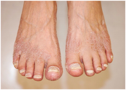
On examination, there were hyperkeratotic, scaly, pruritic plaques on the palmar aspects of her hands. She had cracking and bleeding from areas on the fingers and described pain after applying any topical moisturizer. There were similar plaques on the plantar, medial, and lateral aspects of her feet in a moccasin distribution (). A skin scraping stained with potassium hydroxide (KOH) from the foot revealed branching hyphae and a fungal culture was positive for Trichophyton rubrum. The patient was treated with terbinafine 200 mg daily for two weeks, econazole 1% cream twice daily, and moisturizer as needed. She received her remaining cycles of paclitaxel as planned.
Figure 1. Pink scaly plaques in a moccasin distribution on dorsal surface of feet due to tinea pedis before antifungal treatment.
After completing the two-week oral dose of systemic antifungal therapy, there was clinical improvement of the patient’s tinea pedis and manuum but she continued to have some scaling and erythema of the hands and feet. KOH preparation at this time was once again positive for hyphae. As she had mild elevation in her liver function tests, terbinafine was not restarted. She was treated with econazole 1% cream and urea 40% cream twice daily. Two months after her initial presentation, the patient had significant improvement with just mild scaling on her hands and feet bilaterally.
Case 2
A 47-year-old female with a history of stage III colon cancer with metastases to the pancreas underwent transverse colon resection and distal pancreatectomy in April 2015. After completing five cycles of adjuvant capcitabine chemotherapy, she was referred to a dermatologist in September 2015 for a two-week history of a diffuse itchy rash on her left thigh that spread to the left abdomen. On examination, there were erythematous papules on her abdomen, and bilateral upper extremities. She was started on a two-week trial of topical triamcinolone 0.1% ointment twice daily for contact dermatitis of the left thigh and abdomen.
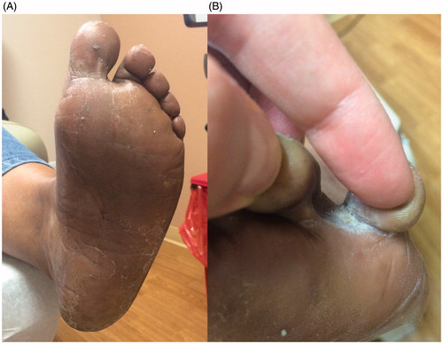
Follow-up two weeks later showed resolution of the rash on the extremities, but new onset development of scaly plaques with interdigital maceration on both feet diagnosed as HFS by her oncologist (). The patient reported that she mistakenly was using the steroid ointment exclusively on her feet. KOH preparation of the feet revealed branching hyphae. She was treated with topical econazole 1% cream twice daily for tinea pedis, likely worsened by her previous exposure to topical corticosteroids on the feet. Capecitabine was continued without interruption. Triamcinolone was continued as needed for residual dermatitis on the left thigh and abdomen.
Figure 2. (A) Scaly plaques on the plantar surfaces of both feet. (B) Interdigital maceration due to tinea pedis.
Case 3
An 85-year-old female with a history of stage I uterine papillary serous carcinoma underwent total abdominal hysterectomy, bilateral salpingooophrectormy, omentectomy, and tumor debulking in 2007. She received subsequent “sandwich” chemotherapy – adjuvant pelvic radiation administered between combination paclitaxel and carboplatin [Citation9]. In March 2014, she presented with metastatic disease and was started on liposomal doxorubicin in July 2014.
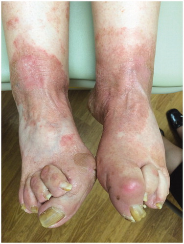
Her first dose was well tolerated, but she developed scale, pruritis, and pain in her hands and feet following her second dose in August 2014. At the time she was referred to a dermatologist, she had elected to skip an infusion of doxorubicin due to these perceived skin side effects. On examination, there were erythematous scaly plaques with fissures on the hands and fingertips. There were similar erythematous scaly annular plaques on the dorsal feet and ankles, along with erythematous papules on the lower legs. The nails appeared thickened and yellow in color (). KOH preparation of skin scrapings from the right ankle was positive for branched hyphae. Fungal culture grew Trichophyton rubrum. The patient was treated with fluconazole 150 mg weekly for three weeks, and econazole 1% cream twice daily. Doxorubicin treatment was resumed. The patient reported improvement but did not return for follow-up care.
Figure 3. Erythematous scaly plaques due to tinea pedis.
Discussion
These cases demonstrate the importance of dermatologic care in patients with skin problems during cancer treatment. Although both HFS and tinea are self-limited and generally non-life-threatening, they are treated very differently and prompt diagnosis and treatment of cutaneous fungal infections can prevent unnecessary reductions and cessations of cancer therapy.
HFS was first described by Zuehlke [Citation10] in 1974 as a cutaneous reaction observed in a group of patients receiving mitotane for hypernephroma. Since then, HFS has been associated with various chemotherapeutic agents, the most common including capecitabine [Citation2,Citation3], fluorouracil (5-FU) [Citation4], cytarabine [Citation5], liposomal doxorubicin [Citation6], and docetaxel [Citation7,Citation8]. Although the pathogenesis of this cutaneous side effect is still poorly understood, proposed mechanisms include capillary damage leading to drug extravasation and, especially in the case of liposomal doxorubicin, drug accrual in the eccrine sweat ducts of the hands and feet [Citation11]. The typical clinical presentation of HFS includes a prodromal paresthesia followed by painful, symmetrical erythema, scale, and edema of the palms and soles that may evolve into blisters and fissures [Citation11,Citation12].
Tinea is a common superficial fungal infection most often caused by one of three pathogens – Trichophyton, Microsporum, and Epidermophyton. Moccasin-type tinea pedis, which is typical of a T. rubrum infection, is characterized by scale, inflammation, and hyperkeratosis that covers the soles and heels of the feet in a pattern that resembles a slipper (as suggested by its name). Tinea manuum, a fungal infection of the hands, presents with erythema, mild scaling, and hyperkeratosis of the palms [Citation13]. Of note, tinea manuum virtually always occurs in a patient who has coexisting tinea pedis, sometimes only involving the dominant hand in a “2 foot 1 hand” infection.
Dermatophyte infections often affect otherwise healthy individuals – up to 20–25% of the world’s general population has skin mycoses [Citation14]. The incidence of dermatophyte infections in patients undergoing chemotherapy has not been quantified, but as can be expected, people with compromised immune systems are especially susceptible. Diagnosis is based on history and clinical presentation, and confirmed by KOH microscopy of lesion scrapings or nail clippings (depending on the affected anatomical system) and/or fungal cultures. Most tinea infections can be managed with topical antifungal cream, such as azoles (econazole, ketoconazole) and allylamines (terbinafine) [Citation15]. Oral treatment, such as terbinafine, itraconozole, or fluconazole, may be required in patients with persistent or widespread infections.
Physical examination findings that suggest tinea infection rather than HFS include spread outside of the palms and soles, a more erythematous, scaly, advancing border, nail thickening and discoloration suggestive of onychomycosis, and finer, powder-like scale rather than thick peeling of the skin. Maceration of the skin between the toes is another common finding in tinea pedis. When fungal infections are suspected, KOH examinations and/or fungal cultures should be performed.
Conclusion
Chemotherapy can lead to multiple cutaneous side effects, ranging from pigmentary changes to exanthematous eruptions. When patients undergoing chemotherapy present with new onset painful, itchy, erythematous scaling of the hands and/or feet, it is important to consider the diagnosis of dermatophyte infection as well as HFS. Accurate diagnosis is important to guide treatment and also to avoid the unnecessary interruptions in chemotherapy.
References
- Nagore E, Insa A, Sanmartin O. Antineoplastic therapy-induced palmar plantar erythrodysesthesia ('hand-foot') syndrome. Incidence, recognition and management. Am J Clin Dermatol 2000;1:225–34.
- Scheithauer W, Blum J. Coming to grips with hand-foot syndrome. Insights from clinical trials evaluating capecitabine. Oncology (Williston Park) 2004;18:1161–8.
- Blum JL, Jones SE, Buzdar AU, LoRusso PM, Kuter I, Vogel C, et al. Multicenter phase II study of capecitabine in paclitaxel-refractory metastatic breast cancer. J Clin Oncol 1999;17:485–93.
- Chiara S, Nobile MT, Barzacchi C, Sanguineti O, Vincenti M, Di Somma C, et al. Hand-foot syndrome induced by high-dose, short-term, continuous 5-fluorouracil infusion. Eur J Cancer 1997;33:967–9.
- Crawford JH, Eikelboom JW, McQuillan A. Recurrent palmar-plantar erythrodysaesthesia following high-dose cytarabine treatment for acute lymphoblastic leukemia. Eur J Haematol 2002;69:315–17.
- Lyass O, Uziely B, Ben-Yosef R, Tzemach D, Heshing NI, Lotem M, et al. Correlation of toxicity with pharmacokinetics of pegylated liposomal doxorubicin (Doxil) in metastatic breast carcinoma. Cancer 2000;89:1037–47.
- Park YH, Ryoo BY, Lee HJ, Kim SA, Chung JH. High incidence of severe hand-foot syndrome during capecitabine-docetaxel combination chemotherapy. Ann Oncol 2003;14:1691–2.
- Zimmerman GC, Keeling JH, Burris HA, Cook G, Irvin R, Kuhn J, et al. Acute cutaneous reactions to docetaxel, a new chemotherapeutic agent. Arch Dermatol 1995;131:202–6.
- Abaid LN, Rettenmaier MA, Brown JV III, Micha JP, Mendivil AA, Wabe MA, et al. Sequential chemotherapy and radiotherapy as sandwich therapy for the treatment of high risk endometrial cancer. J Gynecol Oncol 2012;23:22–7.
- Zuehlke RL. Erythematous eruption of the palms and soles associated with mitotane therapy. Dermatologica 1974;148:90–2.
- Clark AS, Vahdat LT. Chemotherapy-induced palmar-plantar erythrodysesthesia syndrome: etiology and emerging therapies. Support Cancer Ther 2004;1:213–18.
- Lipworth AD, Robert C, Zhu AX. Hand-foot syndrome (hand-foot skin reaction, palmar-plantar erythrodysesthesia): focus on sorafenib and sunitinib. Oncology 2009;77:257–71.
- Hainer BL. Dermatophyte infections. Am Fam Physician 2003;67:101–8.
- Havlickova B, Czaika VA, Friedrich M. Epidemiological trends in skin mycoses worldwide. Mycoses 2008;51 Suppl:2–15.
- Crawford F, Hollis S. Topical treatments for fungal infections of the skin and nails of the foot. Cochrane Database Syst Rev 2007;CD001434.