Abstract
Background. Vertebral fracture assessment (VFA) using dual-energy X-ray absorptiometry can visualize abdominal aortic calcification (AAC). AAC correlates with total atherosclerosis burden. We questioned whether VFA-detected AAC could be used for cardiovascular risk assessment.
Methods. VFA images of 2,500 subjects were evaluated to detect and score AAC (n = 164). A random age- and gender-matched set of subjects (n = 325) without AAC served as control group. Patients with prior cardiovascular disease or procedures were excluded. Base-line cardiovascular risk factors and further cardiovascular events were checked. Design-based Cox regression analysis was used to examine the prognostic value of AAC for cardiovascular outcomes.
Results. AAC-positive subjects were divided into two groups: low-AAC (score 1–3), and high-AAC group (score > 3). Mean age in the groups was 68, 68, and 71 years, percentage of females was 64.4%, 61%, and 66.1%, and the proportion of cardiovascular events within groups was 1.5%, 6.7%, and 11.9% in control, low-AAC, and high-AAC groups, respectively. Age- and gender-adjusted as well as multivariable analysis showed a significant, higher risk for cardiovascular events incidence in AAC-positive, low-AAC, and high-AAC when compared to the control group.
Interpretation. AAC assessed with routine VFA was shown to be a strong predictor for cardiovascular events.
Key messages
Presence of abdominal aortic calcification (AAC) detected by dual-energy X-ray absorptiometry (DEXA) vertebral fracture assessment (VFA) predicts adverse cardiovascular events. Moreover, this method combines the advantages of both low cost and low radiation burden for the patients compared to CT-imaging.
Higher AAC levels, according to an 8-score scale, are associated with higher hazard risk for cardiovascular events.
Introduction
Atherosclerosis can be considered as a systemic disease capable of affecting all arteries in the body. Calcification of atherosclerotic plaques has been well recognized as a risk indicator. It is known that the burden of aortic calcification correlates with the degree of atherosclerosis in other arteries (Citation1,Citation2). Many studies have shown that abdominal aortic calcification (AAC) detected by either lateral lumbar spine radiographs or computed tomography (CT) scan is an independent predictor for cardiovascular disease (CVD) incidence and mortality (Citation3–11). AAC assessment with conventional radiologic techniques has the disadvantage of relatively high radiation burden (1.5 mSv in lateral spinal X-ray (Citation12)) and is thus less desirable for physicians and patients as a screening tool.
Dual energy X-ray absorptiometry (DEXA) is a standard diagnostic technique widely used to detect osteoporosis (Citation13) and vertebral fractures (the hall-mark of osteoporosis (Citation14)) with minimal time and radiation exposure (Citation15,Citation16). In 2002 in the USA a total of 2,195,548 DEXA scans were performed among all Medicare patients (Citation17). Osteoporosis and atherosclerosis have many risk factors in common, such as body mass index, smoking, age, physical activity, and (treatment for) diabetes mellitus. In addition, the coexistence of risk factors for atherosclerosis and osteoporosis in healthy women has been recently reported (Citation18). It may therefore be prudent to evaluate the cardiovascular risk in patients at risk for osteoporosis. Vertebral fracture assessment (VFA) and bone densitometry images intended to detect osteoporosis match reliably with lateral spine radiography results, accompanied by a very low radiation dose of less than 0.01 mSv (Citation19,Citation20). In this context it is a beneficial coincidence that VFA images using DEXA technique can reveal AAC with good sensitivity and specificity compared to standard lateral spine radiographs (Citation19,Citation21,Citation22). However, the relationship between elevated AAC using VFA and risk for the occurrence of cardiovascular events is not well surveyed. A previous study evaluated the association of AAC scored on VFA images in patients with myocardial infarctions or stroke in elderly women (Citation23), because DEXA studies are mainly performed in women. Studies that are focused on the direct risk of cardiovascular events in men and women related to AAC are lacking. Thus routine application of AAC analysis in patients referred for bone mineral densitometry (BMD) and VFA could provide clinicians with an opportunity for cardiovascular risk stratification. The aim of this study is to investigate the prognostic value of AAC in cardiovascular events in men and women using routine VFA images.
Materials and methods
Patients
All consecutive patients referred for BMD between 2005 and 2007 were examined for AAC by two independent physicians blinded to patients’ medical history. The population comprised patients who were tested for BMD at University Medical Center Groningen (UMCG), the Netherlands. Subjects with positive abdominal aortic calcification were identified. A 1:2 age- and sex-matched set of subjects with negative abdominal aortic calcification was chosen as a control group. Patients lacking sufficient information on outcome and risk factors were excluded. This study was approved by the Institutional Ethics Review Board.
Study and AAC scoring procedure
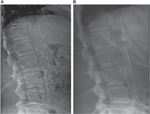
Lateral single-energy images (VFA images) of the lumbar spine were obtained on a Hologic Discovery A densitometer (Hologic, Inc., Bedford, MA, USA). While the patient remained in a supine position the C-arm of the machine moved to the lateral position, and then a lateral fan-beam X-ray image of the spine was obtained. Images that could not be used due to mis-positioning of the aorta in the field were not used for analysis (n = 266; 10.6%). To quantify calcification burden in the abdominal aorta, we applied an 8-score scale system (Citation21). This scale estimates the total length of calcification on the anterior and posterior aortic walls separately in the region anterior to the L1–L4 vertebral bones. In this scale, 0 stands for no calcification, 1 for aggregate length of calcification up to the height of one vertebra, 2 for aggregate length of calcification between one and two vertebrae, 3 for aggregate length of calcification between two and three vertebrae, and 4 for more aggregate length in each of the anterior and posterior aortic walls. Total score was the summation of anterior and posterior calcification scores and ranged from 0 to 8 ().
Figure 1. An example of severe AAC (score 8) with conventional X-ray (A) compared with VFA DEXA (B).

In order to validate VFA images for AAC assessment, 53 patients that underwent VFA as well as conventional lateral X-ray of the spine because of suspected vertebral fracture were selected and compared blinded by an expert reader ().
Cardiovascular risk factors and outcomes
Our medical center maintains a computerized information system of all patients admitted to the hospital. We checked the information system for relevant medical history at the time of the DEXA scan. Subjects with a history of angina pectoris, myocardial infarction (MI), cerebrovascular accident (CVA), or transient ischemic attack (TIA) or past invasive procedure for cardiovascular diseases were excluded from the study. Relevant medical history and family history of cardiovascular diseases (premature coronary heart disease, MI, CVA, or TIA) in first-degree relatives were included according to the medical file of the patients on the hospital information system prior to DEXA imaging at the date nearest to VFA. Hypertension was defined as blood pressure ≥ 140/90 mmHg, current antihypertensive medication including diuretics, or family physician's note. Hypercholesterolemia was defined as total cholesterol levels > 200 mg/dL, current use of lipid-lowering agents, or family physician's note. A fasting plasma glucose level ≥ 126 mg/dL or the use of antidiabetic medications was considered as diabetes mellitus. Subjects who smoked ≥ one cigarette per day within 1 year prior to VFA were considered as smokers. Non-fatal MI, CVA, and TIA were ascertained according to electrocardiogram (ECG), troponin, and creatine kinase (CK MB) levels, available CT scan, or physician's note on patient's file and evaluated by two study experts, blinded to the individuals’ AAC scores. Cardiovascular death was ascertained if a fatal MI/CVA occurred in individuals without known atherosclerotic disease. The patients who did not have an updated medical report on any of risk factors or outcomes were excluded from the study (n = 12; 3 patients in the AAC-positive group and 9 patients in the AAC-negative group. The latter were replaced by age- and gender-matched subjects).
Statistical analysis
All subjects with detectable calcification on VFA were identified. An age- and sex-matched control group was selected in a 2:1 ratio (two controls for each case). Survival was estimated by the Kaplan-Meier product limit method, compared with the log-rank test, and stratified for the three groups. Subjects with calcification burden were over-selected to acquire sufficient subjects with AAC. To overcome this over-sampling of subjects with AAC a design-based analysis was performed. Due to this weighting method our conclusions can be generalized for the original cohort of consecutive patients who underwent VFA. The design-based Cox proportional hazards regression models evaluating the prognostic properties of AAC to the risk of CV events were built with STATA (version 10.0; STATA, Texas). To correct for within-pair correlation of observations among subjects, we applied cluster specification to obtain robust estimates of variance as described previously (Citation24). Data were expressed as mean ± standard deviation (SD). Results are summarized by hazard (risk) ratios with 95% confidence intervals (95% CI). Base-line characteristics were compared within groups using chi-square for categorical variables and Student's t test for continuous variables. Weighted kappa statistics were used to assess agreement in AAC scoring between VFA and conventional lateral spine X-ray. All reported probability values are two-tailed, and P < 0.05 was considered statistically significant.
Results
Altogether 2,500 consecutive patients (mean age ± SD: 52 ± 15; 65% female) underwent VFA imaging between 2005 and 2007. A total of 167 (6.6%) had detectable calcification, and they were divided according to AAC score into two groups: low-AAC (1–3; n = 108, of whom 3 subjects were excluded later due to lack of information) and high-AAC (4–8; n = 59). A further 325 subjects without calcification served as age- and sex-matched control group. The base-line characteristics of 489 subjects according to AAC status are shown in . Statistically significant differences were observed in percentage of cardiovascular risk factors within different groups. Moreover, despite the age-matched control group formation, mean age in the high-AAC group was significantly higher than in the control group.
Table I. Base-line characteristics of study population.
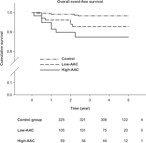
Subjects were followed up for cardiovascular events for 2–60 months (median 31 months). The total number of cardiovascular events was 19. Among the 325 subjects in the control group, 5 subjects had a cardiovascular event. Seven cardiovascular events in the low-AAC group and another seven cardiovascular events in the high-AAC group were observed. The incidence of non-fatal cardiovascular events was 5 (1.5%), 5 (4.8%), and 7 (11.9%) in control, low-AAC, and high-AAC groups, respectively. MI and CVA/TIA incidences among different groups are shown in . The cumulative event rate for total cardiovascular events during the follow-up period was 1.5%, 6.7%, and 11.9% for control, low-AAC, and high-AAC groups, respectively ().
Table II. Number and percentage of cardiovascular events in relation to AAC.
Figure 2. Kaplan-Meier curve for cumulative event-free survival in each group and population at risk at each time point.

In the Cox proportional hazard model, it was confirmed that AAC is associated with higher cardiovascular hazard risk, independent of age, sex, and conventional cardiovascular risk factors, as shown in . The Cox proportional hazard model showed that in comparison with the control group the AAC score was also associated with a higher risk of cardiovascular events. A significantly higher hazard ratio (HR) was found in both the low-AAC (HR 4.7; 95% CI 1.5–15.2; P = 0.009) and the high-AAC groups (HR 8.6; 95% CI 2.7–27.1; P < 0.0001) (). Adjusted analysis showed that this association was independent of age and sex in both low-AAC (HR 4.9; 95% CI 1.5–15.9; P = 0.008) and high-AAC groups (HR 7.3; 95% CI 2.2–23.9; P = 0.001). After the addition of conventional cardiovascular risk factors, the model still showed significant predictive value in both low-AAC (HR 4.2; 95% CI 1.2–15.4; P = 0.027) and high-AAC groups (HR 6.1; 95% CI 1.7–21.6; P = 0.005) compared with the control group ().
Table III. Cox regression analysis for comparing control, low-AAC, and high-AAC groups. Univariate, gender- and age-adjusted, and multivariate-adjusted hazard ratio for AAC in cardiovascular events (95% confidence interval). Control group served as reference. Multivariate analysis involved: age, gender, hypertension, hypercholesterolemia, diabetes mellitus, smoking, and family history of coronary heart disease.
Table IV. Cox regression analysis for comparing AAC-negative versus AAC-positive subjects. Univariate, gender- and age-adjusted, and multivariate-adjusted hazard ratio for AAC in cardiovascular events (95% confidence interval). Control group served as reference. Multivariate analysis involved: age, gender, hypertension, hypercholesterolemia, diabetes mellitus, smoking, and family history of coronary heart disease.
AAC scores in a subselection of 53 patients that underwent VFA as well as conventional lateral X-ray of the spine were compared by an experienced physician blind to the results of other modality, and statistical analysis showed excellent agreement with a kappa of 0.87 (95% CI 0.66–1).
Discussion
In this study we showed the predictive value of AAC on DEXA scans which were performed for routine clinical evaluation of VFA. The strong, independent association between cardiovascular events rate and AAC level gives an opportunity to physicians for early risk stratification of cardiovascular diseases in subjects referred for BMD and VFA. A hazard rate of 11.9% (HR 6.4) in the high-AAC group implies the need for more thorough diagnostic efforts in these subjects.
Abdominal aortic calcification detected by lateral spinal radiography and CT scan has previously been shown to be an independent predictor of cardiovascular diseases (Citation3–11). It is noteworthy that DEXA is able to accurately measure AAC level in comparison with lateral spinal radiographs (Citation19,Citation21) and assess vertebral fractures (Citation14,Citation16) with lower radiation burden, lower costs, and in less time (Citation5,Citation19–21), with a good intra-rater variability (Citation19).
Interestingly, DEXA is able to assess total and regional body fat mass composition (Citation25–27), which has been reported to be associated with cardiovascular risk factors (Citation28–31). According to recommendations for osteoporosis screening, bone densitometry and simultaneous lateral spine imaging for VFA should be performed in a notable subset of the elderly population (Citation13,Citation15,Citation16). Therefore, the strong predictive value of AAC level for cardiovascular events provides influential simultaneous information to practitioners in order to get an estimation of cardiovascular risk in individuals referred for osteoporosis screening by DEXA. Moreover, while there is a hypothesis that body composition analysis and detection of AAC will both improve the utility of DEXA to predict cardiovascular disease outcomes, further research is required to test this.
Thus, due to low radiation exposure in DEXA imaging systems (less than 0.01 mSv versus 2 mSv in lateral spine radiographs (Citation19,Citation22)), and efficiency of DEXA in detecting bone density, vertebral fracture, as well as abdominal aortic calcification, this method seems to be an appropriate tool in patients referred for osteoporosis detection for additional evaluation of cardiovascular risk.
In a previous study, Schousboe et al. compared AAC in 369 cases of women with a mean age of 80 years compared with 363 controls (Citation23). They selected patients with stroke or myocardial infarction. There are several differences between this previous study and our study. Our study did not focus on elderly women but also included younger patients (mean age 68.5 years). More importantly, our study population comprised also 46% males, and to our knowledge it is the first study on cardiovascular predictive value of AAC detected by VFA in both sexes. Furthermore, all cardiovascular events, including TIA and cardiovascular death, were evaluated. Finally, patients in that study were selected in a different way. They selected patients with stroke or MI, whereas we prospectively included all patients with a positive AAC result. The results, however, are comparable, showing a strong predictive value of AAC.
There were limitations in our study. This study represents retrospective data. Our population consists only of patients referred for VFA or osteoporosis. Data should be interpreted cautiously when dealing with other patient groups. Patients were screened for VFA and osteoporosis, and laboratory data relevant for cardiovascular risk (e.g. low-density lipoprotein (LDL) and high-density lipoprotein (HDL) serum levels) were not available for all subjects. Moreover, further relevant variables such as blood pressure, medications, and condition of blood pressure control in hypertensive subjects were lacking in our study. Further studies for other ethnic groups, applying cardiovascular risk prediction systems, and designing cohorts with younger groups, might be needed to obtain a better understanding of the predictive value of AAC measurements. Moreover, investigations comprising more subjects and a longer follow-up period are thus needed. Some other studies (Citation5,Citation11,Citation23) showed a higher percentage AAC in their population than the 6% found in our study. However, our study comprised a younger population (52.8 years versus 60.7 years (Citation11), 69 years (Citation5), and 80 years (Citation23)) and patients without increased risk of cardiovascular disorders, which explains partly the lower detection rate for AAC.
In conclusion, AAC assessed by VFA can predict non-fatal or fatal cardiovascular events among elderly men and women. This finding is independent of conventional risk factors for cardiovascular disease. The routine use of DEXA and VFA for osteoporosis and vertebral fracture screening contributes to a one-stop-shop session with low costs, low radiation burden, and a fast accessible way of risk stratifying for cardiovascular events. Prospective large cohort studies with sequential measurements are needed to further extend these findings to the general population
Declaration of interest: R. Golestani's work is funded by Siemens. The other authors declare no conflicts of interest.
References
- Oei HH, Vliegenthart R, Hak AE, Iglesias del Sol A, Hofman A, Oudkerk M, . The association between coronary calcification assessed by electron beam computed tomography and measures of extracoronary atherosclerosis: the Rotterdam Coronary Calcification Study. J Am Coll Cardiol. 2002;39:1745–51.
- Wu MH, Chern MS, Chen LC, Lin YP, Sheu MH, Liu JC, . Electron beam computed tomography evidence of aortic calcification as an independent determinant of coronary artery calcification. J Chin Med Assoc. 2006;69:409–14.
- Witteman JC, Kok FJ, van Saase JL, Valkenburg HA. Aortic calcification as a predictor of cardiovascular mortality. Lancet. 1986;2:1120–2.
- Blacher J, Guerin AP, Pannier B, Marchais SJ, London GM. Arterial calcifications, arterial stiffness, and cardiovascular risk in end-stage renal disease. Hypertension. 2001;38:938–42.
- Van der Meer IM, Bots ML, Hofman A, del Sol AI, van der Kuip DA, Witteman JC. Predictive value of noninvasive measures of atherosclerosis for incident myocardial infarction: the Rotterdam Study. Circulation. 2004;109:1089–94.
- Reaven PD, Sacks J. Coronary artery and abdominal aortic calcification are associated with cardiovascular disease in type 2 diabetes. Diabetologia. 2005;48:379–85.
- Rodondi N, Taylor BC, Bauer DC, Lui LY, Vogt MT, Fink HA, . Association between aortic calcification and total and cardiovascular mortality in older women. J Intern Med. 2007;261:238–44.
- Hollander M, Hak AE, Koudstaal PJ, Bots ML, Grobbee DE, Hofman A, . Comparison between measures of atherosclerosis and risk of stroke: the Rotterdam Study. Stroke. 2003;34:2367–72.
- Levitzky YS, Cupples LA, Murabito JM, Kannel WB, Kiel DP, Wilson PW, . Prediction of intermittent claudication, ischemic stroke, and other cardiovascular disease by detection of abdominal aortic calcific deposits by plain lumbar radiographs. Am J Cardiol. 2008;101:326–31.
- Walsh CR, Cupples LA, Levy D, Kiel DP, Hannan M, Wilson PW, . Abdominal aortic calcific deposits are associated with increased risk for congestive heart failure: the Framingham Heart Study. Am Heart J. 2002;144:733–9.
- Wilson PW, Kauppila LI, O'Donnell CJ, Kiel DP, Hannan M, Polak JM, . Abdominal aortic calcific deposits are an important predictor of vascular morbidity and mortality. Circulation. 2001;103:1529–34.
- Simpson AK, Whang PG, Jonisch A, Haims A, Grauer JN. The radiation exposure associated with cervical and lumbar spine radiographs. J Spinal Disord Tech. 2008;21:409–12.
- Brown JP, Josse RG; Scientific Advisory Council of the Osteoporosis Society of Canada. 2002 clinical practice guidelines for the diagnosis and management of osteoporosis in Canada. CMAJ. 2002;167 Suppl:S1–34.
- Grigoryan M, Guermazi A, Roemer FW, Delmas PD, Genant HK. Recognizing and reporting osteoporotic vertebral fractures. Eur Spine J. 2003;12 Suppl 2:S104–12.
- Schousboe JT. Cost effectiveness of screen-and-treat strategies for low bone mineral density: how do we screen, who do we screen and who do we treat? Appl Health Econ Health Policy. 2008;6:1–18.
- Schousboe JT, Vokes T, Broy SB, Ferrar L, McKiernan F, Roux C, . Vertebral fracture assessment: the 2007 ISCD official positions. J Clin Densitom. 2008;11:92–108.
- Intenzo CM, Parker L, Rao VM, Levin DC. Changes in procedure volume and service provider distribution among radiologists and nonradiologists in dual-energy x-ray absorptiometry between 1996 and 2002. J Am Coll Radiol. 2005;2:662–4.
- Masse PG, Tranchant CC, Dosy J, Donovan SM. Coexistence of osteoporosis and cardiovascular disease risk factors in apparently healthy, untreated postmenopausal women. Int J Vitam Nutr Res. 2005;75:97–106.
- Schousboe JT, Wilson KE, Hangartner TN. Detection of aortic calcification during vertebral fracture assessment (VFA) compared to digital radiography. PLoS ONE. 2007;2:e715.
- Blake GM, Naeem M, Boutros M. Comparison of effective dose to children and adults from dual X-ray absorptiometry examinations. Bone. 2006;38:935–42.
- Schousboe JT, Wilson KE, Kiel DP. Detection of abdominal aortic calcification with lateral spine imaging using DXA. J Clin Densitom. 2006;9:302–8.
- Toussaint ND, Lau KK, Strauss BJ, Polkinghorne KR, Kerr PG. Determination and validation of aortic calcification measurement from lateral bone densitometry in dialysis patients. Clin J Am Soc Nephrol. 2009;4: 119–27.
- Schousboe JT, Taylor BC, Kiel DP, Ensrud KE, Wilson KE, McCloskey EV. Abdominal aortic calcification detected on lateral spine images from a bone densitometer predicts incident myocardial infarction or stroke in older women. J Bone Miner Res. 2008 Mar;23(3):409–16.
- Lin DY, Wei LJ. The robust inference for the Cox proportional hazards model. J Am Stat Assoc. 1989;84:1074–8.
- Snijder MB, Visser M, Dekker JM, Seidell JC, Fuerst T, Tylavsky F, . The prediction of visceral fat by dual-energy X-ray absorptiometry in the elderly: a comparison with computed tomography and anthropometry. Int J Obes Relat Metab Disord. 2002;26:984–93.
- Svendsen OL, Hassager C, Bergmann I, Christiansen C. Measurement of abdominal and intra-abdominal fat in postmenopausal women by dual energy X-ray absorptiometry and anthropometry: comparison with computerized tomography. Int J Obes Relat Metab Disord. 1993;17: 45–51.
- Glickman SG, Marn CS, Supiano MA, Dengel DR. Validity and reliability of dual-energy X-ray absorptiometry for the assessment of abdominal adiposity. J Appl Physiol. 2004;97:509–14.
- Shaw KA, Srikanth VK, Fryer JL, Blizzard L, Dwyer T, Venn AJ. Dual energy X-ray absorptiometry body composition and aging in a population-based older cohort. Int J Obes (Lond). 2007;31:279–84.
- Wiklund P, Toss F, Weinehall L, Hallmans G, Franks PW, Nordstrom A, . Abdominal and gynoid fat mass are associated with cardiovascular risk factors in men and women. J Clin Endocrinol Metab. 2008;93:4360–6.
- Van Pelt RE, Evans EM, Schechtman KB, Ehsani AA, Kohrt WM. Contributions of total and regional fat mass to risk for cardiovascular disease in older women. Am J Physiol Endocrinol Metab. 2002;282:E1023–8.
- Ito H, Nakasuga K, Ohshima A, Maruyama T, Kaji Y, Harada M, . Detection of cardiovascular risk factors by indices of obesity obtained from anthropometry and dual-energy X-ray absorptiometry in Japanese individuals. Int J Obes Relat Metab Disord. 2003;27:232–7.