Abstract
Essential hypertension is characterized by a left ventricular dysfunction. However, the majority of the studies performed so far investigated patients under drug treatment and/or with concomitant diseases, such as obesity, diabetes, metabolic syndrome or coronary heart disease, which per se may affect diastolic function independently on the blood pressure elevation. The present study aimed at investigating left ventricular diastolic function in untreated, uncomplicated and newly diagnosed hypertensive patients by employing both routine echo-Doppler and pulse tissue-Doppler technique. Data were collected in 86 middle-aged essential hypertensive patients and in 18 sex-matched normotensive controls. At the echo-Doppler approach, about half of the hypertensive patients displayed a diastolic dysfunction (n = 44, E/A: 0.79 ± 0.02). They showed body mass index values slightly greater than hypertensive patients without diastolic dysfunction but superimposable blood pressure values and metabolic variables. When assessed via the pulse tissue-Doppler approach, patients with a reduced E/A displayed an Em/Am ratio significantly lower than patients without diastolic dysfunction and control subjects. This was the case when the data were related to the lateral and septal mitral annulus or averaged together. Furthermore, whereas myocardial systolic peak velocity (Sm) was lower in hypertensive patients than in control subjects, no significant between-groups difference in E/Em ratio was observed. Differently from the data obtained via the echo-Doppler approach, the tissue-Doppler method in patients without diastolic dysfunction showed a significant higher deceleration and isovolumetric relaxation times, with a lower Em velocity compared with the normotensive subjects. At the stepwise multiple regression analysis E/A ratio and E’/A’ values were related with left ventricular mass index and body mass index after correction for age. These data provide evidence that diastolic dysfunction is of frequent detection in the earlier uncomplicated phases of the disease and that tissue Doppler detects an initial impairment of left ventricular relaxation in the patients in which at echo Doppler is still normal.
Introduction
Left ventricular diastolic dysfunction is a common finding in patients with essential hypertension (Citation1–5), with a prevalence ranging from 11% to 56% (Citation1,Citation6,Citation7) in patients with left ventricular hypertrophy (Citation8), according to the different echocardiographic criteria employed in the different published studies.
The majority of the investigations performed so far, however, assessed diastolic function in patients with long-lasting hypertension under antihypertensive drug treatment (Citation1,Citation5–7,Citation8–10) and relatively few studies (Citation11–16) have provided information in newly diagnosed untreated hypertensive patients. Furthermore, some of the above-mentioned investigations enrolled elderly patients with concomitant cardiovascular risk factors, such as obesity, metabolic syndrome and diabetes, i.e. conditions which per se may affect diastolic function independently on the blood pressure elevation (Citation14,Citation17). Furthermore, in the majority of the above-mentioned studies, diastolic function has been assessed in a quite heterogeneous fashion, i.e. by employing different methods, indexes and classifications (Citation13–14,Citation17–20), making difficult a proper comparison between the data collected in the various studies. Finally, to the best of our knowledge, no study has so far provided data evaluating diastolic dysfunction in the single patient employing different echocardiographic methodologies.
With this background in mind, in the present study we assessed diastolic function in newly diagnosed and untreated hypertensive patients, without other cardiometabolic confounders, using an extensive pattern of parameters, derived from both the echo-Doppler and the pulse tissue Doppler techniques.
Methods
Study population
We studied 86 consecutive hypertensive patients and 18 healthy volunteers, matched for age, sex and body mass index (BMI). Hypertensive patients were recruited from those attending our outpatient specialist clinic of the Kocaeli University Hospital, to which they were sent by the general physician for a complete evaluation related to the recent finding of elevated blood pressure values. To be included in the study, patients were required to have a newly diagnosed and untreated mild-to-moderate hypertension (blood pressure values ≥ 140/90 mmHg), according to the European Society of Hypertension and European Society of Cardiology criteria (Citation21). This condition was confirmed by (i) the finding of a stable blood pressure elevation recorded at different medical visits within the previous month; (ii) the lack a blood pressure elevation at previous clinical visits performed before the last month in the general physician office; and (iii) the absence of any antihypertensive drug treatment. Patients with severe hypertension (blood pressure values ≥ 180/100 mmHg), previous antihypertensive therapy, bradycardia (heart rate < 60 beats/ min), atrio-ventricular block, left bundle branch block, atrial fibrillation or other cardiac arrhythmias, valvular heart disease, coronary artery disease, ejection fraction LVEF < 50% or inappropriate echocardiographic window were not eligible. Additional exclusion criteria were diabetes mellitus (fasting plasma glucose > 126 mg/dl), obesity (BMI > 30 kg/m2) or metabolic syndrome, renal impairment (estimated glomerular filtration rate < 60 ml/min/m2), hepatic insufficiency and pregnant or nursing women. The study was carried out according to the Declaration of Helsinki, local ethics committee approval and patients’ written informed consent was individually obtained.
Measurements
Eligible patients underwent standard clinical examination, electrocardiogram (EKG), transthoracic echocardiography, chest radiography and routine laboratory tests. BMI was obtained by dividing body weight in kilograms by the square of the height in meters. Clinic blood pressure was measured three times within the subject having a sitting position for a 10-min period, using a mercury sphygmomanometer and taking the first and fifth Korotkoff to identify for systolic and diastolic blood pressure values, respectively. Measurements were repeated three times at 3-min intervals and the average of the three measurements were taken.
Transthoracic M-mode, two-dimensional and colour Doppler echocardiography was performed via a Toshiba SSA-390A ultrasound machine using a 2.0–3.7-MHz broadband transducer. Measurements of the left atrium and the left and right ventricles were obtained from the parasternal long-axis view as recommended by the American Society of Echocardiography guidelines (Citation22). Left ventricular mass was calculated according to Penn convention method and indexed to body surface area. Left ventricular ejection fraction was determined using the modified Simpson's rule in the apical two- and four-chamber views. Transmitral flow velocity pattern was evaluated from the apical four-chamber view with pulsed-wave Doppler placing the sample volume at the tips of mitral leaflets during diastole. Early (E) and atrial (A) peak velocities, deceleration time of E velocity (DT), isovolumic relaxation time (IVRT), systolic (S), diastolic (D) and atrial reversal pulmonary veins flow velocities (PVA) were measured on three consecutive beats in each patient and averaged for the whole population of the study. The E/A and S/D ratios were calculated. Left ventricular end diastolic and systolic internal diameter (EDD, ESD), interventricular septum and posterior wall thickness (IVST, PWT) were measured according to the American Society of Echocardiography guidelines (Citation22). Relative wall thickness was calculated as 2 × PWT/EDD (Citation19). To enhance the power of our findings and to avoid the influence of preload and afterload, diastolic function was also assessed by the pulse tissue Doppler, performed in the apical four- and two-chamber views, with a 2-mm sample volume placed at the lateral, septal, anterior and inferior mitral annulus. Only the lateral and septal mitral annulus velocities, as well as the average of the two sites, were considered for statistical analysis, because they provide a better evaluation of myocardial diastolic function (Citation23,Citation24). Myocardial systolic (Sm), early (E’) and late (A’) diastolic velocities were measured and the E’/A’ and E/E’ ratios calculated.
All measurements were performed by the same operator, unaware of the patient's blood pressure values. Echocardiographic images were recorded on videotape to allow subsequent offline analysis. Intra-observer variability of the various variables, assessed in 12 patients by repeating the measurements twice (1–5 days apart) was < 5% under the same basal conditions. The primary criteria to define patients with or without diastolic dysfunction was the E/A ratio above or below 1, which is predictive for cardiovascular events in young and elderly hypertensive patients (Citation10,Citation21,Citation25). In addition, the following parameters were considered, as reported by the European Society of Cardiology study group (Citation26): IVRT > 100–105 ms; E/A < 1–0.5 combined with a DT > 220–280 ms, or a S/D ratio > 1.5–2.5, or peak PVA > 35 cm/sec, according to the age.
Data analysis
Data are expressed as means ± SEM. Differences between study groups were analysed in SAS using analysis of variance (ANOVA), Student t-test for quantitative data and chi-square test for categorical variables. All evaluations were performed as two-tailed tests with a p < 0.05 value as criterion for statistical significance. Pearson's correlation coefficient was used to evaluate the relationships between E/A and E’/A’ with various clinical variables. To identify factors independently correlated with these two variables, linear regression models were used with stepwise selection. A p < 0.05 value was considered statistically significant.
Results
General characteristics
Among 125 consecutive patients with newly diagnosed and previously untreated mild-to-moderate hypertension, 39 were excluded for concomitant valvular disease, renal impairment or not evaluable echo Doppler. The general characteristics of the remaining 86 hypertensive patients, subdivided in those without and with diastolic dysfunction, are shown in , which also includes data related to the control group. The hypertensive subjects were slightly, although significantly, older than controls and they had a slightly but not significantly greater BMI. As expected, hypertensive patients displayed significantly greater systo-diastolic blood pressure values, this being the case also for plasma glucose and total cholesterol levels, which were both still within the normal range, however.
Table I. Characteristics of control subjects and hypertensive patients (HT) without or with diastolic dysfunction (DD).
Echocardiographic characteristics
According to the above-mentioned primary criteria, i.e. an E/A ratio value greater or less than 1, the hypertensive population of our study was subdivided in two groups, one without (n = 42, E/A: 1.28 ± 0.03) and the other one with (n = 44, E/A: 0.79 ± 0.02) left ventricular diastolic dysfunction, the corresponding E/A ratio value detected in control normotensive individuals amounting to 1.37 ± 0.05 (p < 0.01 vs patients with left ventricular dysfunction) (data not shown). As illustrated in , patients with an E/A ratio value greater or less than 1 displayed similar values of end-diastolic diameters, end-systolic diameters, ejection fraction, left atrial diameters, left ventricular mass index, posterior wall thickness, relative wall thickness, whereas interventricular septal wall thickness was significantly greater in patients with an E/A ratio < 1. All the above-mentioned parameters were similar to controls, with the exception of left atrial diameter, posterior wall and septal wall thickness, whose values were significantly greater in the two hypertensive patients groups. shows that patients with an E/A ratio < 1 displayed, as expected, IVRT and DT values significantly greater than those detected in control subjects, and in hypertensive patents with an E/A ratio > 1, whereas S/D ratio and endocardial fractional shortening (EFS) did not differ between controls and hypertensive patients. also shows that peak pulmonary vein systolic and diastolic velocity values were significantly lower in hypertensive patients with diastolic dysfunction. In contrast, PVA and filling time values were not statistically different between the two hypertensive groups, but were higher (p < 0.001) compared with controls.
Table II. Echocardiographic data in normotensive controls and in hypertensive patients without or with diastolic dysfunction.
Figure 1. Behaviour of deceleration time of E velocity (DT), isovolumic relaxation time (IVRT), endocardial fractional shortening (EFS), systodiastolic ratio (S/D), peak pulmonary vein systolic (PVs), diastolic (PVd) and average (PVa) velocity values and filling time in normotensive subjects (C, open bars) and in age-matched essential untreated and newly diagnosed hypertensive patients without (HT, grey bars) and with (HTD, black bars) left ventricular diastolic dysfunction. Data are shown as means ± SEM. Asterisks (*p < 0.05, **p < 0.01) refer to the statistical significance between groups.

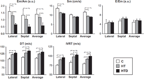
shows the data obtained via the pulse tissue Doppler approach. All patients with left ventricular diastolic dysfunction, as assessed by the M-mode, two-dimensional, colour Doppler echocardiography, had a significantly lower Em/Am ratio than patients without diastolic dysfunction and control subjects. This was the case when the data were related to the lateral or septal mitral annulus or averaged together. Furthermore, whereas myocardial systolic peak velocity (Sm) was lower in hypertensive patients than in control subjects, no significant between-group difference in E/Em ratio was observed. Differently from the data obtained via the echo-Doppler approach, the tissue-Doppler method in patients without diastolic dysfunction showed significant higher deceleration and isovolumetric relaxation times, with a low Em velocity in comparison with the normotensive subjects.
Figure 2. Behaviour of deceleration time of Em/Am ratio, myocardial systolic peak velocity (Sm), E/Em ratio, deceleration time of E velocity (DT), isovolumic relaxation time (IVRT) in normoresive subjects (C, open bars) and in age-matched essential untreated and newly diagnosed hypertensive patients without (HT, grey bars) and with (HTD, black bars) left ventricular diastolic dysfunction. Data are related to the lateral and septal mitral annulus or averaged together Data are shown as means ± SEM. Asterisks (*p < 0.05, **p < 0.01) refer to the statistical significance between groups.
In the group of patients with left ventricular diastolic dysfunction as a whole, there was a significant inverse correlation between E/A ratio value and age, left ventricular mass and left ventricular mass index. In addition to the above-mentioned variables, E’/A’ also correlated with BMI and to a lesser extent with plasma creatinine values (). At the stepwise multiple regression analysis, E/A ratio and E’/A’ values were related with left ventricular mass index and BMI after correction for age, (), whereas in the patients without diastolic dysfunction no significant correlation was found between with E/A ratio and E’/A’ and left ventricular mass index (R = − 0.01 and 0.24, p = NS) or BMI (R = 0.02 and − 0.26, p = NS).
Table III. Person's correlation between E/A and E’/A’ with clinical variables.
Table IV. Linear regression models – stepwise selection.
Discussion
Our study shows that a consistent portion of patients with newly diagnosed and untreated hypertension (about 48%) displays an impairment of left ventricular diastolic function. They also show that this functional cardiac alteration occurs independently of the concomitant presence of other confounding factors frequently associated with left ventricular diastolic dysfunction, such as obesity, metabolic syndrome and diabetes mellitus. They finally show that the detection of left ventricular dysfunction via the echo-Doppler approach is well paralleled by the findings obtained via the tissue-Doppler technique.
Several other results of our study deserve to be briefly discussed. First, the S/D ratio and E/Em ratio, a reliable estimate of left ventricular diastolic pressure (Citation24), were not different between untreated hypertensive patients with and without diastolic impairment and control subjects, suggesting that left ventricular filling pressure was not modified at this presumably early stage of the hypertensive disease. Indeed, a significant increase in left ventricular filling pressure, estimated as E/Em ratio, has been reported in newly diagnosed and untreated hypertensive patients with left ventricular concentric geometry (Citation16). Second, in our study, left ventricular mass index was not significantly different between patients with and without diastolic dysfunction. However, the linear regression analysis showed that, among different variables, left ventricular mass index was independently and significantly correlated with left ventricular diastolic dysfunction (E/A and E’/A’ ratios), confirming that diastolic abnormality may precede the development of left ventricular hypertrophy (Citation8,Citation10,Citation27–29) also in newly diagnosed hypertensive patients (Citation14). Third, left ventricular posterior and interventricular wall thickness were significantly greater in the hypertensive patients than in the normotensive ones, as reported in other studies in newly diagnosed or untreated hypertension (Citation11,Citation27,Citation30). However, the relative wall thickness was not different between patients with and without diastolic dysfunction and was lower than 0.45, the cut-off for concentric remodelling. Fourth, left atrial diameter was slightly increased in hypertensive patients, compared with the control group, as observed in other studies (Citation14,Citation31), but remained into the normal range, confirming that left atrial enlargement is not an early finding in relatively young and never treated hypertensive patients (Citation31,Citation32). Finally, peak systolic tissue velocity was lower in hypertensive patients showing a mild impairment of left ventricular systolic function (Citation33,Citation34). However, the reduction was not accompanied by a decrease in the endocardial shortening, confirming that this parameter is less sensitive than the midwall fractional shortening to assess left ventricular systolic function, as previously reported (Citation1).
Our results also provide other important information about the behaviour of left ventricular diastolic function in hypertension. Differently from the echo Doppler, the tissue Doppler in the patients without diastolic dysfunction showed significant higher deceleration and isovolumetric relaxation times, with a low Em velocity in comparison with the normotensive subjects. These findings demonstrate an initial impairment of left ventricular relaxation is an early stage of hypertension, detected by the tissue-Doppler imaging approach, which is thus more sensitive than the conventional echo-Doppler method. The mechanisms involved in the left ventricular diastolic dysfunction detected in the early clinical phases of hypertension still remain unclear. We can speculate, however, that diastolic dysfunction may be part of a systemic alteration of cardiovascular stiffness. Indeed, in newly diagnosed essential hypertensives, the association between left ventricular diastolic dysfunction and arterial stiffness has been reported (Citation15–16,Citation20,Citation32,Citation35–37), and a significant increase in myocardial fibrosis has been documented (Citation11,Citation30,Citation38,Citation39), even before the development of left ventricular hypertrophy (Citation27).
Our study has some limitations but also a clinical implication. The first limitation refers to the fact that in our patients we did not assess exercise capacity and respiratory function, which may be both altered in this condition (Citation20), and may at least in part participate at the development and/or progression of the diastolic dysfunction (Citation24,Citation26). Such alterations, however, have been reported in more advanced clinical stages of the disease than the ones seen in the present study. Further limitations relate to the fact that (i) the control group was small and (ii) hypertensive patients were slightly but significantly older than controls. This latter limitation may have indeed affect the results, although it is highly unlikely that a difference of 4.7 years may explain the differences we found in the diastolic function between the two groups. The clinical implication is related to the fact that, since tissue Doppler detects an initial impairment of left ventricular relaxation in the patients in which at echo Doppler is still normal, the routine evaluation of the diastolic function by this approach, which unfortunately is performed only in a small percentage of hypertensive patients (Citation40), should be implemented in current clinical practice.
Acknowledgements
None.
Conflicts of interest: None.
References
- Zanchetti A, Cuspidi C, Comarella L, Rosei EA, Ambrosioni E, Chiariello M, . Left ventricular diastolic dysfunction in elderly hypertensives: Results of the APROS-diadys study. J Hypertens. 2007; 25:2158–2167.
- De Simone G, Greco R, Mureddu GF, Romano C, Guida R, Celentano A, . Relation of left ventricular diastolic properties to systolic function in arterial hypertension. Circulation. 2000;101:152–157.
- Liu JE, Palmieri V, Roman MJ, Bella JN, Fabsitz R, Howard BV, . The impact of diabetes on left ventricular filling pattern in normotensive and hypertensive adults: The Strong Heart Study. J Am Coll Cardiol. 2001;37: 1943–1949.
- Gardin JM, Arnold AM, Bild DE, Smith VE, Lima JA, Klopfenstein HS, . Left ventricular diastolic finding in elderly: The Cardiovascular Health Study. Am J Cardiol. 1998;82:345–351.
- Denes M, Kiss I, Lengyel M. Assessment of diastolic dysfunction in elderly hypertensive patients using integrated Doppler echocardiography. Blood Press. 2009;18:135–141.
- Fischer M, Baessler A, Hense HW, Hengstenberg C, Muscholl M, Holmer S, . Prevalence of left ventricular echocardiographic-based survey of a population sample. Eur Heart J. 2003;24:320–328.
- Abhayaratna WP, Marwich TH, Smith WT, Becker NG. Characteristics of left ventricular diastolic dysfunction in the community: An echocardiographic survey. Heart. 2006; 92:1259–1264.
- Wachtell K, Smith G, Gerds E, Dahlof B, Nieminen MS, Papademetriou V, . Left ventricular diastolic filling patterns in patients with hypertension and left ventricular hypertrophy (The Life study). Am J Cardiol. 2000;85:466–472.
- de Simone G, Kitzman D W, Chinali M, Oberman A, Hopkins PN, Rao DC, . Left ventricular concentric geometry is associated with impaired relaxation in hypertension: The HyperGen study. Eur Heart J. 2005;26:1039–1045.
- Schillaci G, Pasqualini L, Verdecchia P, Vaudo G, Marchesi S, Porcellati C, . Prognostic significance of left ventricular diastolic dysfunction in essential hypertension. J Am Coll Cardiol. 2002;39:2005–2011.
- Lindsay MM, Maxwell P, Dunn FG. TIMP-1 a marker of left ventricular diastolic dysfunction and fibrosis in hypertension. Hypertension. 2002;40:136–141.
- Sagie A Benjamin EJ, Galderisi M, Larson MG, Evans JC, Fuller DL, . Echocardiographic assessment of left ventricular structure and diastolic filling in elderly subjects with borderline isolated systolic hypertension (the Framingham Heart Study). Am J Cardiol. 1993;72:662–665.
- Almuntaser I, Brown A, Murphy R, Crean P, King G, Mahmud A, Feely J. Comparison of echocardiographic measures of left ventricular diastolic function in early hypertension. Am J Cardiol. 2007;100:1771–1775.
- Persic V, Ruzic A, Miletic B, Balen S, Jovanovic Z, Vcev A, . Left ventricular diastolic dysfunction in obese patients with newly diagnosed arterial hypertension. Wien Clin Wochenschr. 2007;119:423–427.
- Tsioufis C, Chatzis D, Dimitriadis K, Stougianos P, Kakavas A, Vlasseros I, . Left ventricular diastolic dysfunction is accompanied by increased aortic stiffness in the early stages of essential hypertension: A TDI approach. J Hypertens. 2005;23:1745–1750.
- Galderisi M, de Simone G, D'Errico A, Sidiropulos M, Viceconti R, Chinali M, . Independent association of coronary flow reserve with left ventricular relaxation and filling pressure in arterial hypertension. Am J Hypertens. 2008;21:1040–1046.
- Capra A, Galderisi M, Giannattasio C, Innelli P, Facchetti R, Cesana F, . Early alterations in left ventricular diastolic dysfunction in normotensive diabetic patients. Blood Press. 2012; 21 110–115.
- Ikonomidis I, Tzortis S, Papaioannou T, Protogerou A, Stamatelopoulos K, Papamichael C, . Incremental value of arterial wave reflection in the determination of left ventricular diastolic dysfunction in untreated patients with essential hypertension. J Hum Hypertens. 2008;22:687–698.
- Galderisi M, Tagliamonte MR, D’Errico A, Carella C, Varricchio G, Mondillo S, . Independent association of plasma leptin levels and left ventricular isovolumic relaxation in uncomplicated hypertension. Am J Hypertens. 2001;14: 1019–1024.
- Guven A, Koksal N, Sokmen G, Ozdemir R. Comparison of the echocardiographic and pulmonary function tests findings in elderly treated and untreated essential hypertensive patients. Blood Press. 2003;12:319–325.
- Mancia G, De Backer G, Dominiczak A, Cifkova R, Fagard R, Germano G, . 2007 Guidelines for the Management of Arterial Hypertension: The Task Force for the Management of Arterial Hypertension of the European Society of Hypertension (ESH) and of the European Society of Cardiology (ESC). Blood Press. 2007;16:135–122.
- Sahn DJ, De Maria A, Kisslo J, Weyman A, for the Committee on M-mode Standardization of the American Society of Echocardiography: Recommendations regarding quantitation in M-mode echocardiography: Results of a survey of echocardiographic measurements. Circulation. 1978;58:1072–1083.
- Ommen SR, Nishimura RA, Appleton CP, Miller FA, Oh JK, Redfield MM, . Clinical utility of Doppler echocardiography and tissue Doppler imaging in the estimation of left ventricular filling pressures: A comparative simultaneous Doppler-catheterization study. Circulation. 2000;102:1788–1794.
- Paulus WJ, Tschope C, Sanderson JE, Rusconi C, Flachskampf FA, Rademakers FE, . How to diagnose diastolic heart failure: A consensus statement on the diagnosis of heart failure with normal left ventricular ejection fraction by the Heart Failure and Echocardiography Association of the European Society of Cardiology. Eur Heart J. 2007; 20:2539–2550.
- Aurigemma GP, Gottdiener JS, Shemanski L, Gardin J, Kitzman D. Predictive value of systolic and diastolic function for incident congestive heart failure in elderly: The Cardiovascular Health Study. J Am Coll Cardiol. 2001;37:1042–1048.
- European Study Group on Diastolic Heart Failure. How to diagnose diastolic heart failure. Eur Heart J. 1998;19: 990–1003.
- De Marchi SF, Alleman Y, Seiler C. Relaxation in hypertrophic cardiomyopathy and hypertensive heart disease: Relations between hypertrophy and diastolic function. Heart. 2000;83:678–684.
- Aeschbacher BC, Hutter D, Fuhrer J, Weidmann P, Delacrétaz E, Allemann Y. Diastolic dysfunction precedes myocardial hypertrophy in the development of hypertension. Am J Hypertens. 2001;14:106–113.
- de las Fuentes L, Brown AL, Mathews SJ, Waggoner AD, Soto PF, Gropler RJ, . Metabolic syndrome is associated with abnormal left ventricular diastolic function independent of left ventricular mass. Eur Heart J. 2007;28:553–559.
- Mansoor GA, Suri R, White WB. Determinants of left atrial size in patients with newly diagnosed untreated hypertension. Blood Press Monit. 2003;8:3–7.
- Cuspidi C, Meani S, Valerio C, Fusi V, Catini E, Sala C, . Ambulatory blood pressure, target organ damage and left atrial size in never-treated essential hypertensive individuals. J Hypertens. 2005;23:1589–1595.
- Tsioufis C, Stougiannos P, Taxiarchou E, Skiadas I, Chatzis D, Thomopoulos C, . The interplay between haemodynamic load, brain natriuretic peptide and left atrial size in the early stages of essential hypertension. J Hypertens. 2006; 24:965–972.
- Pavlopoulos H, Grapsa J, Stefanadi E, Philippou E, Dawson D, Nihoyannopoulos P. Is it only diastolic dysfunction? Segmental relaxation pattern and longitudinal systolic deformation in systemic hypertension. Eur J Echocardiogr. 2008; 9:741–747.
- Kobayashi T, Tamano K, Takahashi M, Honda T, Uetake S, Ohrui M. Myocardial systolic function of the left ventricle along the long axis in patients with essential hypertension: A study by pulsed tissue Doppler imaging. J Cardiol. 2003; 41:175–182.
- Abhayaratna WP, Barnes ME, O'Rourke MF, Gersh BJ, Seward JB, Miyasaka Y, . Relation of arterial stiffness to left ventricular diastolic function and cardiovascular risk prediction in patients > = 65 years of age Am J Cardiol. 2006;98:1387–1392.
- Abhayaratna WP, Srikusalanukul W, Budge MM. Aortic stiffness for the detection of preclinical left ventricular diastolic dysfunction: Pulse wave velocity versus pulse pressure. J Hypertens. 2008;26:758–764.
- Erdogan D, Caliskan M, Yildirim I, Gullu H, Baycan S, Ciftci O, . Effects of normal blood pressure, prehypertension and hypertension on left ventricular diastolic function and aortic elastic properties. Blood Press. 2007;16:114–121.
- Cuspidi C, Ciulla M, Zanchetti A. Hypertensive myocardial fibrosis. Nephrol Dial Transplant. 2006;21:20–23.
- Desai A, Fang JC. Heart failure with preserved ejection fraction hypertension, diabetes, obesity, sleep apnea and hypertrophic and infiltrative cardiomyopathy. Heart Fail Clin. 2008;4:87–97.
- Cuspidi C, Valerio C, Sala C, Muiesan ML, Grandi AM, Palumbo G, . The Hyper-Pract Study: A multicentre survey on the accuracy of the echocardiographic assessment of hypertensive left ventricular hypertrophy in clinical practice. Blood Press. 2008;17:124–128.