Abstract
Oxidative stress due to abnormal production of reactive oxygen molecules (ROM) is believed to be involved in the etiology of toxicities of many xenobiotics. Evidence suggested that ROM is involved in the nephrotoxicity of a widely used synthetic anticancer drug cisplatin. The nephroprotective effects of ethanol extract of Aulosira fertilisima Ghose (EEA) was evaluated using cisplatin (5 mg/kg−1 i.p.)-induced renal damage in rats. EEA showed higher significant effect on DPPH radical scavenging activity as compared with methanol extract of A. fertilisima (MEA) and water extract of A. fertilisima (WEA). Thus, EEA was selected for further in vivo studies. The serum urea and creatinine levels in the cisplatin alone-treated group were significantly elevated with respect to normal group of animals. The levels were reduced in the EEA (100 mg/kg, p.o) plus cisplatin-treated groups. Renal oxidative stress was determined by renal TBARS, CD and reduced glutathione levels, and by enzymatic activity of superoxide dismutase (SOD), catalase (CAT), glutathione peroxidase (GPX), and glutathione transferase (GST). A single dose of cisplatin-produced marked renal oxidative and nitrosative stress and significantly deranged renal functions. Chronic EEA treatment significantly and dose-dependently restored renal functions, reduced lipid peroxidation, and enhanced reduced glutathione levels, superoxide dismutase, and catalase activities. The results of the study indicated that A. fertilisima significantly and dose-dependently protected the nephrotoxicity induced by cisplatin. This protection is mediated either by preventing the cisplatin-induced decline of renal antioxidant defense system or by their direct free radical scavenging activity.
INTRODUCTION
Cisplatin (cis-diamminedichloroplatinum II) is one of the most potent chemotherapeutic antitumor drugs. Activity has been demonstrated against a variety of neoplasms, particularly for head and neck, testicular, ovarian, bladder, and small-cell lung cancers.Citation[1] Cisplatin also has been shown to be effective against virally induced, chemically induced, and transplantable tumors in animals.Citation[2] High doses of cisplatin produce hepatotoxicity, but the impairment of kidney function by cisplatin is recognized as the main side effect and the most important dose-limiting factor.Citation[3] The alterations induced by cisplatin in the kidney functions were characterized by signs of injury, such as changes in urine volume; in glutathione status; in an increase of products of lipid peroxidation; and in changes in creatinine clearance.Citation[4]
Cisplatin-induced nephrotoxicity (CIN) is closely associated with an increase in lipid peroxidation in the kidney tissues. This antitumoral was able to generate active oxygen species, such as superoxide anion and hydroxyl radical, and to inhibit the activity of antioxidant enzymes in renal tissue.Citation[5] Furthermore, cisplatin-induced glutathione depletion is a determinant step in oxidative stress in the kidney tissue that leads to nephrotoxicity. Much attention has been given to the possible role that dietary antioxidants play in protecting against CIN.Citation[6] However, few papers have reported on the effects of cyanobacteria in cisplatin-treated rats.Citation[16]
Recent studies have demonstrated that in certain microalgae, a blue protein called phycocyanin belonging to the photosynthetic apparatus has antioxidant and free radical-scavenging properties in both in vivo and in vitro models.Citation[7] Focusing our attention on natural and bioavailable sources of antioxidants, we undertook to investigate the antioxidant properties of the cyanophytes Aulosira fertilisima Ghose, unicellular blue-green alga, which is consumed as a nutrient-dense food source and for its health-enhancing properties. Mostly, the species belonging to the order Nostocales and Oscillatoriales have known hepatoprotective, renoprotective, and biomodulatory effects.Citation[8] A. fertilisima is a cyanobacteria belonging to the order Nostocales. No work in this respect has been carried out with A. fertilisima and perhaps this may be the first report. Therefore, this organism has been selected for the study. This cyanobacteria is an important source of the blue photosynthetic pigment phycocyanin (PC), which has been described as a strong antioxidantCitation[9,Citation10] and anti-inflammatoryCitation[11,Citation12] natural compound, as evidenced by in vitro and in vivo studies on PC from the cyanophyta Spirulina platensis. The objective of the present work is to explore the antioxidant and nephroprotective activity of A. fertilisima against CIN in rats.
MATERIALS AND METHODS
Animals
Male albino rats of Sprague-Dawley strain weighing between 200 ± 10 g were purchased from Small Animal Breeding Section of Kerala Agriculture University, Mannuthy, Trichur, Kerala, India. The animals were maintained in an animal house with standard facilities having CPCEA approval (no. 732). The animals were housed in polypropylene cages and maintained at 25 ± 2°C under 12 h light/dark. They were fed with Amrut Laboratory Animal Feed, manufactured by Nav, Maharashtra Chakan Oil Mills Ltd., Pune, India. Water was provided ad libitum. The animals were acclimatized for one week under laboratory conditions. Ethical clearance for handling the animals was obtained from the ethic committee constituted for the purpose.
Drugs
CIS was suspended in 0.25% carboxymethyl cellulose (CMC), and α,α-diphenyl-β- picrylhydrazyl (DPPH) was purchased from Sigma Chemical Co. (St. Louis, Missouri, USA). Cyanobacteria strain, Aulosira fertilisima, procured from NCCUBGA, IARI, New Delhi, was used for the experiments.
Maintenance of Cyanobacteria Strain
Stock culture of the strains was maintained in our laboratory by using BG-11medium.Citation[13] Cyanobacteria culture (50 ml) from a mid-log phase growth culture was dispersed aseptically into cotton plugged 1000 ml sterilized conical flasks. These were maintained at 25°C ± 2°C under 24 h light in an illuminated chamber at 2.5 Klux. These cultures were thoroughly shaken 2–3 times daily to prevent mat formation.Citation[14] Aeration was provided by using an aerator.
Preparation of Extract
The cyanobacteria culture was grown up to the mid-log phase (10–14 days old culture), and then it was harvested by filter removing the medium through coarse filter paper. Then, the harvested cyanobacteria mass was washed twice with distilled water for completely removing the culture medium.Citation[15] Approximately 8–10 ml water was used for every 1 gm culture harvested. The harvested strains of A. fertilisima were dried at 45–50°C for 48 h. The dried material was in granular form, and this was macerated in liquid nitrogen in a pestle and mortar until fine powder was obtained. The powdered material was mixed with petroleum ether and sonicated to break open the cell wall (25 gm powder was mixed with 125 ml of solvent). Then it was placed on the shaker platform for 24 h for cold extraction. The filtrate was collected by centrifugation, and the material was obtained repeatedly cold extracted with solvents of increasing polarity (chloroform, ethanol, methanol, and distilled water). The filtrate was evaporated by rotary evaporator at temperature 30–35°C, and the mass obtained was dissolved in distilled water and employed for further experiments. The cold extraction procedure was repeated two times. The extract (300 mg) was dissolved in 10 ml of water, and each rat was orally fed with 500 μl of this preparation. Thus, each rat received approximately 15 mg of the drug (i.e., 100 mg/kg body w.t.). The average body weight of each rat was 120–150 g.
Experimental Design
Animals were divided into five groups, each group comprising six animals. Group 1 was kept as normal and received equivalent volume of sodium carboxymethylcellulose for five days. Group 2 animals received single dose of CIS 5 mg kg−1.Citation[16] Animals in Groups 3–5 received ethanol extract of A. fertilisima (EEA) at 25, 50, and 100 mg kg−1, respectively, for two days before and continually until three days after CIS. The animals were kept starved overnight on the sixth day of the CIS treatment. The next day, the animals were sacrificed by decapitation, and the blood was collected by cutting the jugular vein. The kidneys in each case were dissected out, blotted of blood, washed in saline, and stored in a freezer, and were used for various biochemical estimations. Changes in the development of body weight were also determined during the experiments.
Post-Mitochondrial Supernatant Preparation
Kidneys were perfused with ice-cold saline (0.9% sodium chloride) and homogenized in chilled potassium chloride (1.17%) using a homogenizer. The homogenate were centrifuged at 800 g for 5 min at 4°C to separate the nuclear debris. The supernatant so obtained was centrifuged at 10,500 g for 20 min at 4°C to get the post-mitochondrial supernatant, which was used to assay antioxidant enzyme activities.Citation[16]
Biochemical Estimations
A midline abdominal incision was performed, and both kidneys were isolated for enzymatic analysis and histopathological studies. Plasma samples were assayed for blood urea nitrogen (BUN), urea clearance, serum creatinine, and creatinine clearance using standard diagnostic kits (Doctor's Diagnostics, Kerala, India). Assay has done based on the ability of the extracts to inhibit or scavenge the super oxide radical generated from the photo reduction of riboflavin according to the method of McCord and Fridovich.Citation[17] The measurement of thiobarbituric acid reactive substances (TBARS) was done as an index of lipid peroxidation by using the method of Nichans and Samuelson.Citation[18] Conjugated dienes (CD) was estimated according to the method of Beuje and Aust.Citation[19] Activities of superoxide dismutase (SOD),Citation[20] catalase (CAT),Citation[21] reduced glutathione (GSH),Citation[22] glutathione peroxidase (GPX),Citation[23] glutathione transferase (GST),Citation[24] and serum nitrite concentrationCitation[25] was also estimated.
DPPH Free Radical Scavenging Activity
Reduction of 2,2′-diphenyl-1-picrylhydrazyl radical (DPPH) to diphenylpicryl hydrazine by EEAF was measured spectrophotometrically at 517 nm.Citation[26] l-ascorbic acid was used as a reference compound, and data were expressed as the percent decrease in the absorbance compared to the control. The optical density was recorded, and % inhibition was calculated using the formula given belowwhere A = optical density of the blank and B = optical density of the sample.
Histopathological Examination of Kidney
A portion of the kidney in each group was fixed in 10% formasal (formalin diluted to 10% with saline) and protected for histopathology. For histopathology, serial sections of 5 μm thickness were made from the fixed kidney tissues and then studied with hematoxylin and eosin to evaluate the details of renal architecture in each group microscopically. A minimum of 10 fields of kidney slides were examined and assigned for severity of changes by an observer blinded to the treatments of the animals and assigned for severity of changes by using scores on the scale of none (—), mild (+), moderate (++), and severe (+++).
Statistical Analysis
The results are presented as the mean of six animals in each group ± SD. The data obtained by one-way ANOVA followed by Dunnett t- test.Citation[27] The level of significance was set at p < 0.01.
RESULTS
Superoxide Scavenging Activity of A. Fertilisima
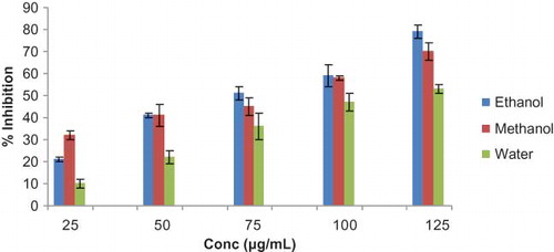
A pilot study was carried out by using the extracts of A. fertilisima in different organic solvents. Among the extracts, extract with ethanol showed maximum antioxidant activity (see ), which was selected for the studies.
Figure 1. Percent superoxide radical scavenging activity of ethanol, methanol, and water extracts of A. fertilisima. Values are mean ± SD of five in vitro experiments.
DPPH Radical Scavenging Assay
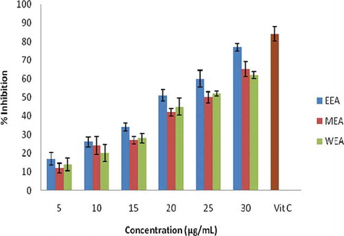
For DPPH radical scavenging assay, shows that the cyanobacteria extract showed a significant effect. At 30 μg/mL, the scavenging abilities on DPPH radicals were 77.14%, 65.23%, and 62.75% for the ethanol, methanol, and water extracts, respectively. l-ascorbic acid was used as positive control and showed 82% inhibition.
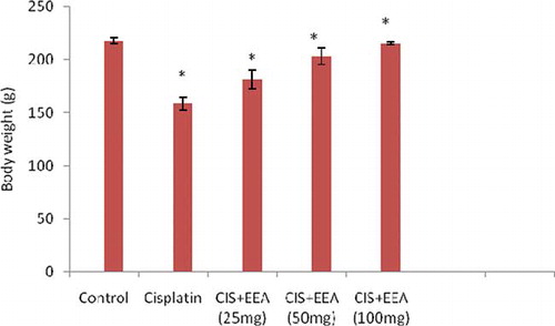
Figure 2. Effect of EEA (50 and 100 mg kg−1 body wt) in development of body weight six days after the administration of cisplatin. Each value represents mean ± SD of six animals. *Statistically significant when compared to control (p < 0.01).
Effect of EEA on Cisplatin-Induced Changes on Body Weight
The results showed that differences in body weight among the groups that received only CIS compared to the normal group of animals (p < 0.01; see ). At the end of the experimental period, the development of body weight was depressed in all animals treated with cisplatin. This reduction was statistically significant when compared to the control (p < 0.01). Decrease in body weight in cisplatin-treated rats was changed with the administration of different doses of EEA.
Figure 3. Percent DPPH radical scavenging capacity of ethanol, methanol, and water extracts of A. fertilisima. Values are mean ± SD of five in vitro experiments.
Effect of EEA on Cisplatin-Induced Nitrosative Stress
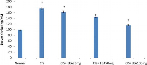
Serum nitrite levels were significantly (p < 0.01) elevated by CIS administration. EEA treatment significantly and dose-dependently improved this increase in serum nitrite levels (see ).
Figure 4. Effect of different doses of EEA in serum nitrite in cisplatin-treated rats. Values are mean ± SD of six animals in each group. Statistical analysis ANOVA followed by Dunnett t-test. *p < 0.01 as compared with group 1, †p < 0.01 as compared with group 2.
Effect of EEA on Cisplatin-Induced Renal Dysfunction
Single administration of CIS significantly (p < 0.01) increased levels of the serum creatinine and blood urea nitrogen (BUN). Chronic EEA treatment significantly and dose-dependently prevented the rise in BUN and serum creatinine (see ). Moreover, the creatinine and urea clearance, which was markedly reduced by CIS administration, was improved dose-dependently by EEA treatment.
Table 1 Effect of ethanol extract of A. fertilisima on CIS-induced nephrotoxicity in rats
Effect of EEA on Cisplatin-Induced Lipid Peroxidation and Changes in Antioxidant Profile
A pilot study was carried out using the extracts of A. fertilisima in different organic solvents. Among the extracts, extract with ethanol showed maximum antioxidant activity (see ), which was selected for the studies. TBARS and CD levels were increased significantly by CIS administration compared with Normal control group of animals. Treatment with EEA produced a significant (p < 0.01) reduction in TBARS and CD in CIS-treated animals (see ). Antioxidant enzyme activities of kidney are presented in . SOD, CAT, GST, and GPX activities were reduced significantly (p < 0.01) in the CIS-intoxicated rats, when compared with normal rats. In CIS+EEA-treated rats, the activities of these enzymes attained a near normalcy. The effect of EEA seems to be dose dependent. Conversely, GSH content in the kidney of group 2 animals showed a significant (p < 0.01) decline when compared with group 1. But in all other group of animals, GSH content was found to attain near normalcy.
Table 2 Effect of ethanol extract of A. fertilisima on lipid peroxidation in CIS-treated rats
Table 3 Effect of ethanol extract of Aulosira fertilisima on activity of antioxidant enzymes in kidney of CIS treated animals
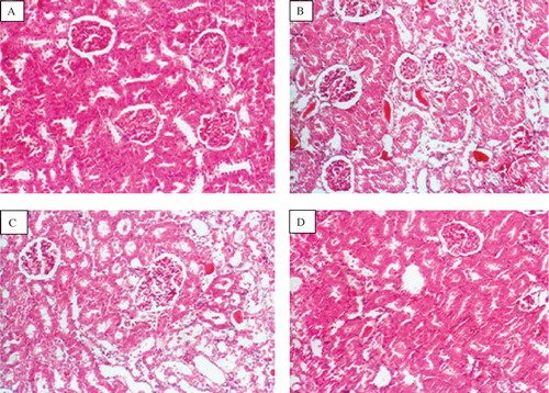
Effect of EEA on Cisplatin-Induced Changes on Renal Morphology
The histological changes in kidneys were evaluated as described in material and methods and results are presented in . Treatment with CIS caused a marked necrosis in proximal tubules (+++) and karyomegaly, hyaline casts in tubular lumen, desquamation, and parenchyma degeneration of the tubular epithelial cells and interstitial nephritis (see ) as compared with normal kidney (see ). Treatment with the EEA decreased the CIS-induced tubular necrosis (see and ).
Table 4 Effect of A. fertilisima treatment on morphological changes as assessed by histopathological examination of kidney in CIS-treated rats
Figure 5. Photomicrographs of kidney sections of rat stained with haematoxylin and eosin (×100). (A) Haematoxylin- and eosin-stained sections of normal rat kidneys. (B) Kidney section of CIS-treated rats showing tubular brush border loss and necrosis of epithelium. (C) Kidney section of CIS + EEA, 50 mg-treated rats showing prevention of CIS-induced alterations. (D) Kidney section of CIS + EEA, 100 mg-treated rats showing prevention of CIS-induced alterations.
DISCUSSION
Different strategies have been proposed to inhibit CIN. The development of therapies to prevent the action or generation of free radicals may influence the progression of oxidative renal damage, along with the appearance of cisplatin-induced acute renal damage. Diet-controlled natural antioxidants such as phycocyanin can be easily and safety increased in the tissues by dietary supplementation. In this study, we investigated the effects of different concentrations of EEA on cisplatin-treated adult rats. Choie et al.Citation[28] observed that cisplatin caused necrosis in the proximal and distal tubes in rat kidneys, with a maximum peak of lesions observed six days after cisplatin injection. In the present study, the animals were killed on the 6th day after the administration of EEA and/or cisplatin. During the experiment, the animals treated with this antitumoral showed a significant reduction in body weight when compared with the control. This weight loss, already reported by other authors, may be due to the gastrointestinal toxicity. Matsushima et al.Citation[29] observed that cisplatin injection caused an increase in urine volume and induced a significant decrease in body weight compared with control rats. The present study showed that the administration of EEA inhibited loss of body weight induced by this antitumoral drug. Even though the exact mechanism of superoxide radical scavenging activity of the extract is not clearly understood, it might be due to the direct scavenging of superoxide anion generated from photo illumination of riboflavin.
Ample experimental and epidemiological studies support the involvement of oxidative stress in the pathogenesis and progression of several chronic diseases.Citation[30] It has been reported that CIS nephrotoxicity is associated with increased lipid peroxidation in renal cortical slices and that antioxidants reverse CIS-induced lipid peroxidation.Citation[31] In the present study, we found that EEA could effectively scavenge the free radicals in a dose-dependent manner. In the present study, elevated level of TBARS and CD observed in CIS-treated rats indicate excessive formation of free radicals and activation of lipid peroxidation system, resulting in kidney damage. The significant decline in the concentration of these constituents in the kidney of CIS+EEA-treated rats indicates anti-lipid peroxidative effect of A. fertilisima. Antioxidant property of Aulosira fertilisima and Nostoc sphaeroides on CCl4-induced hepatic damage in rats had been reported from earlier works conducted in our laboratory.Citation[32,Citation33]. It has been reported that Spirulina fusiformis effectively inhibited CIS-induced lipid peroxidation in rat kidney in vivo.Citation[16] The effect of cisplatin administration on renal excretion, glutathione depletion, increase in substances reactive to thiobarbituric acid, and other parameters analyzed in the present investigation is in accordance with literature data.Citation[34] Lipid peroxidation constitutes a complex chain reaction of free radicals, which leads to a degradation of polyunsaturated fatty acid in cell membranes.Citation[35] In the present investigation, the increase in lipid peroxidation induced by cisplatin was inhibited by the prior administration of EEA. The ability of EEA to significantly decrease renal lipid peroxidation caused by cisplatin supports the idea that supplementation with dietary antioxidants could maintain the integrity of lipids in kidney tissue under oxidative stress induced during chemotherapy. The changes in renal function observed in the rat system correlate well with the nephrotoxic effects of cisplatin in man.Citation[36]
One of the most important intracellular antioxidant systems is the glutathione redox cycle. Glutathione is one of the essential compounds for maintaining cell integrity because of its reducing properties and participation in the cell metabolism.Citation[37] The exact mechanisms of the cisplatin-induced changes in renal glutathione concentrations are not completely elucidated. Thus, glutathione may modulate metal reduction, and the thiol portion is very reactive with several chemical compounds, mainly with alkylating agents such as cisplatin. The depletion of levels of renal glutathione has been observed in rats in response to oxidative stress caused by cisplatin six days after its administration. Although the interaction of cisplatin with the enzymes that contain sulfhydryl groups is fairly well known, some investigations have shown that the nephrotoxicity of cisplatin is not necessarily associated with depletion in renal glutathione content. Results of Braunlich et al.Citation[38] showed that the kidney damage caused by cisplatin is not associated with decreased in renal glutathione. Tian et al.Citation[39] suggested that under oxidative stress conditions or oscillations in the glutathione level, there may occur positive regulation in the biosynthesis of glutathione, contributing to the increase in its intracellular contents. The glutathione uptake appears to be the primary mechanism in tubular cells to maintain intracellular thiol redox status.Citation[40] On the other hand, in the animals that were killed seven days after cisplatin administration, it was observed that the treatment with EEA prevented the depletion of renal glutathione caused by cisplatin, resulting in values close to those observed in the control group. These results suggest the possible involvement of EEA-mediated protection against cisplatin-induced depletion of renal glutathione.
The body has an effective mechanism to prevent and neutralize the free radical-induced damage. This is accomplished by a set of endogenous antioxidant enzymes, such as SOD, CAT, GPX, and GST. When the balance between ROS production and antioxidant defenses is lost, oxidative stress results, which through a series of events deregulates the cellular functions leading to various pathological conditions.Citation[41] Nevertheless, the expression of superoxide dismutase and glutathione peroxidase genes is down-regulated by CIS.Citation[42] Any compound, natural or synthetic, with antioxidant properties might contribute toward the partial or total alleviation of this type of damage. In the present study, decline in the level of antioxidant enzymes like SOD, CAT, GPX, and GST observed in the CIS-treated rat is a clear manifestation of excessive formation of free radicals and activation of lipid peroxidation system resulting in tissue damage. The significant increase (p < 0.01) in the concentration of these constituents in the kidney of CIS+EEA-treated animals indicates an antioxidant effect of EEA. The antioxidant activity (in vitro) of Nostoc sphaeroides and Spirulina maxima has been reported.Citation[43,Citation44] It was found that Dunaliella salina, a green marine algae, has the ability to protect against oxidative stress in vivo using animal models.Citation[15] It has been established that carotenoids from microalgae exert their action against lipid peroxidation, either through decreased production of free radical derivatives or due to the antioxidant activity of the protective agent itself.Citation[45]
Alterations in values of creatinine clearance and serum creatinine levels observed in the treatment with cisplatin are taken as indications of an abnormal glomerular function.Citation[46] EEA pre-treatment inhibited the enhancement of serum creatinine and the reduction of creatinine clearance induced by cisplatin. EEA, as an antioxidant agent, may have inhibited the chain reactions of the cisplatin-generated free radicals or scavenged the free radicals before they reached the cell targets, damaging the glomerular kidney function. Under the experimental condition of the present study, EEA showed protection in a dose-dependent manner on cisplatin-induced oxidative damage on adult rat kidneys. Experimental evidence has suggested that CIS deteriorates renal functions in a dose-dependent manner.Citation[47] Glomerular filtration rate (GFR) of both kidneys represents the integration of GFR in all functioning nephrons. The cause of the decrease in glomerular filtration is afferent vasoconstriction and possibly an altered ultra filtration coefficient.Citation[48] At the single nephron level, ROS may decrease effective GFR by adversely affecting the determinants of single nephron glomerular filtration rate (SNGFR) and by predisposing to back leak of ultrafiltrate across the tubular epithelium. Thus, ROS may contribute to the reduction of GFR in acute tubular necrosis (ATN) by adversely affecting the determinants of glomerular hemodynamics, by injuring tubular epithelial cells, and by provoking tubular cast formation.Citation[49]
In the present study, the single administration of CIS caused marked renal dysfunction, as evidenced by decreased creatinine and urea clearance. CIS predictably lowers GFR in a dose-dependent manner, the cause for which is afferent vasoconstriction and possibly an altered ultrafiltration coefficient.Citation[48] Results of the present study showed that EEA significantly improved creatinine and BUN. It may be possible that A. fertilisima, due to its potential antioxidant properties, improved renal function via attenuating oxidative stress-mediated decline in GFR and renal hemodynamics. Our previous studies have reported that ethanol extract of Aphanizomenon flos-aquae showed significant antioxidant and renoprotective effect on CIN.Citation[50] The vasodilating property of A. fertilisima on rat aortic rings possibly depends on a cyclo-oxygenase-dependent product of arachidonic acid and nitric oxide.
Nitrite, the stable end product of nitric oxide metabolism, reacts with superoxide radicals, which finally leads to nitrosative stress. Large amounts of nitric oxide can lead to the depletion of cellular ATP, which can in activate enzymes that contain iron-sulphur clusters, such as the TCA cycle enzyme aconitase, and enzymes involved in mitochondrial electron transport. Nitric oxide damages DNA, and this in turn stimulates the DNA repair enzyme poly-ADP-ribose synthetase, which further exacerbates ATP depletion and reduces cellular levels of NAD.Citation[51] By depleting ATP and promoting ADP ribosylation, nitric oxide may impair cytoskeleton integrity. In the present study, CIS produced severe nitrosative stress as assessed by marked increase in serum nitrite levels, which was significantly and dose-dependently attenuated by EEA, probably due to the inhibition of inducible nitric oxide synthase.
It has been reported that ROS may lead to tubular damage in CIS-treated rats. ROS can induce either sub-lethal or lethal cell injury, which culminates in either necrosis or apoptosis. The kidneys of CIS-treated rats showed marked histological alterations, especially in the outer cortex and medullary region of the kidneys, compared with kidneys of normal control group. EEA significantly regressed these structural changes in the kidney, suggesting possible involvement of ROS in mediating these histological alterations. A beneficial effect on kidney function was found when adult rats treated with cisplatin received different amounts of EEA. Under the experimental condition of the present study, EEA showed protection in a dose-dependent manner on cisplatin-induced oxidative damage on adult rat kidneys. The renoprotective effect of EEA may be due to the presence of phycocyanin pigment present in EEA. In this regard, it is relevant to point out that phycocyanin has been suggested to act as an antioxidant and exert its antioxidant activity by scavenging lipid peroxidation. Thus, the plausible mechanism of the renoprotective effect of EEA may be due to its antioxidant effect. Further study is needed to identify and isolate the active principle of EEA, which can have offer antioxidant and renoprotective properties.
ACKNOWLEDGMENT
We owe our thanks to Professor Ramesh, Department of Statistics, Rubber Research Institute, Kottayam, India, for giving valuable support for statistical analysis. Financial assistance from University Grand Commission, India, is acknowledged.
The authors report no conflicts of interest. The authors alone are responsible for the content and writing of the paper.
Related Research Data
REFERENCES
- Rosenberg B. Fundamental studies with cisplatin. Cancer. 1985;55:2303–2315.
- Via LD, Noto VD, Vidali M, Scomazzon F, Ni D, Deana R. Action of antitumoral platinum complexes on in vitro platelet functions. Chem-Biol Interact. 1998;110:203–220.
- Meyer KB, Madias ME. Cisplatin nephrotoxicity. Miner Electrolyte Metab. 1994;20:201–213.
- Kersten L, Braunlich H, Keppler BK, Gliesing C, Wendelin M, Westphal J. Comparative nephrotoxicity of some antitumor-active platinum and ruthenium complexes in rats. J Appl Toxicol. 1998;18:93–101.
- Liu J, Liu Y, Habeebu SM, Klaassen CD. Metal-lothionein MT. Null mice are sensitive to cisplatin-induced hepatotoxicity. Toxicol Appl Pharmacol. 1998;149:24–31.
- Sadzuka Y, Shoji T, Takino Y. Effect of cisplatin on the activities of enzymes which protect against lipid peroxidation. Biochem Pharmacol. 1992;43:1873–1875.
- Chu HJ, Tsang CT. Research and utilization of cyanobacteria in China: A report. Arch Hydrobiol Suppl.1988;80:1–6.
- Romay C, Armesto J, Remirez D, Gonzalez R, Ledon L, Garcia I. Antioxidant and anti-inflammatory properties of C-phycocyanin from blue-green algae. Inflamm Res. 1998;47: 36–43.
- Bhat VB, Madyastha KM. C-phycocyanin: A potent peroxyl radical scavenger in vivo and in vitro. Biochem Biophys Res Commun. 2000;275:20–25.
- Romay C, Gonzalez R. Phycocyanin is an antioxidant protector of human erythrocytes against lysis by peroxyl radicals. J Pharm Pharmacol. 2000;52:367–368.
- Bhat VB, Romay C, Armesto J, Remirez D, Gonzalez R, Ledon N, Garcia I. Antioxidant and anti-inflammatory properties of C-phycocyanin from blue-green algae. Inflamm Res. 1998;47:36–41.
- Reddy CM, Bhat VB, Kiranmai G, Reddy MN, Reddanna P, Madyastha KM. Selective inhibition of cyclooxygenase-2 by C-phycocyanin, a biliprotein from Spirulina platensis. Biochem Biophys Res Commun. 2000;277:599–603.
- Stainer RY, Kunisawa R, Mandel M, Cohen Bazire G. Purification and properties of unicellular cyanobacteria (Order: Chroococcales). Bacteriol Rev. 1971;35:171–176.
- Rippka R, Deruelles J, Waterbury JB, Herdeman M, Stainer RY. Generic assignments, strain histories and properties of pure cultures of cyanobacteria. J Gen Microbiol. 1979;111:1–7.
- Chidambara Murthy KN, Rajesha J, Mahadeva Swamy M, Ravishankar GA. Comparative evaluation of hepatoprotective activity of carotenoids of microalgae. J Med Food. 2005;8:523–527.
- Anurag K, Naveen T, Sangeeta P, Kanwaljit C. Renoprotective effect of Spirulina fusiformis on cisplatin-induced oxidative stress and renal dysfunction in rats. Ren Fail. 2006; 28:247–253.
- McCord JM, Fridovich I. Superoxide dismutase, an enzymatic function for erythrocuprein. J Biol. Chem.1969;224: 6049–6054.
- Nichans WG, Samuelson B. Formation of MDA from phospholipids arachidonate during microsomal lipid peroxidation. Eur J Biochem. 1968;6:126–132.
- Beuje JA, Aust SD. Antioxidants. In: Clowick SP, Kaplan NO ( eds.). Methods in enzymology. New York: Academic Press; 1978:302–305.
- Kakkar P, Das B, Vishwanath DN. A modified spectro-photometric assay of superoxide dismutase. Indian J Biochem Biophys. 1984;21:130–134.
- Calibrone AL. Handbook of methods for oxygen radical research. Boca Raton, Fla.: CRC Press;1985:283–287.
- Bentler E, Kelly BM. The effects of sodium nitrate on red cell glutathione. Experientia. 1963;19:96–97.
- Rotruck JT, Pope AL, Gantter HE. Selenium: Biochemical roles as a component of glutathione peroxidase. Science. 1979;186:588–593.
- Beutler E. Red cell metabolism: A manual of biochemical methods. New York: Gruene FP Stratton;1986:57–59.
- Green LC, Wagner DA, Glogowski J, Skipper PL. Analysis of nitrate, nitrite and [15N] nitrate in biological fluids. Anal Biochem. 1982;126:131–138.
- Kordali S, Kotan R, Mavi A, Cakir A, Ala A, Yildirim A. Determination of the chemical composition and antioxidant activity of the essential oil of Artemisia dracunculus and of the antifungal and antibacterial activities of Turkish Artemisia absinthium, A. dracunculus, Artemisia santonicum, and Artemisia spicigera essential oils. J Agri Food Chem. 2005;53:9452–9458.
- Girish AS, Sudhir WG, Avinash DK. Evaluation of hepatoprotective effect of Amalkadi ghrita against carbon tetrachloride induced hepatic damage in rats. J Ethnopharmacol. 2004;90:229–335.
- Choie DD, Longnecker DS, Del Campo AA. Acute and chronic cisplatin nephrotoxicity in rats. Lab Int. 1981;44: 397–402.
- Matsushima H, Yonemura K, Ohishi K, Hishida A. The role of oxygen free radicals in cisplatin-induced acute renal failure in rats. J Lab Clin Med. 1998;131:518–526.
- Lynch ED, Gu R, Pierce C, Kil J. Reduction of acute cisplatin ototoxicity and nephrotoxicity in rats by oral administration of allopurinol and ebselen. Hear Res. 2005; 201:81–84.
- Anurag K, Sangeeta P, Sameer S, Naveen T, Kanwaljit C. 6-Gingerol prevents cisplatin-induced acute renal failure in rats. [My paper]Biofactors. 2006;26:189–192.
- Gini KC, Agi TM, Muraleedhara KG. Antioxidant effect of Nostoc sphaeroides against carbon tetrachloride induced liver dysfunction in rats. Indian J Bot Res. 2006;2:83–86.
- Gini KC, Muraleedhara KG. Antioxidant activity of Aulosira fertilisima on CCl4 induced hepatotoxicity in rats. Ind J Exp Biol. 2008;46:52–56.
- Appenroth D, Kersten L, Splinter EK, Winnefeld K. Protective effects of vitamin E and C on cisplatin nephrotoxicity in developing rats. Arch Toxicol. 1997;71:677–683.
- Zhang JG, Lindup WE. Role of mitochondria in cisplatin-induced oxidative damage exhibited by rat renal cortical slides. Biochem Pharmacol. 1993;45:2215–2222.
- Segelov E, Mann G, DeFazio A, Harnett PR. Mechanisms determining sensitivity to cisplatin in three mutant Chinese hamster ovary cell lines. Mutat Res. 1998;407:243–252.
- Campos R, Garrido A, Valenzuela A, Guerra R. Silybin dihemisuccinate protects against glutathione depletion and lipid peroxidation induced by acetaminophen on rat liver. Planta Med. 1989;55:417–421.
- Braunlich H, Appenroth D, Fleck C. Protective effects of methimazole against cisplatin-induced nephrotoxicity in rats. J Appl Toxicol. 1996;17:41–45.
- Tian L, Shi MM, Forman HJ. Increased transcription of the regulatory subunit of gamma glutamylcysteine synthetase in rat lung ephitelial L2 cells exposed to oxidative stress or glutathione depletion. Arch Biochem Biophys. 1997;342:126–133.
- Weijl NI, Cleton FJ, Osanto S. Free radicals and antioxidants in chemoterapy-induced toxicity. Cancer Treat Res. 1997; 23:209–240.
- Castro JA, Ferrya GC, Castro CR, Sasamett OM, Gillette JR. Prevention of carbon tetrachloride-induced necrosis by inhibitors of drug metabolism. Further studies on the mechanism of their action. Biochem Pharmacol. 1974;23:295–301.
- Ferrari R, Ceconi C, Curello S, Cargnoni A, Paini E, Visloli O. The occurrence of oxidative stress during reperfusion in experimental animals and men. Cardiovasc Ther. 1991;52:77–82.
- Gini KC, Muraleedhara KG. Antioxidant activity (in vitro) of Nostoc sphaeroides. Ind J Bot Res. 2008;4:54–59.
- Miranda MS, Cintra RG, Barros SM, Mancini-Filho J. Antioxidant activity of the microalga Spirulina maxima. Braz J Med Biol Res.1998;31:1075–1081.
- Kerfeld CA. Structure and function of water soluble carotenoid-binding protein of cyanobacteria. Photosyn Res. 2004; 81:215–219.
- Nagai J, Takano M. Molecular aspects of renal handling of amino glycosides and strategies for preventing the nephrotoxicity. Drug Metab Pharmacokinet. 2004;19: 159–163.
- Taguchi T. Cisplatin-associated nephrotoxicity and pathological events. Contrib Nephrol. 2005;148:107–111.
- Rossert J. Drug-induced acute interstitial nephritis. Kidney Int. 2001;60:804–809.
- Daugaard G, Abildggaad U. Cisplatin nephrotoxicity. Cancer Chemother Pharmacol. 1989;25:1–6.
- Gini KC, Muraleedhara KG. Evaluation of renoprotective effect of Aphanizomenon flos-aquae on cisplatin induced renal dysfunction in rats. Ren Fail. 2008;30:717–725.
- Rao M, Rao MN. Protective effects of cystone, a polyherbal ayurvedic preparation, on cisplatin induced renal toxicity in rats. J Ethnopharmacol. 1998;62:1–6.