Abstract
Introduction: Preimplantation biopsy provides a window on the state of the renal allograft. In this study, the prognostic value of frozen section preimplantation graft biopsy was estimated and compared to regularly processed formalin-fixed biopsy. Materials and Methods: Seventy-four renal allograft recipients were studied. The degree of glomerulosclerosis, acute tubular necrosis, interstitial fibrosis, arteriosclerosis, and arteriolosclerosis was rapidly estimated in frozen sections and correlated to the renal function in the immediate posttransplantation period and 3 months thereafter. The histological changes were also examined in paraffin-embedded sections. Results: The histological changes observed in rapidly processed frozen sections were comparable to those observed on regularly processed sections and their differences did not reach statistical significance. Glomerulosclerosis and arteriolosclerosis were underestimated, whereas acute tubular necrosis and interstitial fibrosis were overestimated, in the frozen sections compared to permanent ones, but those differences were not statistically significant. Immediate graft function was observed in 45 patients (61%). Delayed graft function was more frequently observed among recipients with donor age above 60 years (57% vs. 32%). Serum creatinine 3 months after transplantation was above 2 mg/dL in 33 recipients (44.5%) and was positively correlated to the degree of tubular necrosis (p = 0.04) and donor age (p = 0.03). Donor age was correlated to the degree of arteriolosclerosis (p < 0.01). Conclusions: Frozen section preimplantation biopsy gives reliable information for the situation of the graft that is related to the outcome of renal transplantation.
INTRODUCTION
Kidney transplantation represents the ideal treatment of patients with end-stage renal failure. Although the short-term renal allograft survival has been improved with the development of new immunosuppressive drugs, the long-term outcome remains unsatisfactory.Citation1,Citation2 Several immunological and nonimmunological factors have been identified as predictive. Advanced donor age, time of cold and warm ischemia, and presence of delayed graft function are considered important determinants of the long-term graft survival.Citation2,Citation3 The widening gap between the number of renal transplants performed and the number of patients waiting for transplantation makes the expansion of potential donor pool necessary.Citation4,Citation5 An increasing number of kidneys are recovered from marginal donors (age > 60 years old, history of hypertension or diabetes), whereas dual kidney transplantation programs are also ongoing.Citation5–7 However, problems with the quality and size of the kidney or with the age of the donor are the most common reasons for discarding kidneys offered for transplantation.Citation5 Such marginal kidneys having fewer functional nephrons initiate a self-perpetuating process of progressive renal function deterioration, similar to that observed in animal models in which renal mass is reduced surgically.Citation5
Preimplantation biopsies may be an important tool in donor selection since pathologic examination of the kidney tissue provides an objective window on the state of the kidney that cannot be deduced from clinical records and renal function tests.Citation4 The severity of histological lesions, estimated semiquantitatively on preimplantation biopsies, correlates with the incidence of delayed graft function and long-term allograft function and survival.Citation8–10 Chronic tubulointerstitial damage, arteriolar hyaline changes, fibrous intimal thickening, and percentage of globally sclerosed glomeruli represent the main histological lesions associated with graft outcome.Citation9,Citation10 In addition to the estimation of the state of the implanted graft, a preimplantation biopsy serves as a baseline for comparison with future biopsies that might be performed to identify chronic allograft nephropathy or calcineurin inhibitor (CNI) toxicity.Citation4 Chronic allograft nephropathy causes most kidney allograft losses and, despite improvement in immunosuppression, remains the central clinical challenge.Citation11 Protocol biopsies show that allograft damage is common, time dependent, and progressive, whereas a preimplantation biopsy represents the basis for this estimation.Citation11,Citation12 However, the regular processing of paraffin-embedded sections is time consuming, whereas rapid processing of frozen sections can be more easily performed. In this study, the prognostic value of frozen section preimplantation biopsy to the immediate and long-term renal allograft function is estimated and compared to that of regularly processed biopsy.
MATERIALS AND METHODS
Patients
Seventy-four renal allograft recipients (49 males and 25 females) aged 44 ± 13 years old who underwent a preimplantation graft biopsy were included in the study. Out of the 74 donors, 10 were living and 64 cadaveric. The mean age of the donors was 49 ± 17 years old, whereas 53 were below and 21 were above 60 years. Thirteen donors were between 50 and 59 years and 6 of 13 fulfilled the criteria to be classified as expanded criteria donors.Citation6 The estimated glomerular filtration rate (eGFR) was 65 ± 15 mL/min for donors above 50 years and 73 ± 12 mL/min for donors below 50 years. The mean time of cold ischemia was 20 ± 7 hours. The immunosuppressive regimen used in all patients was induction therapy with basiliximab and combination of methylprednisolone, (CNI) [cyclosporine (target trough blood levels of 150–200 ng/mL) or tacrolimus (target trough levels 8–10 ng/mL)], and mycophenolate mofetil (2 g/day). In recipients with severe acute tubular injury, the dose of (CNI) was lower targeting to the lower end of the previously reported trough levels. No rejection episodes, infections, or other complications that could potentially affect renal function were observed in patients included in the study.
Graft biopsy and histological evaluation
A needle biopsy (16 G) was obtained at the ‘back table’ during the preparation of the graft for implantation at the end of the cold ischemia time. We chose to perform a needle biopsy to provide assurance that the deep cortex has been adequately sampled. After tissue was obtained, a frozen section was performed, stained with a rapid hematoxylin and eosin (H&E) protocol. At this point, we should explain why we chose to perform a frozen section and we did not use a rapid processing paraffin protocol of 2-hour duration. The latter requires a technologist to be on call 24 hours a day, 7 days a week.Citation8 In our institution in the vast majority (98%) of the cases, the allograft biopsy procedure was performed between 9.00 pm and 3.00 am. Since the transplant surgeons needed to be aware of the allograft status and no technician is on call during those hours to utilize the rapid paraffin process, we chose to perform a frozen section.
On each frozen section biopsy, the following parameters were evaluated over 1 hour from the time of biopsy performance: number of glomeruli included in the biopsy and percentage of sclerotic (partially or totally) glomeruli, degree of acute tubular necrosis, interstitial fibrosis, and inflammation as well as presence of arteriosclerosis or arteriolosclerosis.
Thereafter, renal tissues were fixed in 10% neutral formalin for regular processing purposes: tissues were embedded in paraffin and 4-μm thick sections were obtained. In each case, the following stains were performed: H&E, periodic acid Schiff (PAS), Masson trichrome, and methenamine silver trichrome (MST). In these biopsies, the percentage of globally sclerosed glomeruli was counted and the severity of acute tubular injury and interstitial fibrosis was evaluated according to previously published protocolsCitation13 and quantitated as mild (affecting <25% of cortical area), moderate (25–50%), and severe (>50% of cortical area). As arteriolosclerosis or arteriosclerosis was considered the presence or absence of fibrous hyperplasia of the vascular intima,Citation14 arteriosclerosis or arteriolosclerosis was recorded as absent or present according to the ratio of wall thickness/arterial lumen diameter (absent: ratio <0.5, present: ratio >0.5).Citation15 Arterial hyalinosis (PAS-positive hyaline thickening in arteriolar wallCitation13) was identified only in the permanent sections and not during the frozen section biopsy procedure.
Estimation of the prognostic value of the rapidly processed preimplantation graft biopsy
The histological lesions observed in the fresh frozen sections of preimplantation graft biopsy were compared to those observed in the regularly processed formalin-fixed and paraffin-embedded sections. The relation of the severity of histological lesions observed in the frozen sections with the renal function in the immediate posttransplantation period and 3 months thereafter, a period necessary for stabilization of renal function, as well as with other clinical parameters such as donor age and time of cold ischemia was also investigated.
Statistical analysis
Continuous variables are described by their mean value and standard deviation (SD), whereas categorical variables are described by the frequencies of their respective categories. The chi-square test is used to examine the independence of two categorical variables. A p-value of less than 5% (p < 0.05) is considered as statistically significant. All aforementioned analyses were carried out using the SPSS® for Windows, version 14.
RESULTS
Histological lesions in frozen sections and regularly processed graft biopsies
shows the pathologic features evaluated in graft biopsies in frozen sections and regularly processed specimens. The percentage of globally sclerosed glomeruli in the transplanted kidneys ranged from 0 to 18%. Frozen section preimplantation biopsies showed absence of globally sclerosed glomeruli in 65 out of 74 grafts (88%) and presence of glomerulosclerosis in less than 20% of the glomeruli in nine grafts (12%). Mild acute tubular necrosis (affecting <25% of cortical area) was observed in 35 grafts (47%) and moderate to severe (affecting >25% of cortical area) in 39 (53%). As shows, the degree of acute tubular necrosis was overestimated in the frozen sections compared to permanent ones. Arteriolosclerosis was present in 19 (26%) and absent in 55 (74%) grafts.
TABLE 1. Pathologic features of grafts in frozen section and regularly processed specimens
Mild chronic inflammation was present in 3 of 74 biopsies. Mild interstitial fibrosis was present in 12 of 74 biopsies. Arterial branches were included in 6 of 74 biopsy specimens and arteriosclerosis was present in 2 of 6 biopsies. Mild arterial hyalinosis (mild-to-moderate PAS-positive hyaline thickening in at least one arteriole) was identified in 9 of 74 biopsy specimens.
The percentage of glomerulosclerosis was underestimated in the frozen section specimens (compared to regularly processed specimens) in 5 of 74 cases. This could potentially be attributed to the fact that in the regularly processed specimens multiple tissue levels were examined. Similarly, the presence or absence of arteriolosclerosis was underestimated in the frozen section specimens in 6 of 74 cases. On the contrary, interstitial fibrosis and acute tubular necrosis were overestimated in the frozen section specimens in 3 of 74 and 5 of 74 cases, respectively.
In general, as shows, there were no statistically significant differences in the evaluation of pathologic features, when the findings of frozen sections were compared to those of permanent sections.
Renal function after transplantation
Excellent graft function in the immediate posttransplantation period was observed in 45 patients (61%). Delayed graft function was more frequently observed among recipients of kidneys from older donors (above 60 years) (57 vs. 32%).
The renal function, 3 months after transplantation, was stabilized to serum creatinine levels below 2 mg/dL in 41 (55.5%) and above 2 mg/dL in 33 (44.5%) allograft recipients.
Clinical and histological features in frozen section preimplantation graft biopsies related to outcome
Donor age and time of cold ischemia inversely related to the immediate posttransplantation renal function (p < 0.01 and p = 0.05, respectively), whereas donor age, severity of acute tubular necrosis, and presence of arteriolosclerosis in the preimplantation graft biopsies related to the renal function 3 months after transplantation (p < 0.05).
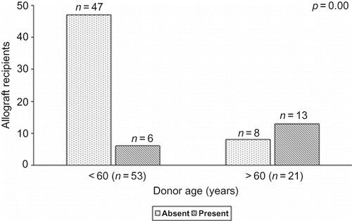
Patients who received grafts from older donors and patients with more severe acute tubular necrosis in the preimplantation biopsies had higher serum creatinine 3 months after transplantation (p = 0.03 and p = 0.04, respectively) ( and ). Presence of arteriolosclerosis, a poor prognostic feature for the long-term outcome of renal transplantation, was more frequently observed in the preimplantation biopsies of kidneys from older donors (p < 0.01) ().
FIGURE 1. Donor age and serum creatinine 3 months after transplantation.
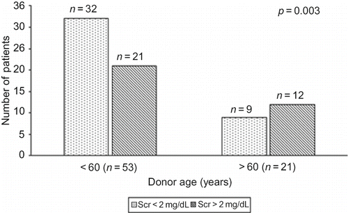
FIGURE 2. Severity of acute tubular necrosis and serum creatinine 3 months after transplantation (Tx).
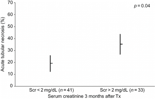
FIGURE 3. Donor age and presence of arteriolosclerosis in preimplantation graft biopsies.
DISCUSSION
In this study, the prognostic value of frozen section preimplantation graft biopsy, in the outcome of renal transplantation, was estimated. Important information of the situation of the implanted kidney concerning the severity of acute tubular necrosis and arteriolosclerosis that was related to the donor age and to the obtained renal function after transplantation was received from the frozen section preimplantation biopsies.
Although many have argued that donor screening should include a pretransplant graft biopsy, estimation of the condition of the graft at the time of implantation is important since histological examination provides more objective data than the renal function tests. The knowledge of the situation of graft at the time of implantation is very crucial, particularly for marginal donors (older individuals or donors with elevated serum creatinine due to prerenal failure or acute tubular necrosis).Citation4 Pretransplant donor biopsy is particularly valuable in older individuals because the rate at which the kidney ages is highly variable and reliable decisions about donor suitability cannot be made on the basis of age alone.Citation4 It was recently shown that 69% of older donors (above 55 years old) had either no glomerulosclerosis at all or less than 10% sclerosed glomeruli and showed excellent graft survival after 4 years of follow-up.Citation16 Furthermore, allocation of the kidneys from older donors, according to the biopsy findings before transplantation as single or dual transplants, has been proven very important for the long-term allograft survival.Citation5,Citation17 In addition, combined evaluation of donor glomerulosclerosis and chronic vascular and interstitial damage allows a precise prediction of graft outcome.Citation10 We noticed delayed graft function more frequently in patients transplanted with kidneys from older donors (above 60 years) with higher incidence of arteriolosclerosis. Renal biopsy findings of donors could be used as a tool to identify high-risk grafts that, although they may have lower half-life, could still be used in patients with an urgent need of transplantation.
It is well known that glomerulosclerosis is a poor prognostic feature for the long-term outcome of renal transplantation, and kidneys with greater than 20% sclerotic glomeruli should not be used.Citation16 However, a large number of glomeruli should be present in the biopsy (at least 15 glomeruli) to identify the degree of glomerulosclerosis with relative accuracy. Tubulointerstitial injury and arteriolosclerosis that are more diffuse lesions can be estimated even in small samples of renal tissue and can give valuable information about the situation of the graft.Citation18 In addition, tubulointerstitial lesions are more widespread within the kidney and thus they are more reproducible in small biopsy samples. Arteriolar hyalinosis has been found to be the best predictor of transplant failure and shows good reproducibility independent of the sample size.Citation18 Similar results were observed in our study where the presence of arteriolosclerosis was related to the long-term outcome of allograft recipients. Thus, it has been proposed that preimplantation biopsies may be an important tool not only in donor selection but also in prediction of recipient outcome.Citation9 The evaluation of interstitial fibrosis, tubular atrophy, and arteriolar hyalinosis in preimplantation biopsies is very important and represents the basis for protocol biopsies performed to estimate the evolution of chronic allograft nephropathy.Citation11 In addition, the estimation of the severity of acute tubular injury caused by cold ischemia is crucial since it can be a marker of the immediate graft function after transplantation.Citation19 The latter was also confirmed in this study where a correlation of the severity of acute tubular necrosis with the short- and long-term outcomes was observed. The estimation of acute tubular injury also contributes to the selection of the suitable immunosuppressive regimen. Thus, in cases with marked acute tubular injury and expected delayed graft function, the use of CNIs is better to be avoided or restricted to lower doses over the immediate posttransplantation period. At this point, we should mention that frozen and formalin-fixed tubules show different morphology. Thus, the overestimation of the degree of acute tubular necrosis that occurred in the current study could be attributed to the aforementioned factor.
A previous study reported that attempts to quantitate interstitial fibrosis or identify mild acute tubular necrosis are frequently beyond the capabilities of the usual frozen tissue section.Citation20 However, as it has been stated earlier, in Starzl Transplantation Institute, transplant surgeons need to know, before graft implantation, the degree of interstitial fibrosis, since they hesitate to use any kidney with more than mild interstitial fibrosis (>25% of cortical area affectedCitation8).
In another studyCitation15, the authors evaluated the impact of glomerulosclerosis, glomerular size and periglomerular fibrosis, vascular pathology with arterial wall-to-lumen ratio, and arteriolar hyalinosis and interstitial pathology with measurement of cumulative fibrosis and presence of scar on donor kidney biopsies. They found that biopsy features independently associated with an increased risk of graft failure were glomerulosclerosis (≥15%), interlobular arterial wall-to-lumen ratio (≥0.5), and the presence of periglomerular fibrosis, arteriolar hyalinosis, or scar. Thus, they developed the Maryland Aggregate Pathology Index based on these parameters. Finally, they concluded that the Maryland Aggregate Pathology Index may help transplant physicians to estimate graft survival from the preimplantation biopsy findings, in clinical situations similar to this study population (cold ischemia over 24 hours, glomerulosclerosis (GS) < 25%).
Regarding arterial hyalinosis, a previous studyCitation15 reported that the presence of any degree of arterial hyalinosis in pretransplant kidney biopsies carried an increased risk for graft loss by 293% [heart rate (HR) = 3.93, 95% confidence interval (CI) = 2.02–7.64, p ≤ 0.0001]. The 5-year graft survival was 48% for presence and 85% for absence of arteriolar hyalinosis (p < 0.0001). Also another previous study showed that arteriolar hyalinosis was the best pathologic predictor of transplant failure.Citation18 However, other studies reported that there was no correlation of graft function and the presence of arterial hyalinosis in the pretransplant graft biopsies.Citation19 Furthermore, another study showed that arterial hyalinosis is more pronounced in pretransplant kidney biopsies compared to posttransplant ones. It was suggested that this difference resulted from a propensity of vascular lesions to affect deeper vessels, which are more likely to be sampled when a biopsy gun is pointed directly at the surface of a donor kidney.Citation21 In this study, only mild arterial hyalinosis (mild-to-moderate PAS-positive hyaline thickening in at least one arteriole) was identified in 9 of 74 biopsy specimens. No biopsy showed moderate (moderate-to-severe PAS-positive hyaline thickening in more than one arteriole) or severe (severe PAS-positive hyaline thickening in many arterioles).Citation13 In addition, the presence of mild arterial hyalinosis in those nine biopsies did not affect the graft function. We tend to believe that mild arterial hyalinosis is unlikely to contribute significantly to graft dysfunction.
In this study, needle-obtained biopsies were used to provide assurance that the deep cortex has been adequately sampled. We did not use wedge biopsies because those biopsies may not include juxtamedullary renal tissue that contains the larger arteries, more likely to display the arteriosclerosis of aging. A wedge biopsy of less than 30 glomeruli may not be fully representative since globally sclerotic glomeruli caused by arteriosclerosis tend to be clustered in the subcapsular area of the cortex and are often overrepresented in superficial wedge biopsies.Citation4 On the other hand, arteriosclerosis is by nature patchy in distribution and so may be under- or overrepresented in a small biopsy. For this reason, some centers advocate performance of a needle biopsy as well as a wedge biopsy to minimize sampling error and to allow assessment of both juxtamedullary and outer cortex.Citation4 Another important issue is the processing of the urgent preimplantation biopsy. Renal morphology after frozen section process is adequate to demonstrate glomerulosclerosis, advanced interstitial fibrosis, and arteriolosclerosis in the donor kidney.
In conclusion, a preimplantation graft biopsy gives important information for the state of the transplanted kidney that is related to the outcome. A frozen section preimplantation biopsy can give reliable information for the situation of the renal allograft and can be performed as a routine.
Declaration of interest: The authors report no conflicts of interest. The authors alone are responsible for the content and writing of the paper.
REFERENCES
- Pascual M, Theruvath T, Kawai T, Tolkoff-Rubin N, Cosimi B. Medical progress: Strategies to improve long-term outcomes after renal transplantation. N Engl J Med. 2002;346(8):580.
- Afzali B, Taylor AL, Goldsmith JA. What we can do about chronic allograft nephropathy: Role of immunosuppressive modulations. Kidney Int. 2005;68:2429.
- Chapman JR, O’Connell PJ, Nankivell BJ. Chronic renal allograft dysfunction. J Am Soc Nephrol. 2005;16:3015.
- D’Agati VD, Cohen DJ. Pre-implantation renal biopsy: Structure does predict function. Transplantation. 2003;75:264.
- Remuzzi G, Cravedi P, Perna A, Long-term outcome of renal transplantation from older donors. N Engl J Med. 2006;354:343.
- Metzger RA, Delmonico FL, Feng S, Port FK, Wynn JJ, Merion RM. Expanded criteria donors for kidney transplantation. Am J Transplant. 2003;3(Suppl. 4):114.
- Bunnapradist S, Gritsch HA, Peng A, Jordan SC, Cho YW. Dual kidneys from marginal adult donors as a source for cadaveric renal transplantation in the United States. J Am Soc Nephrol. 2003;14:1031.
- Randhawa P. Role of donor kidney biopsies in renal transplantation. Transplantation. 2001;71:1361.
- Malaise J, Cosyns JP, Lallier M, Baseline biopsy for the quality assessment of kidney graft. Curr Opin Organ Transplant. 1999;4:139.
- Lopes JA, Moreso F, Riera L, Evaluation of pre-implantation kidney biopsies: Comparison of Banff criteria to a morphometric approach. Kidney Int. 2005;67:1595.
- Nankivell BJ, Chapman JR. Chronic allograft nephropathy: Current concepts and future directions. Transplantation. 2006;81:643.
- Seron D, Moresco FW. Protocol biopsies and risk factors associated with chronic allograft nephropathy. Transplant Proc. 2002;34:331.
- Racusen L, Solez K, Colvin RB, The Banff 97 working classification of renal allograft pathology. Kidney Int. 1999;55:713.
- Karpinski J, Lajoie G, Cattran D, Outcome of kidney transplantation from high-risk donors is determined by both structure and function. Transplantation. 1999;67:1162.
- Munivenkatappa RB, Schweitzer EJ, Papadimitriou JC, The Maryland aggregate pathology index: A deceased donor kidney biopsy scoring system for predicting graft failure. J Transplant. 2008;8:2316.
- Escofet X, Osman H, Griffiths DF, Woydag S, Jurewicz WA. The presence of glomerular sclerosis at time zero has significant impact on function after cadaveric renal transplantation. Transplantation. 2003;75:344.
- Remuzzi G, Grinyo J, Ruggenenti P, Early experience with dual kidney transplantation in adults expanded donor criteria. J Am Soc Nephrol. 1999;10:2591.
- Wang HJ, Kjellstrand CM, Cockfield SM, Solez K. On the influence of sample size on the prognostic accuracy and reproducibility of renal transplant biopsy. Nephrol Dial Transplant. 1998;13:165.
- Sulikowski T, Tejchman K, Domanski L, Histopathologic evaluation of pre-transplant biopsy as a factor influencing graft function after kidney transplantation: A 1-year observation. Transplant Proc. 2007;39:943.
- Walker PD, Cavallo T, Bonsib SM, Ad Hoc Committee on Renal Biopsy Guidelines of the Renal Pathology Society. Practice guidelines for the renal biopsy. Mod Pathol. 2004; 17:1555.
- Sund S, Reisaeter AV, Fauchald P, Living donor kidney transplants: A biopsy study 1 year after transplantation, compared with baseline changes and correlation to kidney function at 1 and 3 years. Nephrol. Dial. Transplant. 1999;14:2445.