Abstract
Paraneoplastic manifestations in malignant pleural mesothelioma are rare. We report a case of malignant pleural mesothelioma associated with minimal change disease (MCD). A 58-year-old man with occupational exposure to asbestos presented with severe peripheral edema, heavy proteinuria, and acute renal failure shortly after the diagnosis of mesothelioma had been confirmed. The renal biopsy demonstrated MCD. The underlying pathogenesis of this association remains unknown.
INTRODUCTION
Minimal change disease (MCD) has been associated with hematological malignancies such as Hodgkin's disease, non-Hodgkin's lymphoma, and leukemia. However, rare cases of MCD associated with solid tumor have been reported. Solid tumors are more commonly associated with membranous nephropathy.
CASE REPORT
A 58-year-old man presented with a 1-month history of progressive dyspnea and was found to have a right-sided pleural effusion. He was a builder and had been exposed to asbestos. He underwent a thoracotomy and biopsy which revealed epithelial-type diffuse malignant pleural mesothelioma. Two weeks later, he developed peripheral edema, fatigue, worsening dyspnea, and reduction in urine output. He was reviewed on the day of the planned first cycle of chemotherapy. Routine blood tests prior to planned chemotherapy revealed acute renal failure with life-threatening hyperkalemia and he was admitted to the intensive care unit for urgent hemodialysis. Chemotherapy was not administered at this time. On physical examination, he was breathless at rest, apyrexial, and hypertensive (180/90 mmHg), and had widespread edema. He had decreased right chest wall movement with reduced air entry, and basal lung crepitations were audible on the left side.
Laboratory investigations revealed acute renal failure with serum creatinine concentration of 330 μmol/L (60–120 μmol/L) and urea 49 mmol/L (3–8 mmol/L). His renal function had been normal at the time of thoracotomy (creatinine 95 μmol/L). There was laboratory evidence of nephrotic syndrome with serum albumin 15 g/L (33–46 g/L) and urinalysis revealing >3 g/L of proteinuria. A 24 h urinary protein estimation was not done in the setting of oliguric renal failure. Urine microscopy revealed hyaline and granular casts but no dysmorphic red blood cells or red cell casts. He was mildly anemic with hemoglobin of 123 g/L (130–170 g/L) and the liver function tests were normal. Additional laboratory studies revealed normal serum levels of C3 and C4 complement and plasma levels of IgA, IgG, and IgM were within the normal reference range. Antinuclear antibody, dsDNA antibody, antineutrophil cytoplasmic antibody, and antiglomerular basement membrane antibody were all negative.
A chest X-ray showed an extensive right pleural effusion, whereas the left lung field appeared clear. Renal ultrasound showed two normal-sized unobstructed kidneys with preserved corticomedullary differentiation. Duplex ultrasound demonstrated normal renal arterial and venous blood flows.
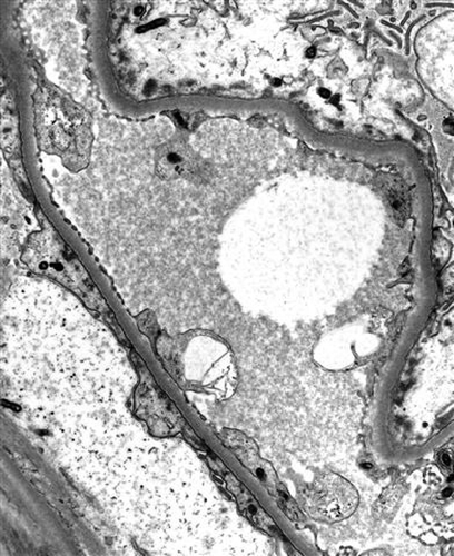
A renal biopsy demonstrated preserved glomeruli with mild mesangial matrix excess on light microscopic examination. There was focal tubular luminal dilatation with variable epithelial flattening. Interstitial edema and patchy interstitial mononuclear cell infiltrate was noted. These features were consistent with acute tubular necrosis. Subtle IgA deposition was seen in the mesangial area with immunofluorescence. Electron microscopy showed diffuse foot process effacement associated with basement membrane thinning, the pattern being characteristic of MCD ().
He received urgent continuous hemodialysis to control his hyperkalemia, uremia, and fluid overload. He was initially treated with dexamethasone 4 mg daily; gemcitabine 1500 mg and carboplatin 150 mg were then administered as specific therapy for the mesothelioma and this led to pancytopenia requiring regular blood and platelet transfusions. A bone marrow examination revealed megaloblastoid changes consistent with myelodysplastic syndrome with no evidence of malignancy. Although the hyperkalemia and edema were controlled, the renal failure and pancytopenia persisted (). His condition continued to deteriorate and he received palliative care with dialysis being discontinued. He died of sepsis 6 weeks after initial presentation.
TABLE 1. Summary of physiological and biochemical parameters during the course of admission
DISCUSSION
Glomerular disease in the setting of malignancy has been recognized for several decades. Most commonly, the glomerular pathology is membranous nephropathy. MCD has been associated with Hodgkin's lymphoma as well as other hematological malignancies and has rarely been associated with carcinomas such as renal cell carcinoma and bronchogenic carcinoma.Citation1,Citation2
Glomerular disease associated with malignant mesothelioma is extremely rare with only seven cases having been reported in the English medical literature ().Citation3–9 Three cases were associated with MCD, two with membranous nephropathy and one each with focal segmental glomerulosclerosis and segmental mesangial proliferative glomerulonephritis. Six of these cases had pleurally based mesothelioma and most were epithelial, with only one having sarcomatoid histology.
TABLE 2. Reported cases of mesothelioma associated with nephrotic syndrome
In most of the cases reported thus far, the nephrotic state either preceded (within 6 months) or occurred close to the time of diagnosis of the malignancy. The diagnosis of paraneoplastic nephrotic syndrome may be considered if the following criteria are present: (1) no evidence of other etiology; (2) nephrotic syndrome develops either 6 months before or after the diagnosis of malignancy; (3) tumor treatment is associated with a decrease in proteinuria; (4) tumor recurrence is associated with an increase in proteinuria.Citation10 The present case fulfills the first two criteria with the patient dying of sepsis shortly after treatment was given. The pathophysiology of the association between MCD and mesothelioma remains unknown. With MCD and Hodgkin's lymphoma, clinical observations and experimental data suggest that both diseases are associated with an expansion of T cells polarized toward a Th2 phenotype.Citation1 However, such an explanation seems unlikely with other malignancies such as mesothelioma. Cytokines, chemokines, and other related factors produced by tumor cells or lymphoid tissues may be implicated. The general inflammation caused by the tumor may evolve into granulomas and aggregated lymphoid tissues with cytokine production.
Mesangial IgA deposition in MCD, as seen in this case, has been reported previously. In one study, IgA deposition was found in 60 of 363 nephrotic patients with MCD (16.8%).Citation11 There has been no agreement on the classification or nomenclature of this entity. It has been suggested that IgA deposition is a risk factor for renal impairment and may indicate a frequently relapsing course.Citation12
Serum creatinine in this reported series () ranged from normal to 330 μmol/L. Acute renal failure in the setting of MCD has been well described. In one study, it occurred in 25% of the adult MCD patients and 4% of cases required dialysis.Citation13 This case is the first to have required dialysis in the setting of renal failure associated with MCD and mesothelioma. The cause of acute renal failure was most likely related to acute tubular necrosis in the setting of MCD. The prognosis here was poor and the requirement for hemodialysis may well have contributed to the patient's limited survival with death occurring because of leucopenic sepsis. In five of the previously reported cases, the proteinuria improved with treatment. This followed chemotherapy and/or corticosteroid treatment and in one of these cases pneumonectomy was also performed. Nevertheless, the mesothelioma did not respond to chemotherapy in these cases and recurrence of mesothelioma occurred following pneumonectomy. It is plausible to suggest that chemotherapy altered the immunologic state responsible for the nephrotic syndrome. The development of nephrotic syndrome in the setting of mesothelioma is associated with poor prognosis. Only one patient in the published literature survived more than 1 year.
In summary, we present a rare case of MCD associated with malignant pleural epithelial mesothelioma. In addition, the MCD not only revealed itself as a nephrotic presentation but was also accompanied by acute renal failure requiring dialysis support. The nephrotic syndrome and acute renal failure contributed significantly to the patient's complexity of care and most likely contributed to his poor outcome. Treatment of the mesothelioma may improve this renal paraneoplastic complication but is unlikely to improve the overall prognosis of mesothelioma.
Declaration of interest: We have had no involvements that might raise the question of bias in the work reported or in the conclusions, implications, or opinions stated. This manuscript has not been published or submitted for publication elsewhere.
REFERENCES
- Audard V, Larousserie F, Grimbert P, Minimal change nephrotic syndrome and classical Hodgkin's lymphoma: Report of 21 cases and review of the literature. Kidney Int. 2006;69:2251–2260.
- Glassock RJ. Secondary minimal change disease. Nephrol Dial Transplant. 2003;18(Suppl. 6):vi52–vi58.
- Schroeter NJ, Rushing DA, Parker JP, Beltaos E. Minimal-change nephrotic syndrome associated with malignant mesothelioma. Arch Intern Med 1986;146:1834–1836.
- Absy M, Gazzawi B, Amoah E. Focal and segmental glomerulosclerosis associated with malignant mesothelioma. Nephron 1992;60:250.
- Tanaka S, Oda H, Satta H, Nephrotic syndrome associated with malignant mesothelioma. Nephron 1994;67:510–511.
- Sakamoto K, Suzuki H, Jojima T. Membranous glomerulonephritis associated with diffuse malignant pleural mesothelioma: Report of a case. Surg Today. 2000;30:1124–1126.
- Galesic K, Bozic B, Heinzi R, Scukanec-Spoljar M, Bozikov V. Pleural mesothelioma and membranous nephropathy. Nephron. 2000;84:71–74.
- Farmer CKT, Goldsmith DJA. Nephrotic syndrome and mesenteric infarction secondary to metastatic mesothelioma. Postgrad Med J. 2001;77:333–334.
- Bacchetta J, Ranchère D, Dijoud F, Droz JP. Mesothelioma of the testis and nephrotic syndrome: A case report. J Med Case Reports. 2009;3:7248.
- Burstein DM, Korbet SM, Schwartz MM. Membranous glomerulonephritis and malignancy. Am J Kidney Dis. 1993; 9:23–26.
- Choi J, Jeong HJ, Lee HY, Significance of mesangial IgA deposition in minimal change nephrotic syndrome: A study of 60 cases. Yonsei Med J. 1990;31:258–263.
- Westhoff TH, Waldherr R, Loddenkemper C, Mesangial IgA deposition in minimal change nephrotic syndrome: Coincidence of different entities or variant of minimal change disease? Clin Nephrol. 2006;65:203–207.
- Waldman M, Crew RJ, Valeri A, Adult minimal-change disease: Clinical characteristics, treatment, and outcomes. Clin J Am Soc Nephrol. 2007;2:445–453.
