Abstract
Obstructive uropathy in sarcoidosis can result from retroperitoneal involvement, retroperitoneal fibrosis (RPF), renal stones, or ureteral involvement. We report acute kidney injury (AKI) from obstructive uropathy because of RPF in a female patient, 2 years following the resolution of pulmonary sarcoidosis. We propose that RPF may occur in sarcoidosis even when a lag of years separates the presentations of the illnesses.
INTRODUCTION
Although two-thirds of the cases are idiopathic,Citation1 retroperitoneal fibrosis (RPF) may result from malignancies, medications, hemorrhage, surgery, infections, irradiation, inflammatory bowel diseases, periarteritis secondary to autoimmune diseases, arteriosclerosis, or abdominal aortic aneurysm.Citation2,Citation3 Genetic and environmental factors may predispose to RPF, as well.Citation3 We report acute kidney injury (AKI) resulting from obstructive uropathy because of RPF, presenting 2 years following the resolution of pulmonary sarcoidosis.
CASE REPORT
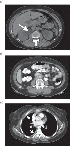
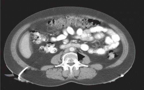
A 49-year-old woman presented with abdominal pain, nausea, and AKI. Before 2 years, she suffered from sarcoidosis manifested as pulmonary infiltrates, for which recovery, assessed both clinically and radiologically, was complete following 6 months of steroid therapy. Her current physical examination was unremarkable and vital signs were normal. A urinary catheter drained 100 mL of clear urine. Blood tests were abnormal for serum creatinine (7.2 mg/dL), blood urea nitrogen (BUN) (63 mg/dL), calcium (7.8 mg/dL), phosphorous (7.9 mg/dL), potassium (6.5 mEq/L), and hemoglobin (10.7 g%). Both ultrasonography and an abdominal CT scan without contrast media demonstrated severe bilateral hydroureteronephrosis with medial deviation of ureters, extrinsically compressed by a retroperitoneal mass at the level of L4 (,b). The serum creatinine normalized following an urgent decompression of the obstruction by the insertion of bilateral percutaneous nephrostomies. A contrast CT scan and an MRI revealed retroperitoneal soft tissue mass, enveloping the great vessels and ureters. No lymphadenopathy was noted. In addition, right pleural thickening that was minimal in a chest CT scan of 2 years earlier () was now prominent. A pleural biopsy performed by thoracoscopy confirmed fibrosis, with no evidence of granulomatous process. Arterial blood gases, serum angiotensin-converting enzyme, and a gallium scan were normal. Chest CT and spirometry did not show lung abnormalities. Bone marrow aspiration and biopsy and purified protein derivative (PPD) skin test were negative. Once RPF was diagnosed and 60 mg of oral prednisone was administered, the patient was discharged. Within 4 months, prednisone was tapered to 5 mg. After 6 months of steroid treatment, poor blood glucose control led to its replacement with 2 mg/kg of daily azathioprine. The latter was later discontinued because of nonadherence, pruritus, and marked elevation of liver enzymes. Both intravenous pyelogram and CT continued to demonstrate the moderate bilateral hydroureteronephrosis and smoothly tapered ureters at the level of L4–L5, although the diameter of the fibrotic plaque decreased from 14 mm to 8 mm (). A year after diagnosis, following medical therapy failure, open bilateral ureterolysis, and reposition of both ureters within the peritoneal cavity, while wrapped within a sleeve of omentum, were performed. Deep biopsies of the fibrotic mass revealed the foci of lymphocytes and fibrosis with no evidence of granulomas. The postoperative course was uneventful and nephrostomies were removed. Normal blood tests and renal DTPA scan confirmed intact kidney function.
DISCUSSION
Patient medical history, imaging, laboratory, and histopathological findings did not indicate a cause of RPF. Because the patient suffered from sarcoidosis with pulmonary involvement that resolved before 2 years, we assume the association between the sarcoidosis and the RPF. Clinical evidence attests to renal involvement in 11% of sarcoidosis patients.Citation4 Renal manifestations are usually consequent to hypercalcemia or hypercalciuria. Nephrolithiasis, nephrocalcinosis, and kidney insufficiency are prevalent, with granulomatous interstitial nephritis, glomerular disease, and obstructive uropathy also presenting. The latter may result from retroperitoneal involvement, RPF, renal stones, and ureteral involvement.Citation4 Obstruction in sarcoidosis patients, without evidence of nephrolithiasis, as in the present case, is rarely documented. Ureteral sarcoidosis presented in a female patient treated with β-blockers, who had left flank pain, left hydronephrosis, and irregular soft tissue surrounding the ureter. Following ureterolysis and omental wrapping, noncaseating granulomas were detected in the resected portion of the ureter.Citation5 In another female, fibrosis surrounding kidneys and proximal ureters caused obstruction and renal insufficiency 10 years following a primary diagnosis of tuberculous cervical lymphadenitis.Citation6 Detection of noncaseating granulomas upon reevaluation of an earlier cervical lymph node biopsy led to the diagnosis of sarcoidosis.Citation6
We suggest that the bilateral hydroureteronephrosis in our patient was a late manifestation of the sarcoidosis, diagnosed 2 years earlier. Perhaps the RPF progressed insidiously, parallel to the healing of pulmonary sarcoidosis with underlying retroperitoneal involvement, and to the progression of pleural fibrosis.Citation7 Lack of abdominal CT scans at pulmonary sarcoidosis diagnosis and follow-up defers validation of this hypothesis. Alternatively, pleural fibrosis could have resulted from an extraperitoneal manifestation of idiopathic RPF,Citation8 without any association to sarcoidosis. Albeit, it seems unlikely that two uncommon and unrelated conditions occurred consecutively in the same patient.
Ureteral obstruction occurs in 80–100% of patients with RPF.Citation8 Although usually developing in the later stages of the disease, it sometimes parallels constitutional symptoms.Citation2,Citation4 Ultrasonography, CT scanning, and MRI facilitate the diagnosis of RPF. Typically, fibrosis appears as a periaortic confluent mass and a low-intensity T1 signal in MRI. As in our case, imaging may demonstrate proximal hydroureteronephrosis, medial deviation of the ureters, mainly at the level of L3–L4, and extrinsic compression of the ureters.Citation1,Citation9 Recently, fluorodeoxyglucose–positron emission tomography served as an additional, though not specific, modality for the assessment of extraperitoneal tissue involvement and response to therapy.Citation3 A definitive tissue diagnosis is recommended when secondary RPF, because of malignancy, is suspected.Citation3,Citation10 Furthermore, biopsies should be routinely performed when surgical intervention is required.Citation3 Histopathological findings usually demonstrate the invasion of tissue by inflammatory cells and myofibroblasts, followed by fibrous scarring.Citation11
Well-controlled, prospective studies comparing treatments for RPF are lacking. Currently, the combination of steroids with either a ureteral stent or a nephrostomy is suggested. Administration of an immune-suppressive agent such as azathioprine or antiestrogen preparation is optional. Medical treatment demonstrates particular effectiveness for systemic complaints and for fibrosis involving multiple sites, although relapse may occur.Citation12 Ureterolysis with lateral or intraperitoneal displacement and omental wrapping is usually indicated when the above failsCitation3 or are poorly tolerated, as with our patient.
In summary, a 49-year-old woman with a history of sarcoidosis presented with obstructive uropathy and AKI apparently because of RPF. We propose sarcoidosis as a possible cause of RPF even when a lag of years separates the presentations of the illnesses. Studies evaluating the extent and markers of retroperitoneal involvement in patients with active sarcoidosis, or up to several years subsequent to such, are needed to determine effective treatment.
Declaration of interest: The authors report no conflicts of interest. The authors alone are responsible for the content and writing of the paper.
REFERENCES
- Vivas I, Nicolas AI, Velazquez P, Elduayen B, Fernandez-villa T, Martinez-Cuesta A. Retroperitoneal fibrosis: Typical and atypical manifestations. Br J Radiol. 2000;73:214–220.
- Demko TM, Diamond JR, Groff J. Obstructive nephropathy as a result of retroperitoneal fibrosis: A review of its pathogenesis and associations. J Am Soc Nephrol. 1997;8:684–688.
- Vaglio A, Salvarani C, Buzio C. Retroperitoneal fibrosis. Lancet. 2006;367:241–251.
- Godin M, Fillastre J, Ducastelle T, Hemet J, Morere P, Nouvet G. Sarcoidosis. Retroperitoneal fibrosis, renal arterial involvement and unilateral focal glomerulosclerosis. Arch Int Med. 1980;140:1240–1242.
- Mariano RT, Sussman SK. Sarcoidosis of the ureter. Am J Roentgenol. 1998;171:1431.
- Ullmann AS, Daco MR. Multifocal idiopathic fibromatosis. Dis Chest. 1968;53:656–659.
- Baughman RP, Lower EE, du Bois RM. Sarcoidosis. Lancet. 2003;361:1111–1118.
- Swartz RD. Idiopathic retroperitoneal fibrosis: A review of the pathogenesis and approaches to treatment. Am J Kidney Dis. 2009;54(3):546–553.
- van Bommel EFH, Jansen I, Hendriksz TR, Aarnoudse ALHJ. Idiopathic retroperitoneal fibrosis: Prospective evaluation of incidence and clinico radiologic presentation. Medicine. 2009;88(4):193–201.
- Gilkeson GS, Allen NB. Retroperitoneal fibrosis. A true connective tissue disease. Rheum Dis Clin North Am. 1996;22: 23–28.
- Corradi D, Maestri R, Palmisano A, Idiopathic retroperitoneal fibrosis: Clinicopathologic features and differential diagnosis. Kidney Int. 2007;72:742–753.
- Moroni G, Gallelli B, Banfi G, Sandri S, Messa P, Ponticelli C. Long-term outcome of idiopathic retroperitoneal fibrosis treated with surgical and/or medical approaches. Nephrol Dial Transplant. 2006;21:2485–2490.

