Abstract
Dominant or codominant IgA deposits in the setting of proliferative glomerulonephritis usually indicate IgA nephropathy, Henoch–Schönlein purpura nephritis, or, sometimes, lupus nephritis. However, a new type of poststaphylococcal glomerulonephritis with predominantly IgA deposition has been increasingly reported. Herein, we report an unusual case of rapidly progressive glomerulonephritis following methicillin-resistant Staphylococcus aureus infection. Renal biopsy showed crescentic IgA nephropathy. The renal function improved after eradication of infection and administration of immunosuppressive therapy. Although the limited data support the use of immunosuppressive agents in this setting, one must proceed with caution. We suggest that immunosuppressive therapy should only be an option if the underlying infection has definitely been well controlled while the renal disease still progresses.
INTRODUCTION
The glomerular histological changes of postinfectious glomerulonephritis are variable and are dependent on a number of factors related to both the host and the infectious organism. The association between group A streptococcal infection and the subsequent development of acute glomerulonephritis are well recognized. Although poststreptococcal glomerulonephritis is now relatively uncommon in developed countries, it has become apparent that other infections may produce a variety of clinical syndromes and glomerular lesions.Citation1–3 Staphylococcal infections have also been identified as causal agents in the genesis of glomerulonephritis. Most reports linking staphylococcal infection and glomerulonephritis emphasize two clinical settings: Staphylococcus epidermidis bacteremia in patients with ventriculojugular shunt and Staphylococcus aureus bacteremia in patients with endocarditis.Citation4,Citation5 These staphylococcal infection-related glomerulonephritis are associated with immune complex deposits, but glomerular IgA deposition is usually not predominant. In recent years, a new type of poststaphylococcal glomerulonephritis has been increasingly reported, in which glomeruli contain IgA-dominant deposition, resembling IgA nephropathy. Herein, we report an unusual case of crescentic IgA nephropathy associated with methicillin-resistant S. aureus (MRSA) infection and review the clinicopathological characteristics of another nine cases reported in the literature.
CASE REPORT
A 57-year-old man was referred for investigation of edema and rapidly declining renal function. He had no particular past or family history of illness and denied alcohol and injection drug use. Two weeks before referral, the patient had been admitted to the local hospital because of a 2-week history of fever and right groin pain with restricted movement. Initial laboratory examinations revealed leukocytosis (a leukocyte count of 23,300/μL), normal renal function (a serum creatinine of 1.1 mg/dL), and hematuria (a urinary red blood cell count greater than 100 per high-power field). The C‐reactive protein (CRP) value was 10.65 mg/dL (normal = <0.5 mg/dL). The kidneys were normal in size by ultrasound (right kidney = 12.3 cm; left kidney = 12.1 cm). The cultures from blood and synovial fluid all grew MRSA. He was diagnosed as having septic arthritis of the right hip. In addition to antibiotic treatment with vancomycin, open surgical drainage was performed. His body temperature had gradually decreased thereafter. However, after 2 weeks in hospital, the patient began to present with constitutional symptoms, with increasing lethargy, anorexia, and gradual onset of peripheral edema. At this time, his serum creatinine had increased from a baseline level of 1.1 to 2.8 mg/dL. The patient was then referred to our hospital. On admission physical examination, systolic blood pressure was elevated at 160 mmHg and marked pitting edema was present in the lower extremities. There was no purpuric rash. Examination of the heart, chest, and abdomen was unremarkable. Laboratory evaluation showed a leukocyte count of 7200/μL with a normal differential count; hemoglobin, 10.8 g/dL; platelet count, 386,000/μL; urea nitrogen, 94.3 mg/dL; creatinine, 2.8 mg/dL; glucose, 90 mg/dL; uric acid, 12.5 g/dL; and albumin, 1.4 g/dL. Liver function tests were normal. Urinalysis showed active sediment with greater than 100 red blood cells per high-power field and sterile pyuria. Urinary protein excretion was 2.9 g/24 h. Results of serological tests for antinuclear antibody, anti-DNA antibody, anti-neutrophil cytoplasmic antibody, anti-glomerular basement membrane antibody, rheumatoid factor activity, hepatitis B surface antigen, and hepatitis C virus antibody were all negative. Serum IgG was 4.396 mg/dL (normal = 700–1600 mg/dL); IgA, 554 mg/dL (normal = 70–400 mg/dL); IgM, 447 mg/dL (normal = 40–230 mg/dL); C3, 89 mg/dL (normal = 90–180 mg/dL); and C4, 14.6 mg/dL (normal = 10–40 mg/dL).
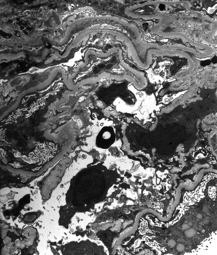
In the first week of hospitalization, his serum creatinine rose to 6.4 mg/dL. At this time, there was no fever and the CRP value declined to 0.77 mg/dL. Furthermore, repeated blood cultures showed no growth. In view of still progressive renal failure after elimination of infection, a diagnostic renal biopsy was performed. The specimen contained 17 glomeruli for light microscopic examination, of which 2 were globally sclerotic. The glomeruli showed mild increase in mesangial matrix and cellularity. No endocapillary proliferation was noted. Of the remaining 15 non-sclerotic glomeruli, 8 had cellular to fibrocellular crescents (). There were focal tubular atrophy and mild interstitial fibrosis. Focal mononuclear cell infiltration in the interstitium was observed. No granuloma or vasculitis was seen. Immunofluorescence studies revealed granular staining for IgA (2+), IgG (1+), IgM (1+), and C3 (2+) in the mesangium and along the glomerular capillary walls (). Electron microscopy showed electron-dense deposits in the mesangium (). No subepithelial hump-shaped deposition was found.
Figure 1. Glomerulus showing mesangial proliferation with cellular crescent formation (periodic acid–Schiff stain, ×400).
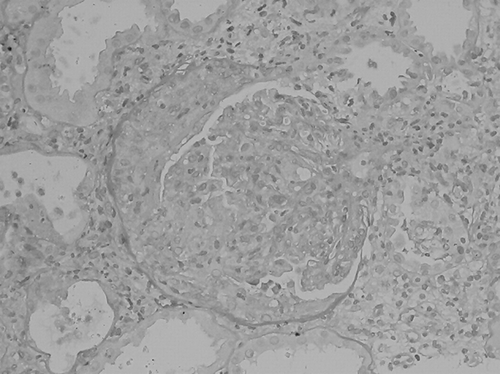
Figure 2. Immunofluorescence staining for IgA showing 2+ granular deposition in mesangium and glomerular capillary loops (original magnification ×400); there is also 2+ staining for C3 in a similar pattern (not shown).
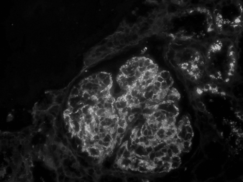
Figure 3. Electron microscopy showing electron-dense deposits in the mesangium (original magnification ×8000).
In addition to anti-MRSA therapy with vancomycin, the patient was treated with three doses of pulse methylprednisolone (500 mg/day), followed by oral prednisolone (60 mg/day). Thereafter, his renal function gradually improved. Oral prednisolone was gradually tapered in an outpatient clinic. At a follow-up approximately 2 months after renal biopsy, his serum creatinine was 1.9 mg/dL. Microscopic hematuria and mild proteinuria were still present.
DISCUSSION
Since staphylococcal infections were first identified as causal agents of glomerulonephritis by Powell in 1961,Citation6 two pathological patterns of glomerulonephritis associated with staphylococcal infection are well defined. Glomerulonephritis secondary to S. aureus infection, whether occurring after skin infection, deep-seated abscesses, or endocarditis, exhibits a histological pattern identical to acute poststreptococcal glomerulonephritis. Typical findings include diffuse endocapillary proliferative and exudative glomerulonephritis on light microscopy, C3-dominant deposits with or without IgG co-deposition on immunofluorescence, and subepithelial hump-shaped deposits on electron microscopy. In contrast, ventriculojugular shunt nephritis associated with S. epidermidis most commonly displays a pattern indistinguishable from type I membranoproliferative glomerulonephritis, with glomerular deposition of C3, IgG, and IgM.
In recent years, a third pattern of Staphylococcus-associated glomerulonephritis was described, which generally occurs in patients infected with MRSA. A distinguishing feature of this form of poststaphylococcal glomerulonephritis is IgA-dominant or IgA-codominant mesangial staining, resembling IgA nephropathy. In 1995, Koyama et al. first reported 10 cases of glomerulonephritis in patients infected with MRSA.Citation7 Clinical features included abrupt or insidious onset of rapidly progressive renal failure, hematuria, proteinuria, and, occasionally, purpura. Studies of these patients showed polyclonal increases in serum IgA and IgG, with high levels of circulating immune complexes and marked increased T-cell activation. Renal biopsies were performed in 6 of 10 patients; 5 had IgA-codominant deposits. The spectrum of glomerular histological findings included mesangial and/or endocapillary proliferative glomerulonephritis with varying degrees of crescent formation. They speculated that bacterial enterotoxins produced by MRSA may serve as superantigens, leading to T-cell activation and release of cytokines, which contribute to this type of glomerulonephritis.Citation8 Since then, there have been 34 cases of this entity reported in the English literature.Citation7–19 All patients were adults with a mean age of 59 years (range = 21–89 years). Twenty-six patients (76%) had MRSA, 5 (15%) had methicillin-sensitive S. aureus, and 3 (9%) had S. epidermidis. The clinical onset of infection varied from less than 1 week to 16 weeks (mean = 6.9 weeks) before the onset of renal manifestations. All biopsies in eight patients with underlying diabetes mellitus showed features of overlying diabetic glomerulosclerosis. Importantly, serum complements were depressed in only 12 patients (35%) and ultrastructurally characteristic subepithelial hump-shaped deposits were present in only 6 of 28 (21%) patients with available data.
However, a number of questions remain unanswered. The cause of the selective IgA deposition in patients with poststaphylococcal glomerulonephritis is unknown. In addition, one might argue that these patients may have preexisting IgA nephropathy, which is only incidentally discovered because of the superimposed postinfectious glomerulonephritis. As a result, it is important to differentiate IgA-dominant poststaphylococcal glomerulonephritis from primary IgA nephropathy. In our experience, in addition to hypocomplementemia and subepithelial hump-shaped deposits on electron microscopy characteristic of postinfectious glomerulonephritis, the findings that favor IgA-dominant poststaphylococcal glomerulonephritis over primary IgA nephropathy include intercurrent culture-documented staphylococcal infection, older age, acute renal failure at presentation, diffuse endocapillary proliferation with prominent neutrophil infiltration on light microscopy, and capillary loop deposits on immunofluorescence.Citation20
Glomerular crescent formation may complicate this new type of poststaphylococcal glomerulonephritis and there are nine cases of this entity reported in the literature.Citation9,Citation12,Citation15,Citation18,Citation19 The clinical characteristics of patients with at least 10% crescents on renal biopsy are summarized in . All patients were infected with MRSA. The origin of infection was variable. The mean value of serum creatinine at the time of renal biopsy was 2.6 mg/dL. The mean percentage of glomeruli affected by crescents was 35% (range = 10–67%). In addition to antibiotic treatment, immunosuppressive therapy was administrated in five patients. All patients got partial recovery of renal function, but two patients finally died of infectious complications.
Table 1. Clinical characteristics of patients with crescentic IgA nephropathy associated with staphylococcal infection described in the literature
In conclusion, staphylococcal infection has emerged as one of the leading causes of postinfectious glomerulonephritis in adults. A new type of poststaphylococcal glomerulonephritis has the distinct feature of containing IgA-dominant or IgA-codominant deposits, which generally occur in patients infected with MRSA. Crescent formation may complicate this type of glomerulonephritis. Although the prognosis of crescentic glomerulonephritis is generally thought to be poor, a review of the literature and our experience show a favorable renal prognosis. The natural history of infection-associated glomerulonephritis perhaps contributed to the favorable outcome. Although the limited evidence supports the use of immunosuppressive agents in this setting, serious consideration must be given to the fact that the renal disease might respond to antimicrobial therapy alone. Furthermore, the risk of overwhelming underlying infection precipitated by immunosuppression might abrogate the benefit for renal function. We suggest that immunosuppressive therapy should be an option only if the underlying MRSA infection has definitely been well controlled while the renal disease still progresses. Even in such instances, one must proceed with caution and simultaneous antibiotic treatment should be considered.
Declaration of interest: The authors report no conflicts of interest. The authors alone are responsible for the content and writing of the paper.
REFERENCES
- Montseny JJ, Meyrier A, Kleinknecht D, Callard P. The current spectrum of infectious glomerulonephritis: Experience with 76 patients and review of the literature. Medicine (Baltimore). 1995;74:63–73.
- Nasr SH, Markowitz GS, Stokes MB, Said SM, Valeri AM, D'Agati VD. Acute postinfectious glomerulonephritis in the modern era: Experience with 86 adults and review of the literature. Medicine (Baltimore). 2008;87:21–32.
- Wen YK. The spectrum of adult postinfectious glomerulonephritis in the new millennium. Ren Fail. 2009;31:676–682.
- Dobrin RS, Day NK, Quie PG, The role of complement, immunoglobulin and bacterial antigen in coagulase-negative staphylococcal shunt nephritis. Am J Med. 1975;59:660–673.
- Pertschuk DO, Vuletin JC, Sutton AL, Velazquez LA. Demonstration of antigen and immune complex in glomerulonephritis due to Staphylococcus aureus. Am J Clin Pathol. 1976;66:1027.
- Powell DE. Non-supportive lesions in staphylococcal septicemia. J Pathol Bacteriol. 1961;82:141–149.
- Koyama A, Kobayashi M, Yamaguchi N, Glomerulonephritis associated with MRSA infection: A possible role of bacterial superantigen. Kidney Int.1995;47:207–216.
- Yoh K, Kobayashi M, Hirayama A, A case of superantigen-related glomerulonephritis after methicillin-resistant Staphylococcus aureus (MRSA) infection. Clin Nephrol. 1997; 48: 311–316.
- Nagaba Y, Hiki Y, Aoyama T, Effective antibiotic treatment of methicillin-resistant Staphylococcus aureus-associated glomerulonephritis. Nephron. 2002;92:297–303.
- Nasr SH, Markowitz GS, Whelan JD, IgA-dominant acute post staphylococcal glomerulonephritis complicating diabetic nephropathy. Hum Pathol. 2003;34:1235–1241.
- Handa T, Ono T, Watanabe H, Takeda T, Muso E, Kita T. Glomerulonephritis induced by methicillin-sensitive Staphylococcus aureus infection. Clin Exp Nephrol. 2003;7:247–249.
- Kai H, Shimizu Y, Hagiwara M, Post-MRSA infection glomerulonephritis with marked Staphylococcus aureus cell envelope antigen deposition in glomeruli. J Nephrol. 2006; 19:215–219.
- Long JA, Cook WJ. IgA deposits and acute glomerulonephritis in a patient with staphylococcal infection. Am J Kidney Dis. 2006;48:851–855.
- Satoskar AA, Nadasdy G, Plaza JA, Staphylococcus infection-associated glomerulonephritis mimicking IgA nephropathy. Clin J Am Soc Nephrol. 2006;1:1179–1186.
- Hashimoto M, Nogaki F, Oida E, Glomerulonephritis induced by methicillin-resistant Staphylococcus aureus infection that progressed during puerperal period. Clin Exp Nephrol. 2007;11:92–96.
- Nasr SH, Share DS, Vargas MT, D'Agati VD, Markowitz GS. Acute post staphylococcal glomerulonephritis superimposed on diabetic glomerulosclerosis. Kidney Int. 2007;71:1317–1321.
- Okuyama S, Wakui H, Maki N, Successful treatment of post-MRSA infection glomerulonephritis with steroid therapy. Clin Nephrol. 2008;70:344–347.
- Zeledon JI, McKelvey RL, Servilla KS, Glomerulonephritis causing acute renal failure during the course of bacterial infections. Histological varieties, potential pathogenetic pathways and treatment. Int Urol Nephrol. 2008;40:461–470.
- Riley AM, Wall BM, Cooke CR. Favorable outcome after aggressive treatment of infection in a diabetic patient with MRSA-related IgA nephropathy. Am J Med Sci. 2009;337: 221–223.
- Wen YK, Chen ML. Discrimination between postinfectious IgA-dominant glomerulonephritis and idiopathic IgA nephropathy. Ren Fail. 2010;32:572–577.